Fc Engineering for Enhanced Antibody Therapies: A Comprehensive Guide to Optimizing Effector Functions
This article provides a detailed overview of Fc engineering strategies to modulate antibody effector functions for therapeutic applications.
Fc Engineering for Enhanced Antibody Therapies: A Comprehensive Guide to Optimizing Effector Functions
Abstract
This article provides a detailed overview of Fc engineering strategies to modulate antibody effector functions for therapeutic applications. It explores the fundamental biology of Fcγ receptors and complement, surveys cutting-edge methodologies for Fc domain modification, addresses common challenges in functional optimization, and compares the performance of next-generation Fc variants. Targeted at researchers and drug development professionals, this guide synthesizes current knowledge to inform the design of more potent and tailored biologic therapeutics.
The Fc Domain Decoded: Understanding the Core Mechanisms of Antibody Effector Functions
Within the broader thesis of Fc engineering to optimize antibody effector functions, understanding the core mechanisms of Fc-mediated activities is paramount. The fragment crystallizable (Fc) region of an antibody, particularly IgG, is the primary mediator of effector functions by engaging specific Fc gamma receptors (FcγRs) on immune cells or components of the complement system. These functions are critical for the therapeutic efficacy of monoclonal antibodies (mAbs) in oncology, infectious diseases, and autoimmunity. This Application Note details the three primary effector functions—Antibody-Dependent Cellular Cytotoxicity (ADCC), Antibody-Dependent Cellular Phagocytosis (ADCP), and Complement-Dependent Cytotoxicity (CDC)—providing current protocols and data analysis frameworks to support Fc engineering research.
Key Effector Functions: Mechanisms and Pathways
Antibody-Dependent Cellular Cytotoxicity (ADCC)
Mechanism: ADCC is mediated primarily by Natural Killer (NK) cells. The Fc region of a target-bound IgG antibody engages the activating FcγRIIIa (CD16a) on the NK cell surface. This cross-linking triggers intracellular signaling cascades leading to NK cell degranulation and the release of perforin and granzymes, inducing apoptosis in the target cell.
Antibody-Dependent Cellular Phagocytosis (ADCP)
Mechanism: ADCP is executed by professional phagocytes like macrophages, monocytes, and dendritic cells. Target-bound antibody Fc regions engage activating FcγRs (e.g., FcγRI, FcγRIIa, FcγRIIIa) on the phagocyte, promoting the engulfment and internalization of the antibody-opsonized target into a phagosome for destruction.
Complement-Dependent Cytotoxicity (CDC)
Mechanism: The Fc region of cell surface-bound antibodies (IgM or IgG1/3) recruits and activates the C1q protein, initiating the classical complement cascade. This leads to the formation of membrane attack complexes (MAC) that pore the target cell membrane, causing osmotic lysis.
Diagram Title: Core Signaling Pathways for ADCC, ADCP, and CDC
Quantitative Comparison of Effector Functions
Table 1: Comparative Overview of Fc-Mediated Effector Functions
| Feature | ADCC | ADCP | CDC |
|---|---|---|---|
| Primary Effector Cell | NK Cells | Macrophages, Monocytes, DCs | Complement Proteins (C1q→C9) |
| Key Fc Receptor | FcγRIIIa (CD16a) | FcγRI, FcγRIIa, FcγRIII | C1q (binds Fc, not an FcγR) |
| IgG Subclass Potency | IgG1 > IgG3 > IgG4 >> IgG2 | IgG1, IgG3 > IgG2, IgG4 | IgG1 > IgG3 > IgG2 >> IgG4 |
| Kinetics | Hours (2-24h) | Minutes to Hours (0.5-24h) | Minutes (0.5-2h) |
| Key Readout | % Target Cell Lysis (LDH, 51Cr) | % Phagocytosis (Flow Cytometry) | % Cytolysis (PI Uptake, LDH) |
| Primary Signaling Molecule | Syk/ZAP-70, ITAM | Syk, ITAM | C1r, C1s (Serine Proteases) |
| Engineered Fc Variants (Examples) | G236A/S239D/A330L (ADCC ↑) | S267E/L328F (FcγRIIb binding ↓, ADCP ↑) | E345R/E430G/S440Y (Hexamerization ↑, CDC ↑) |
Table 2: Common In Vitro Assay Parameters and Typical Results
| Assay Type | Effector:Target Ratio | Incubation Time | Common Positive Control | Typical Max Efficacy Range* |
|---|---|---|---|---|
| ADCC (NK Cell-Based) | 5:1 to 10:1 | 4 - 6 hours | Rituximab (anti-CD20) + CD20+ cells | 40-80% Specific Lysis |
| ADCP (Macrophage-Based) | 5:1 to 10:1 | 2 - 4 hours | Trastuzumab (anti-HER2) + HER2+ cells | 20-60% Phagocytic Index |
| CDC (Serum-Based) | N/A (Use 10-50% Serum) | 1 - 2 hours | Rituximab + CD20+ cells | 50-90% Specific Lysis |
*Ranges are highly dependent on target antigen density, cell line, and donor serum/cells.
Detailed Experimental Protocols
Protocol 4.1: ADCC Reporter Bioassay (Luminescence-Based)
This protocol uses engineered Jurkat T cells stably expressing FcγRIIIa (V158 high-affinity variant) and an NFAT-response element driving luciferase.
I. Materials & Reagent Preparation
- Target Cells: CHO-K1 or other adherent cells stably expressing target antigen.
- Effector Cells: Frozen ADCC Reporter Bioassay Effector Cells (e.g., Promega).
- Test Articles: Serial dilutions of IgG antibodies (engineering variants).
- Assay Medium: RPMI-1640 + 1% HI-FBS.
- Luminescent Substrate: Bio-Glo Luciferase Assay Reagent.
II. Procedure
- Day 0: Seed target cells in white-walled, clear-bottom 96-well tissue culture plates at 10,000 cells/well in 100 µL complete growth medium. Incubate overnight (37°C, 5% CO₂).
- Day 1:
- Prepare 3X serial dilutions of antibody test articles in assay medium (typically 9 points from 10 µg/mL).
- Thaw effector cells rapidly, wash once, and resuspend in assay medium to 1.5 x 10⁶ cells/mL.
- Remove medium from target cell plate.
- Add 50 µL of antibody dilution per well.
- Add 50 µL of effector cell suspension (75,000 cells) per well, achieving a 7.5:1 E:T ratio.
- Include controls: Background (target + effector), Target Max Lysis (target + lysis buffer), Effector Control (effector only).
- Incubate plate for 6 hours (37°C, 5% CO₂).
- Measurement: Equilibrate plate and Bio-Glo reagent to room temperature (RT) for 20 min. Add 75 µL of reagent per well. Incubate in dark for 5-10 min, then measure luminescence on a plate reader.
III. Data Analysis
- Calculate Relative Luminescence Units (RLU).
- % Specific Lysis = (RLU
Sample– RLUBackground) / (RLUTarget Max– RLUBackground) * 100. - Plot % Specific Lysis vs. antibody concentration to determine EC₅₀.
Protocol 4.2: Flow Cytometry-Based ADCP Assay
I. Materials
- Target Cells: Suspension cells (e.g., Raji B-cells) expressing target antigen.
- Effector Cells: THP-1 monocytes differentiated into macrophages with PMA, or primary monocyte-derived macrophages (MDMs).
- pHrodo BioParticles Conjugation Kit or pHrodo-labeled target cells (fluorescence increases in acidic phagosome).
- Flow Cytometry Buffer: PBS + 2% FBS + 1mM EDTA.
- Antibodies: Anti-human CD11b (for gating phagocytes).
II. Procedure
- Label Target Cells: Label target cells with pHrodo SE dye per manufacturer's protocol. Quench with complete medium, wash, and count.
- Opsonization: Incubate pHrodo-labeled target cells (2 x 10⁵ cells/mL) with test antibodies (1-10 µg/mL) for 30 min at 37°C. Wash twice.
- Phagocytosis: Co-culture opsonized target cells with differentiated macrophages at a 5:1 target:phagocyte ratio in a U-bottom 96-well plate. Centrifuge at 300 x g for 1 min to initiate contact. Incubate for 2-4 hours at 37°C.
- Stop & Stain: Place plate on ice. Wash cells with cold buffer. Resuspend in buffer containing anti-CD11b antibody and viability dye. Incubate 30 min at 4°C in the dark. Wash twice.
- Acquisition: Analyze by flow cytometry. Gate on live, single CD11b+ macrophages.
- Analysis: Measure the percentage of pHrodo+ macrophages (indicating phagocytosis) and the Median Fluorescence Intensity (MFI) of pHrodo within the positive population (phagocytic index).
Protocol 4.3: CDC Assay Using Propidium Iodide (PI) Uptake
I. Materials
- Target Cells: Adherent or suspension cells expressing high levels of target antigen (critical for CDC).
- Human Complement Serum: Pooled normal human serum (NHS). Heat-inactivated serum (56°C, 30 min) serves as negative control.
- Dilution Buffer: HBSS with Ca²⁺/Mg²⁺ + 0.1% BSA.
- Propidium Iodide (PI) Solution: 1 µg/mL in PBS.
- Lysis Buffer: 2% Triton X-100 (for Max Lysis control).
II. Procedure
- Seed target cells in 96-well plates (adherent: 20,000/well overnight; suspension: 50,000/well just before assay).
- Prepare serial dilutions of test antibody in dilution buffer.
- Remove medium from cells and add 50 µL antibody dilution per well. Incubate 15-30 min at RT.
- Prepare complement source: Add NHS to dilution buffer for a final concentration of 20-40% in the well.
- Add 50 µL of the NHS/buffer mix to each well (final NHS 10-20%). For Max Lysis, add 50 µL lysis buffer.
- Incubate plate for 60-90 min at 37°C.
- Add 20 µL of PI solution per well. Incubate 5-15 min at RT in dark.
- Read fluorescence immediately (Ex/Em ~535/617 nm). For suspension cells, centrifugation may be needed before reading.
- Data Analysis: Calculate % Specific Lysis = (Fluorescence
Sample– FluorescenceBackground) / (FluorescenceMax Lysis– FluorescenceBackground) * 100.
Diagram Title: Generalized Workflow for Fc Effector Function Assays
The Scientist's Toolkit: Research Reagent Solutions
Table 3: Essential Reagents for Fc Effector Function Research
| Reagent / Material | Primary Function in Research | Example Vendor/Product |
|---|---|---|
| FcγR Blocking Antibodies | To confirm FcγR-specificity in cellular assays by inhibiting receptor engagement. | BioLegend (anti-human CD16, CD32, CD64) |
| ADCC Reporter Bioassay Kits | Standardized, off-the-shelf kits for high-throughput, robust ADCC potency measurement without primary NK cells. | Promega (ADCC Reporter Bioassay, NFAT) |
| Recombinant Human FcγR Proteins | For surface plasmon resonance (SPR) or ELISA to measure binding affinity/kinetics of engineered Fc variants. | ACROBiosystems, Sino Biological |
| Pooled Normal Human Serum (NHS) | Source of active complement proteins for standardized CDC assays. | Complement Technology, Innovative Research |
| pHrodo Dyes (SE, BioParticles) | pH-sensitive fluorescent probes for quantitative, kinetic measurement of phagocytosis without quenching steps. | Thermo Fisher Scientific |
| Engineered Cell Lines | Stable antigen-expressing target cells or FcγR-expressing effector cells (e.g., Jurkat NFAT-luc CD16a) for consistent, defined assays. | ATCC, GenScript (gene editing services) |
| Glycoengineered Antibody Controls | Afucosylated IgG controls (e.g., produced in POTELLIGENT cells) as high-ADCC benchmark comparators. | Lonza (POTELLIGENT Platform) |
| Complement-Depleted Serum | Negative control for CDC assays to confirm complement-dependent mechanism. | Complement Technology (C1q-, C2-, etc.) |
| High-Affinity FcγRIIIa (V158) Mutant | Recombinant protein/cell line expressing the high-affinity allotype, critical for assessing clinical relevance. | Multiple vendors (R&D Systems, etc.) |
| Hexamerization-Enhancing Fc Mutants | Positive control antibodies (e.g., with E430G, E345R mutations) for CDC optimization studies. | Available through academic labs or custom protein expression. |
1. Introduction: The Role of FcγRs in Therapeutic Antibody Function Within the broader thesis of Fc engineering to optimize antibody effector functions, a detailed understanding of Fc Gamma Receptors (FcγRs) is paramount. These receptors, expressed on the surface of immune cells, are the critical mediators that transduce the Fc domain's "signal" into diverse biological outcomes, including antibody-dependent cellular cytotoxicity (ADCC), antibody-dependent cellular phagocytosis (ADCP), and modulation of inflammation. The net therapeutic effect of an antibody is dictated by the balance of activating (e.g., FcγRIIIa, FcγRI) and inhibitory (FcγRIIb) signals, which in turn is heavily influenced by the cell-type-specific expression profiles of these receptors. This application note provides a quantitative summary of human FcγR expression and detailed protocols for its experimental assessment.
2. Quantitative Overview of Human FcγR Expression Across Immune Cells The following tables consolidate current data on the expression patterns and key characteristics of human FcγRs.
Table 1: Human Fc Gamma Receptor Classes, Affinities, and Signaling
| Receptor | Gene | IgG Affinity (KD) | Signaling Motif | Primary Cell Expression | Key Function in Therapy |
|---|---|---|---|---|---|
| FcγRI | FCGR1A | ~10⁻⁸ - 10⁻⁹ M (high) | ITAM (via γ-chain) | Monocytes, Macrophages, DCs, IFNγ-activated Neutrophils | Phagocytosis, Antigen Presentation, Pro-inflammatory cytokine release. |
| FcγRIIa (H131) | FCGR2A | ~10⁻⁶ M (low) | ITAM (intracellular) | Monocytes, Macrophages, Neutrophils, Platelets, DCs | Phagocytosis, Respiratory burst, Platelet activation. |
| FcγRIIb | FCGR2B | ~10⁻⁶ M (low) | ITIM (intracellular) | B cells, Monocytes, Macrophages, Basophils, DCs | Inhibitory receptor; modulates activation thresholds, critical for IVIg effect. |
| FcγRIIIa (V158) | FCGR3A | ~10⁻⁶ M (low) | ITAM (via ζ/γ-chain) | NK cells, Monocytes, Macrophages, Subset of T cells | Principal mediator of ADCC by NK cells. |
| FcγRIIIb | FCGR3B | ~10⁻⁶ M (low) | GPI-anchor (non-signaling) | Neutrophils | Decoy receptor, aids in immune complex clearance, neutrophil activation. |
Table 2: Representative Surface Expression Levels (Antibodies Bound per Cell, ABC)
| Cell Type | FcγRI (CD64) | FcγRII (CD32) | FcγRIII (CD16) | Notes |
|---|---|---|---|---|
| Classical Monocyte | 20,000 - 40,000 | 10,000 - 20,000 (IIa) | 5,000 - 15,000 (IIIa) | High phagocytic potential. |
| NK Cell | Negligible | Negligible | 10,000 - 30,000 (IIIa) | Primary ADCC effector. |
| Neutrophil | Low (inducible) | 20,000 - 40,000 (IIa) | 100,000 - 200,000 (IIIb) | Dominated by FcγRIIIb. |
| B Cell | Negligible | 1,000 - 5,000 (IIb) | Negligible | Exclusively inhibitory FcγRIIb. |
| Macrophage (M1) | High | High (IIa) | Moderate (IIIa) | Pro-inflammatory phenotype. |
3. Experimental Protocols
Protocol 1: Multi-Parameter Flow Cytometry for FcγR Profiling in PBMCs Objective: To simultaneously quantify FcγR surface expression across defined immune cell subsets in human peripheral blood mononuclear cells (PBMCs). Materials: See "The Scientist's Toolkit" below. Procedure:
- PBMC Isolation: Isolate PBMCs from fresh human blood using density gradient centrifugation (e.g., Ficoll-Paque).
- Cell Staining: Resuspend 1x10⁶ PBMCs in 100 µL of FACS Buffer (PBS + 2% FBS).
- Add Human TruStain FcX (Fc receptor blocking reagent) and incubate for 10 minutes on ice.
- Add a pre-titrated antibody cocktail for 30 minutes on ice in the dark. A representative panel:
- Lineage/Discrimination: CD45 (BV510), CD3 (FITC) for T cells, CD19 (FITC) for B cells, CD56 (APC-Cy7) for NK cells, CD14 (PerCP-Cy5.5) for monocytes.
- FcγRs: CD64 (FcγRI, PE), CD32 (FcγRII, PE-Cy7), CD16 (FcγRIII, APC).
- Include fluorescence-minus-one (FMO) and isotype controls.
- Wash & Acquisition: Wash cells twice with 2 mL FACS Buffer, resuspend in 300 µL, and acquire data on a flow cytometer capable of detecting 8+ colors (e.g., 3-laser BD Fortessa).
- Gating & Analysis:
- Gate on single, live CD45⁺ lymphocytes or monocytes.
- Subset gating: T cells (CD3⁺), B cells (CD19⁺), NK cells (CD56⁺ CD3⁻), Monocytes (CD14⁺).
- Analyze median fluorescence intensity (MFI) or calculate Antibodies Bound per Cell (ABC) using quantification beads for each FcγR within subsets.
Protocol 2: FcγR-Specific Cellular Binding Assay (SPR or Cell-Based) Objective: To measure the kinetic and affinity parameters of an engineered antibody variant for specific recombinant or cell-expressed FcγRs. Materials: Biacore T200/8K SPR system or plate-based flow cytometer, recombinant human FcγR proteins, Fc-engineered IgG samples. SPR Procedure:
- Surface Preparation: Immobilize a monoclonal anti-human Fc antibody (~10,000 RU) on a CM5 sensor chip using amine coupling to capture IgG variants uniformly.
- Binding Analysis: Dilute purified FcγR ectodomains (FcγRI, FcγRIIa/b, FcγRIIIa-158V/F) in HBS-EP+ buffer.
- Cycle: Inject IgG variant (1 µg/mL) for 60s to achieve capture (~200 RU), followed by FcγR analyte injection (0.78-100 nM) for 120s association, then dissociation for 300s. Regenerate with 10 mM Glycine pH 1.5.
- Data Processing: Double-reference sensograms (subtract blank flow cell and buffer injection). Fit data to a 1:1 Langmuir binding model to derive ka, kd, and KD.
4. Visualizations
Diagram 1: FcγR Signaling Pathways in Effector Functions
Diagram 2: FcγR Profiling Experimental Workflow
5. The Scientist's Toolkit: Key Research Reagent Solutions
| Reagent/Material | Function & Application |
|---|---|
| Recombinant Human FcγR Proteins (monomeric) | Used in surface plasmon resonance (SPR) or ELISA to measure binding affinity/kinetics of antibody variants in a cell-free system. |
| Fc-Blocking Reagent (e.g., Human TruStain FcX) | Blocks non-specific, Fc-mediated binding of staining antibodies to FcγRs on immune cells, critical for clean flow cytometry data. |
| Fluorophore-Conjugated Anti-FcγR Antibodies (clone-specific) | Essential for detecting surface expression of specific receptors (CD64, CD32, CD16) in multi-parameter flow cytometry. |
| Quantitative Bead Standard (e.g., QIFIKIT) | Enables conversion of flow cytometry Median Fluorescence Intensity (MFI) to absolute Antibody Binding Capacity (ABC) for cross-experiment comparison. |
| FcγR-Expressing Reporter Cell Lines (e.g., NFAT-luciferase) | Engineered cells providing a functional readout (luminescence) upon FcγR cross-linking and signaling, used for high-throughput screening of Fc variants. |
| Allele-Specific Reagents (e.g., anti-FcγRIIIa-V158/F158) | Tools to distinguish between functionally distinct genetic polymorphisms, crucial for stratified analysis in research and development. |
The classical complement pathway, initiated by the binding of the C1 complex (C1q-C1r2-C1s2) to antibody-antigen immune complexes, is a critical effector mechanism for therapeutic antibodies. In Fc engineering, modulating C1q affinity is a primary strategy to enhance or fine-tune Complement-Dependent Cytotoxicity (CDC). This application note details the molecular basis of C1q binding and provides protocols for its quantitative assessment in antibody development pipelines.
Key Quantitative Data on C1q Binding & Activation
Table 1: Binding Affinities (KD) of Human IgG Subclasses to C1q
| IgG Subclass | Approximate KD for C1q (M)* | Relative CDC Potency | Key Fc Residue Influencing Binding |
|---|---|---|---|
| IgG1 | 1-3 x 10^-7 | High (Reference) | E318, K320, K322 |
| IgG2 | Very weak (>10^-5) | Negligible | V318, G320, G322 |
| IgG3 | 0.5-1 x 10^-7 | Very High | Same as IgG1, longer hinge |
| IgG4 | Very weak (>10^-5) | Negligible | F318, R/R/S at 320/322/331 |
Note: Affinities are for hexamerized IgG/immune complexes, not monomeric IgG.
Table 2: Engineered Fc Variants with Altered C1q Binding
| Variant Name | Amino Acid Modifications (EU numbering) | Reported Effect on C1q Binding (vs IgG1) | Impact on CDC |
|---|---|---|---|
| E345K | E345K | ~10-fold increase | Enhanced |
| E430G | E430G | ~3-fold increase | Enhanced |
| S267E/H268F | S267E, H268F | Significant increase | Enhanced |
| K322A | K322A | Abolished | Abolished |
| G236A/S239D | G236A, S239D (2xAA) | Promotes hexamerization; Enhanced | Greatly Enhanced |
Experimental Protocols
Protocol 3.1: Surface Plasmon Resonance (SPR) for C1q-Antibody Binding Kinetics
Objective: Determine the kinetic parameters (KD, ka, kd) of C1q binding to immobilized immune complexes. Key Reagents:
- Human purified C1q protein.
- Anti-human Fab antibody (e.g., Goat F(ab')2 anti-human IgG F(ab')2).
- Running Buffer: HBS-EP+ (10mM HEPES, 150mM NaCl, 3mM EDTA, 0.05% v/v Surfactant P20, pH 7.4). Procedure:
- Ligand Immobilization: Dilute the anti-human Fab antibody to 10 µg/mL in 10 mM sodium acetate (pH 4.5). Inject over a CMS sensor chip using amine coupling to achieve ~5000 RU.
- Immune Complex Capture: Inject the monoclonal antibody of interest (10 µg/mL) for 60s at 10 µL/min to capture a consistent level (~100 RU) via its Fab region.
- Analyte Binding: Inject a 2-fold dilution series of C1q (e.g., 0.5 – 32 µg/mL) at a flow rate of 30 µL/min for 180s association, followed by 600s dissociation.
- Regeneration: Regenerate the surface with two 30s pulses of 10 mM Glycine-HCl (pH 1.5).
- Data Analysis: Double-reference data (buffer blank & reference flow cell). Fit to a 1:1 binding model. Note: Report as apparent affinity due to avidity effects.
Protocol 3.2: CDC Functional Assay Using Luminescent Readout
Objective: Quantify complement-mediated killing of target cells by an antibody. Key Reagents:
- Target cells expressing antigen of interest.
- Human complement serum (pooled normal human serum, or C1q-depleted/reconstituted serum for specificity).
- Cell viability reagent (e.g., luminescent ATP detection assay). Procedure:
- Seed target cells in a 96-well white plate at 5 x 10^3 cells/well in 50 µL complete medium.
- Add 50 µL of serially diluted antibody (in triplicate) and incubate for 15-30 minutes at room temperature.
- Add 20 µL of human complement serum diluted in CDC buffer (e.g., RPMI with 1% BSA) to a final concentration of 5-20%. Include controls: No Antibody + Complement (background), No Complement (max viability).
- Incubate plate for 1-2 hours at 37°C in 5% CO2.
- Equilibrate plate to room temperature, add 80 µL of luminescent viability substrate, mix, and read luminescence.
- Data Analysis: Calculate % Cytotoxicity = 100 x [1 - (RLUsample - RLUblank)/(RLUnoab - RLU_blank)]. Determine EC50 or maximum lysis.
Visualizations
Diagram 1: Classical Complement Pathway Activation (94 chars)
Diagram 2: Fc Engineering Workflow for CDC (74 chars)
The Scientist's Toolkit: Research Reagent Solutions
Table 3: Essential Reagents for C1q/Complement Research
| Item | Function & Rationale | Example Supplier/Product |
|---|---|---|
| Human C1q Protein | Purified ligand for direct binding studies (SPR, ELISA). Essential for measuring intrinsic affinity/avidity. | Complement Technology, Inc.; Merck. |
| C1q-Depleted Human Serum | Validates C1q-specific effects in functional assays. Reconstitution with purified C1q confirms mechanism. | Complement Technology, Inc. |
| Normal Human Serum (NHS) | Source of intact complement for functional CDC assays. Must be batch-tested for activity. | Commercial donors; BioreclamationIVT. |
| Anti-human CH2 Domain mAb | Detects IgG in complex formation assays. Some clones are C1q-binding sensitive (conformational). | e.g., Mouse anti-human IgG (clone #). |
| SPR Sensor Chips (CM5/CM4) | Gold standard for label-free kinetics. Anti-Fab capture method mimics immune complex presentation. | Cytiva. |
| Luminescent Viability Assay | High-sensitivity, ATP-based readout for CDC. Superior signal-to-noise over colorimetric (LDH, MTT). | Promega (CellTiter-Glo). |
| Fc Gamma R Blocking Antibody | Controls for specificity in CDC; blocks ADCC/ADCP confounding effects, isolating complement lysis. | e.g., anti-CD16/32. |
| C1q Binding ELISA Kit | Semi-quantitative, high-throughput screen for C1q-Fc interaction of antibody variants. | Various commercial kits. |
Application Notes
Within the broader thesis on Fc engineering for optimizing antibody effector functions, this document details the structural and biophysical principles governing the interaction between the antibody Fragment crystallizable (Fc) region and Fc gamma receptors (FcγRs). The affinity and specificity of this interaction directly dictate critical immune effector functions such as Antibody-Dependent Cellular Cytotoxicity (ADCC), Antibody-Dependent Cellular Phagocytosis (ADCP), and modulation of inflammation. A precise understanding of glycosylation patterns and binding site architecture is fundamental for rational Fc engineering strategies aimed at enhancing therapeutic efficacy, tuning immune activation, or creating silent Fc backbones.
Key Structural and Quantitative Data
Table 1: Affinity of Human IgG Subclasses for Human FcγRs
| FcγR | IgG1 KD (nM)* | IgG2 KD (nM)* | IgG3 KD (nM)* | IgG4 KD (nM)* | Primary Binding Site on Fc | Key Residues |
|---|---|---|---|---|---|---|
| FcγRI (CD64) | 1-10 | >1000 | 1-10 | 10-100 | Lower Hinge/CH2 | L234, L235, G236, D265, N297 |
| FcγRIIa (H131) | 100-1000 | ~5000 | 100-1000 | 1000-5000 | Lower Hinge/CH2 | L234, L235, P331, I332 |
| FcγRIIb (I232) | 2000-10000 | >10000 | 1000-5000 | >10000 | Lower Hinge/CH2 | L234, L235, P331, I332 |
| FcγRIIIa (V158) | 50-200 | >5000 | 20-100 | >5000 | Lower Hinge/CH2 | F241, V264, D265, N297, E269, A327, P329 |
| FcγRIIIb (NA1) | 500-2000 | >5000 | 200-1000 | >5000 | Lower Hinge/CH2 | F241, D265, N297 |
KD values are approximate ranges from surface plasmon resonance (SPR) studies and can vary based on glycosylation and experimental conditions. *N297 is the canonical glycosylation site.
Table 2: Impact of Fc Glycan Composition on FcγRIIIa Binding
| Glycoform | Core Fucosylation | Terminal Galactose (G2F vs G0F) | Bisecting GlcNAc | Sialylation | Relative ADCC Activity (vs G0F) |
|---|---|---|---|---|---|
| G0F | Yes | 0 | No | No | 1.0 (Baseline) |
| G2F | Yes | 2 | No | No | ~1.0 - 1.2 |
| G0 | No | 0 | No | No | ~10 - 50x increase |
| G0 + Bisecting | No | 0 | Yes | No | ~10 - 100x increase |
| Sialylated (G2FS2) | Yes | 2 | No | Yes (α2,6) | ~0.1 - 0.5 (Anti-inflammatory) |
Research Reagent Solutions Toolkit
Table 3: Essential Materials for Fc-FcγR Interaction Studies
| Item | Function/Application | Example/Notes |
|---|---|---|
| Recombinant Human FcγRs (extracellular domains) | Binding partners for SPR, BLI, or ELISA. Crucial for affinity measurements. | His-tagged or biotinylated monomers or dimers. |
| Glycoengineered Antibody Panels | To study the specific effect of glycan structures (afucosylated, sialylated, etc.) on binding and function. | Produced in CHO, HEK, or engineered cell lines (e.g., FUT8 KO). |
| Surface Plasmon Resonance (SPR) Chip (e.g., CMS, SA) | Immobilization surface for kinetic analysis (KD, ka, kd). | Protein A/G for capturing IgG; Streptavidin for capturing biotinylated FcγR. |
| Biolayer Interferometry (BLI) Biosensors (e.g., Anti-Human Fc, Streptavidin) | Alternative label-free kinetic analysis platform. | For rapid screening of Fc variant libraries. |
| ADCC/ADCP Reporter Bioassays | Functional cell-based readouts for engineered Fc variants. | Use of engineered effector cells (e.g., Jurkat NFAT-luc with FcγR) for standardized measurement. |
| Crystallization Screening Kits | For determining high-resolution co-crystal structures of Fc:FcγR complexes. | Commercial sparse matrix screens. |
| PNGase F | Enzyme to completely remove N-linked glycans for aglycosylated Fc control experiments. | |
| EndoS / EndoS2 | Glycosidase that cleaves Fc glycans with specificity; useful for probing glycan accessibility. | |
| Fc Engineering Mutant Libraries (e.g., Site-directed mutagenesis kits) | To generate specific point mutations at key binding residues (L234A, L235A, etc.). |
Experimental Protocols
Protocol 1: Surface Plasmon Resonance (SPR) for Fc-FcγR Binding Kinetics
Objective: Quantify the binding affinity (KD) and kinetics (ka, kd) between an IgG Fc variant and a recombinant human FcγR.
Materials:
- SPR instrument (e.g., Biacore, Sierra SPR)
- Series S Sensor Chip CMS
- Running Buffer: HBS-EP+ (10 mM HEPES, 150 mM NaCl, 3 mM EDTA, 0.05% v/v Surfactant P20, pH 7.4)
- Recombinant Protein A or G
- Recombinant human FcγR (extracellular domain, biotinylated or as a Fc-fusion)
- Glycoengineered IgG samples (analytes)
Procedure:
- System Setup: Prime the SPR instrument with running buffer.
- Ligand Immobilization (Capture Method):
- Activate the CM5 chip surface with a 7-minute injection of a 1:1 mixture of 0.4 M EDC and 0.1 M NHS.
- Inject recombinant Protein A (50 µg/mL in 10 mM sodium acetate, pH 4.5) over the flow cell to achieve a capture level of ~5000-8000 Response Units (RU).
- Deactivate excess reactive groups with a 7-minute injection of 1 M ethanolamine-HCl, pH 8.5.
- Analyte Binding Assay:
- Dilute IgG samples (ligand) in running buffer (e.g., 0.5-200 nM range for a high-affinity interaction like IgG1:FcγRI).
- For each cycle: a. Capture: Inject the IgG over the Protein A surface for 60 seconds to achieve a consistent capture level (~100-200 RU). b. Association: Inject the FcγR (analyte) at a series of concentrations (e.g., 1.56, 3.125, 6.25, 12.5, 25, 50 nM) for 120-180 seconds. c. Dissociation: Monitor dissociation in running buffer for 300-600 seconds. d. Regeneration: Regenerate the Protein A surface with two 30-second pulses of 10 mM Glycine, pH 1.5, to remove bound IgG and FcγR without damaging Protein A.
- Data Analysis:
- Subtract the reference flow cell signal and buffer blank injections.
- Fit the resulting sensograms to a 1:1 Langmuir binding model using the instrument's software to calculate ka (association rate constant), kd (dissociation rate constant), and KD (kd/ka).
Protocol 2: Cell-Based ADCC Reporter Bioassay
Objective: Functionally assess the impact of Fc engineering or glycosylation on FcγRIIIa signaling and effector cell activation.
Materials:
- ADCC Reporter Bioassay Kit (e.g., Promega, BioLegend) or components: Engineered Jurkat cells stably expressing FcγRIIIa (V158 or F158) and an NFAT-response element driving luciferase.
- Target cells expressing the antigen for the test antibody.
- Glycoengineered or Fc-mutated test antibodies.
- White-walled, clear-bottom 96-well tissue culture plates.
- Luciferase detection reagent.
- Luminometer.
Procedure:
- Plate Target Cells: Harvest and count target cells. Seed them in the assay plate at 10,000 cells per well in 75 µL of growth medium. Incubate overnight.
- Antibody Dilution: Prepare 3- or 10-fold serial dilutions of the test antibodies in medium in a separate plate.
- Add Antibody and Effector Cells: Add 25 µL of each antibody dilution to the target cell plate. Then, add 100 µL of ADCC Reporter Effector Cells (resuspended to the recommended density, e.g., 75,000 cells/well) to each well. Final effector:target ratio is typically 7.5:1. Include controls (target+effector cells only, target+antibody only, etc.).
- Incubation: Incubate the plate at 37°C, 5% CO2 for 6-24 hours (as optimized).
- Signal Detection: Equilibrate plate and Bio-Glo Luciferase Assay Reagent to room temperature. Add 100 µL of reagent to each well. Mix briefly on an orbital shaker and incubate for 5-10 minutes to stabilize the luminescent signal.
- Measurement: Read luminescence on a plate-reading luminometer.
- Data Analysis: Plot luminescence (Relative Light Units, RLU) vs. antibody concentration. Calculate EC50 values using a 4-parameter logistic curve fit to compare the potency of different Fc variants.
Visualizations
Title: Determinants of Fc-FcγR Binding and Signaling
Title: SPR Protocol Workflow for Fc-FcγR Kinetics
Title: Core Fucose Impact on FcγRIIIa Affinity and ADCC
1. Introduction and Clinical Relevance Within the thesis on Fc engineering to optimize antibody effector functions, a critical translational component is understanding the impact of natural genetic variation in human Fc gamma receptors (FcγRs). These receptors, expressed on immune cells, are the primary mediators of antibody-dependent cellular cytotoxicity (ADCC), antibody-dependent cellular phagocytosis (ADCP), and antibody-dependent neutrophil phagocytosis (ADNP). Single nucleotide polymorphisms (SNPs) and copy number variations (CNVs) in FCGR genes lead to differential binding affinities for IgG Fc domains, resulting in substantial inter-individual variability in therapeutic antibody efficacy and safety. This document provides application notes and standardized protocols for characterizing these allelic forms in preclinical and clinical research.
2. Key Allelic Variants: Quantitative Data Summary Table 1: High-Impact Human FcγR Polymorphisms Affecting IgG1 Binding and Clinical Outcomes
| Receptor | Gene | Key Allele/SNP | Amino Acid Change | Effect on IgG1 Affinity | Associated Clinical Response (Example) |
|---|---|---|---|---|---|
| FcγRIIIA (CD16A) | FCGR3A | V158F (rs396991) | Valine → Phenylalanine at 158 | V/V: High > V/F: Intermediate > F/F: Low | Enhanced efficacy of rituximab (NHL), trastuzumab (HER2+ BC) in V carriers. |
| FcγRIIA (CD32A) | FCGR2A | H131R (rs1801274) | Histidine → Arginine at 131 | H/H: High for IgG1/IgG2 > H/R: Intermediate > R/R: Low | H allele linked to better response to IVIG, mAbs requiring phagocytosis. |
| FcγRIIIB (CD16B) | FCGR3B | NA1/NA2 (rs447536, rs448740) | Multiple differences | NA1: Higher affinity than NA2 | NA1 allele and CNV linked to autoimmune disease risk and mAb neutropenia. |
| FcγRIIB (CD32B) | FCGR2B | I232T (rs1050501) | Isoleucine → Threonine in transmembrane | Alters inhibitory signaling potency | T allele associated with SLE; impacts ITIM-dependent therapeutic window. |
Table 2: Genotype Frequency Distribution in Major Populations (Approximate %)
| Genotype | European | Asian | African |
|---|---|---|---|
| FCGR3A V/V | ~10-15% | ~5-10% | ~20-25% |
| FCGR3A F/F | ~40-45% | ~50-55% | ~20-25% |
| FCGR2A H/H | ~25% | ~40-45% | ~35% |
| FCGR2A R/R | ~20% | ~10-15% | ~15-20% |
3. Core Experimental Protocols
Protocol 3.1: Genotyping of FCGR Polymorphisms via TaqMan qPCR Application: Determine SNP genotypes (e.g., FCGR3A V158F, FCGR2A H131R) from human genomic DNA. Reagents: Genomic DNA (10-20 ng/µL), TaqMan Genotyping Master Mix, validated TaqMan SNP Genotyping Assay (FAM/VIC probes), nuclease-free water. Procedure:
- Prepare reaction mix per sample: 5.0 µL Master Mix, 0.5 µL 20X TaqMan Assay, 3.5 µL nuclease-free water.
- Aliquot 9 µL of mix into a 96-well PCR plate. Add 1 µL of genomic DNA per well. Include no-template controls (NTC).
- Seal plate and centrifuge briefly. Run on a real-time PCR system using standard TaqMan genotyping cycling conditions.
- Analyze endpoint fluorescence (FAM vs. VIC) using the instrument's allelic discrimination software to assign genotypes (V/V, V/F, F/F).
Protocol 3.2: Functional Assessment of FcγR Variants via ADCC Reporter Bioassay Application: Quantify the impact of FcγR allelic variation on effector function in a standardized, cell-based system. Reagents: Engineered ADCC Reporter Bioassay cells (stably expressing either FcγRIIIA-V158 or -F158), target cells expressing target antigen, therapeutic antibody serially diluted, assay medium, luciferase detection substrate. Procedure:
- Seed target cells in a white-walled 96-well plate at 10,000 cells/well in 75 µL assay medium. Incubate overnight.
- Prepare 3X serial dilutions of the test antibody in a separate plate.
- Harvest ADCC effector reporter cells (select V158 or F158 variant line), resuspend in assay medium at 0.5-1x10^6 cells/mL.
- Add 25 µL of antibody dilution to target cells, followed by 50 µL of effector cell suspension (effector:target ratio ~5:1). Include antibody-only and effector cell-only controls.
- Incubate plate at 37°C, 5% CO2 for 6-24 hours.
- Equilibrate Bio-Glo Luciferase Assay Substrate, add 75 µL to each well. Shake, incubate 5-10 minutes, measure luminescence. Calculate EC50 values for each FcγR variant.
Protocol 3.3: Surface Plasmon Resonance (SPR) for Affinity Measurement Application: Directly measure kinetic binding parameters (Ka, Kd, KD) of IgG variants to recombinant soluble FcγR allelic proteins. Reagents: CMS Series S Sensor Chip, recombinant human FcγR (e.g., FcγRIIIA-V158, -F158), anti-His antibody (for capture), HBS-EP+ running buffer, therapeutic IgG as analyte. Procedure:
- Dock sensor chip and prime system with HBS-EP+ buffer.
- Using amine coupling, immobilize anti-His antibody to a reference and sample flow cell.
- Dilute His-tagged soluble FcγR in running buffer. Capture the receptor on the sample flow cell to a consistent RU level (~50-100 RU). Use the reference flow cell for background subtraction.
- Prepare a 2-fold serial dilution series of the IgG analyte (e.g., 100 nM to 0.78 nM).
- Inject analyte concentrations over reference and sample flow cells at 30 µL/min for 180s association, followed by 600s dissociation.
- Regenerate surface with 10 mM Glycine pH 1.5. Fit double-referenced sensorgrams to a 1:1 Langmuir binding model to determine kinetics.
4. Visualization of Concepts and Workflows
Title: Genetic Variants Impact Therapeutic Antibody Response
Title: Workflow for Characterizing FcγR Allelic Impact
5. The Scientist's Toolkit: Research Reagent Solutions Table 3: Essential Reagents for FcγR Variant Research
| Reagent/Material | Function/Application | Example Supplier/Format |
|---|---|---|
| Recombinant Human FcγR Proteins (Allelic Forms) | Essential for in vitro binding studies (SPR, ELISA), blocking assays, and standardization. | R&D Systems, Sino Biological; His-tagged or Fc-fused monomers. |
| Genotyping Assays (TaqMan, rhAmp) | Accurate, high-throughput SNP determination from low-input genomic DNA. | Thermo Fisher (TaqMan), IDT (rhAmp SNP); pre-validated for FCGR loci. |
| ADCC Reporter Bioassay Kits (Isogenic Variant Cells) | Standardized, reproducible functional assessment without primary cells. | Promega (FcγRIIIA V158 & F158 effector cells). |
| FcγR-Specific Monoclonal Antibodies (for Flow Cytometry) | Quantify receptor surface expression on primary immune cell subsets. | BioLegend (clone 3G8 for CD16), BD Biosciences (clone 2E1 for CD32A). |
| Reference Therapeutic Antibodies (Rituximab, Trastuzumab) | Positive controls for functional assays and binding studies. | Commercial clinical-grade formulations. |
| Next-Generation Sequencing Panels (Immunogenetics) | Comprehensive variant detection across all FCGR genes, including CNVs. | Illumina TruSight, custom hybrid-capture panels. |
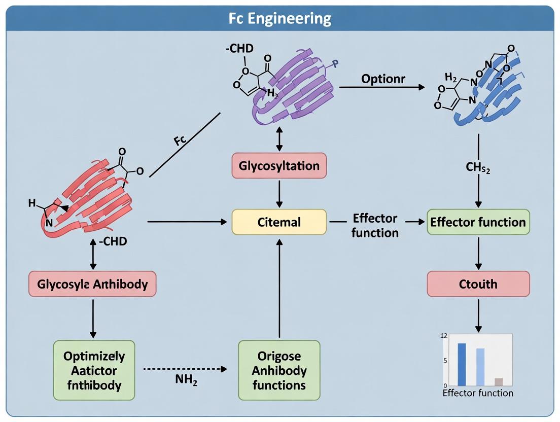
Engineering the Fc Domain: Cutting-Edge Techniques and Therapeutic Applications
Application Notes: Strategic Mutagenesis for Fc Function Optimization
Within the broader thesis of Fc engineering for optimized antibody therapeutics, site-directed mutagenesis at specific hotspots in the IgG constant region (Fc) is a fundamental strategy to fine-tune effector functions such as Antibody-Dependent Cellular Cytotoxicity (ADCC), Antibody-Dependent Cellular Phagocytosis (ADCP), and Complement-Dependent Cytotoxicity (CDC). These functions are mediated by interactions with Fc gamma receptors (FcγRs) and the C1q complement protein. The goal is to design next-generation antibodies with enhanced potency for oncology or reduced effector function for inflammatory applications.
Key Functional Hotspots:
- Lower Hinge Region (e.g., L234, L235, G236, P331): Critical for FcγR and C1q binding. Mutations here (e.g., L234A/L235A, "LALA" variant) dramatically reduce ADCC/CDC.
- FcγR Interface (e.g., S239, I332, E333): Targeted for enhanced affinity to activating FcγRIIIa (F158/V158 variants). The S239D/I332E/A330L ("DELL") variant significantly boosts ADCC.
- Glycosylation Site (N297): The conserved N-linked glycan is essential for FcγR binding. Aglycosylated mutations (N297Q/A) ablate effector function, while engineered glycoforms enhance ADCC.
- C1q Binding Site (e.g., K326, E333, K322): Residues influencing CDC. Mutations like E333S/K322A can selectively reduce CDC while sparing ADCC.
Table 1: Quantitative Impact of Key Fc Mutations on Receptor Affinity and Effector Function
| Mutation/Hotspot | Target Receptor/Function | Key Change (vs. Wild-Type) | Primary Application |
|---|---|---|---|
| L234A/L235A (LALA) | FcγRI/II/III, C1q | ~1000-fold reduction in FcγR binding; ablated ADCC/CDC | Anti-inflammatory, block effector function |
| G236A/S239D/I332E (GASDALIE) | FcγRIIIa | ~400-fold increased affinity for FcγRIIIa-V158; enhanced ADCC | Oncology, enhanced cytotoxic activity |
| S239D/I332E/A330L (DELL) | FcγRIIIa | ~2 orders of magnitude increased affinity; potent ADCC/ADCP | Oncology, enhanced macrophage phagocytosis |
| N297Q/A | All FcγRs | Abolishes FcγR binding; no ADCC/ADCP/CDC | Anti-inflammatory, pure blocking/signaling |
| E333S/K322A | C1q (CDC) | Selective reduction in CDC; modest impact on FcγR | Applications requiring ADCC without CDC |
| F243L/R292P/Y300L/P396L (Variant 18) | FcγRn (pH-dependent) | Enhanced half-life (~2-3x increase in mice) | All applications, improved pharmacokinetics |
Protocols for Fc Mutagenesis and Functional Validation
Protocol 1: Site-Directed Mutagenesis of Fc Region in IgG Expression Vector Objective: Introduce specific point mutations into the CH2 domain of an IgG1 antibody expression plasmid. Materials: Wild-type IgG1 heavy chain plasmid, high-fidelity DNA polymerase (e.g., PfuUltra II), DpnI restriction enzyme, competent E. coli, mutagenic primers. Procedure:
- Design complementary primers (25-45 bases) containing the desired mutation in the center with ~15 bp flanking sequences.
- Set up a PCR reaction (50 µL): 10-50 ng plasmid template, 125 ng of each primer, 1x reaction buffer, 200 µM dNTPs, 1 µL PfuUltra II polymerase.
- Cycle: 95°C 2 min; 18 cycles of [95°C 30s, 55-60°C 1min, 68°C 2 min/kb]; 68°C 10 min.
- Cool reaction to 37°C and add 1 µL DpnI (10 U) directly to digest methylated parental template. Incubate 1 hour.
- Transform 2-5 µL into competent E. coli, plate on selective agar, and incubate overnight.
- Pick colonies for sequencing to confirm the mutation.
Protocol 2: Production and Purification of Mutant IgG Antibodies Objective: Express and purify mutant antibodies from mammalian cells for functional testing. Materials: Expi293F or CHO cells, PEI transfection reagent, heavy and light chain plasmids, Protein A affinity resin, dialysis/PBS buffer. Procedure:
- Co-transfect exponentially growing Expi293F cells with mutant heavy chain and corresponding light chain plasmids using PEI.
- Harvest cell culture supernatant 5-7 days post-transfection by centrifugation.
- Filter supernatant and load onto a Protein A column. Wash with 10 column volumes (CV) of PBS.
- Elute IgG with 5 CV of low-pH elution buffer (e.g., 0.1 M Glycine, pH 2.7-3.0) and immediately neutralize with 1/10 volume 1 M Tris-HCl, pH 9.0.
- Dialyze into PBS, determine concentration (A280), and assess purity by SDS-PAGE.
Protocol 3: Surface Plasmon Resonance (SPR) for FcγRIIIa Binding Affinity Objective: Quantify binding kinetics (KD) of mutant IgGs to human FcγRIIIa (V158). Materials: Biacore/SPR instrument, CMS chip, anti-human Fc capture antibody, mutant IgG samples, recombinant FcγRIIIa. Procedure:
- Immobilize anti-human Fc antibody on a CMS chip via amine coupling to ~5000 RU.
- Dilute mutant IgGs to 2 µg/mL in HBS-EP+ buffer. Inject for 60s to capture a consistent level (~50 RU).
- Inject a 2-fold dilution series of FcγRIIIa (e.g., 200 nM to 3.125 nM) at 30 µL/min for 120s association, followed by 300s dissociation.
- Regenerate the surface with two 30s pulses of 10 mM Glycine, pH 1.5.
- Process data: double-reference subtraction, fit to a 1:1 Langmuir binding model to determine ka, kd, and KD.
Protocol 4: In Vitro ADCC Reporter Bioassay Objective: Measure the potency of mutant antibodies to elicit ADCC. Materials: ADCC Reporter Bioassay Kit (e.g., Promega), target cells expressing antigen, mutant IgG antibodies. Procedure:
- Harvest and count target cells. Seed at 10,000 cells/well in a white-walled 96-well plate.
- Prepare 3-fold serial dilutions of mutant antibodies in assay buffer.
- Thaw ADCC effector cells (FcγRIIIa NFAT-luciferase Jurkat cells), resuspend, and add to wells (effector:target ratio = 6:1).
- Incubate plate at 37°C, 5% CO2 for 6 hours.
- Add Bio-Glo Luciferase reagent, incubate 5-20 min, and measure luminescence. Plot dose-response curves and calculate EC50 values.
Visualizations
Title: Functional Outcomes of Mutagenesis at Key Fc Hotspots
Title: Workflow for Fc Mutagenesis and Functional Assays
The Scientist's Toolkit: Key Research Reagents and Materials
Table 2: Essential Reagents for Fc Engineering Studies
| Item | Function in Research | Example/Note |
|---|---|---|
| High-Fidelity DNA Polymerase | Introduces point mutations with minimal error rates during PCR. | PfuUltra II, KAPA HiFi. Critical for accurate SDM. |
| Competent E. coli Cells | For plasmid propagation after mutagenesis. | High-efficiency strains (e.g., NEB 5-alpha, Stbl3). |
| Mammalian Expression System | Produces properly folded, glycosylated IgG for testing. | Expi293F cells, Freestyle 293, CHO cells. |
| Polyethylenimine (PEI) | Cost-effective transfection reagent for mammalian cells. | Linear PEI, MW 25,000. |
| Protein A Affinity Resin | Standard capture and purification of IgG from culture supernatant. | Agarose or magnetic bead formats. |
| Recombinant FcγRs | For binding affinity and kinetics measurement (SPR, ELISA). | FcγRI, FcγRIIa/b, FcγRIIIa (V158/F158). |
| ADCC Reporter Bioassay Kit | Standardized, cell-based assay to measure ADCC potency. | Promega, BioLegend. Uses engineered Jurkat effector cells. |
| Surface Plasmon Resonance (SPR) Instrument | Gold-standard for label-free, real-time kinetics analysis. | Biacore 8K/S200, Nicoya OpenSPR. |
| Anti-Human Fc Capture Antibody | For immobilizing IgGs on SPR chips in consistent orientation. | Mouse anti-human IgG Fc, recombinantly produced. |
Within the broader thesis of Fc engineering for optimizing antibody effector functions, glycoengineering of the Fc N-linked glycan at asparagine 297 (N297) represents a critical, clinically validated strategy. Afucosylation, the intentional reduction or elimination of core fucose from this glycan, enhances antibody-dependent cellular cytotoxicity (ADCC) by up to 100-fold. This effect is achieved through significantly increased affinity for FcγRIIIa (CD16a) on natural killer (NK) cells and macrophages, thereby potentiating the antitumor efficacy of therapeutic monoclonal antibodies (mAbs). This application note details current strategies and protocols for generating afucosylated antibodies.
Key Afucosylation Strategies: Mechanisms and Quantitative Outcomes
Table 1: Comparison of Primary Glycoengineering Strategies for Afucosylated Antibody Production
| Strategy | Mechanism of Action | Typical Afucosylation Level Achieved | Relative ADCC Potency Increase (vs. Fucosylated) | Key Advantages | Key Challenges |
|---|---|---|---|---|---|
| FX-KO Cell Line Engineering | Genetic knockout of the FUT8 gene encoding α-1,6-fucosyltransferase. | >95% | 50-100x | Stable, consistent production; no process changes. | Potential for clonal variation; need for new cell line development. |
| GDP-6-Deoxy-D-lyxo-4-hexulose Reductase (GDR) Knock-In | Competitive inhibition of the GDP-fucose biosynthesis pathway by overexpressing GDR. | 85-99% | 30-80x | High efficiency; can be combined with other knockouts. | Metabolic burden on host cell. |
| Potentiation with Small Molecule Inhibitors | Addition of fucosylation inhibitors (e.g., 2F-Peracetyl-fucose) to culture media. | 70-95% | 20-50x | Applicable to standard CHO cells; flexible. | Cost, potential cytotoxicity, removal from final product. |
| Fucosyltransferase (FUT8) mRNA Silencing | siRNA or shRNA-mediated knockdown of FUT8 expression. | 60-90% | 10-40x | Tunable level of knockdown. | Transient effect; requires co-transfection. |
| Glycosyltransferase Overexpression (GnTIII) | Overexpression of β-1,4-N-acetylglucosaminyltransferase III to add bisecting GlcNAc. | 50-80% (with reduced fucose) | 10-30x | Also increases serum half-life. | Can create glycan heterogeneity. |
Protocols
Protocol 1: Generation of a StableFUT8Knockout CHO-S Cell Line Using CRISPR-Cas9
Objective: To create a clonal host cell line deficient in α-1,6-fucosyltransferase for stable production of afucosylated antibodies.
Materials (Research Reagent Solutions Toolkit):
- CHO-S Cells: Chinese Hamster Ovary suspension cells, adapted for serum-free culture.
- CRISPR-Cas9 Ribonucleoprotein (RNP) Complex: Composed of recombinant Cas9 protein and synthetic gRNA targeting the FUT8 gene exon.
- Electroporation Buffer: Optimized, low-conductivity buffer for CHO cell transfection.
- Nucleofector/Electroporator: Device for delivering RNP into cells.
- Cloning Dilution Media: Selective media with antibiotics (e.g., Puromycin) or lacking key metabolites for selection.
- 96-Well Limiting Dilution Plates: For single-cell cloning.
- Glycan Analysis Buffer Set: For rapid extraction and labeling of N-glycans.
- Liquid Chromatography-Mass Spectrometry (LC-MS) System: For confirmatory glycan structural analysis.
Procedure:
- Design & Complex Formation: Design a gRNA targeting an early exon of the CHO FUT8 gene. Complex 10 µg of purified Cas9 protein with 5 µg of gRNA to form the RNP complex at room temperature for 10 minutes.
- Cell Preparation: Harvest 1x10^6 logarithmically growing CHO-S cells by centrifugation. Wash once with PBS and resuspend in 100 µL of pre-warmed electroporation buffer.
- Electroporation: Mix the cell suspension with the RNP complex. Transfer to a certified electroporation cuvette. Electroporate using a pre-optimized CHO-specific pulse code (e.g., "CM-137" on a Nucleofector 2b).
- Recovery & Selection: Immediately add 500 µL of pre-warmed culture media. Transfer cells to a 6-well plate with 2.5 mL media. After 48 hours, apply appropriate selection pressure (e.g., puromycin at 5 µg/mL) for 7-10 days.
- Single-Cell Cloning: Perform limiting dilution to 0.5 cells/well in a 96-well plate. Monitor for single colonies. Expand positive clones.
- Screening & Validation:
- Perform PCR on genomic DNA from expanded clones to detect indel mutations at the target site.
- Transiently transfect top clones with an IgG expression vector.
- After 7 days, purify antibodies via Protein A chromatography.
- Analyze glycan composition using HILIC-UPLC or LC-MS to confirm >95% afucosylation.
Protocol 2: Production of Afucosylated Antibodies Using a Commercially Available FUT8 Inhibitor
Objective: To produce afucosylated antibodies from standard CHO cells by adding a fucosylation inhibitor to the bioreactor.
Materials (Research Reagent Solutions Toolkit):
- Standard CHO DG44 or CHO-K1 Cell Line: Expressing the mAb of interest.
- 2F-Peracetyl-Fucose (2F-PAF): Cell-permeable small molecule inhibitor of cellular fucosylation.
- Fed-Batch Bioreactor System: Controlled for pH, DO, and temperature.
- Protein A Affinity Resin: For high-purity mAb capture from harvested cell culture fluid (HCCF).
- Titer Measurement Kit: e.g., Protein A HPLC or SoloVPE system.
Procedure:
- Inoculum and Bioreactor Setup: Expand mAb-expressing CHO cells in a seed train. Inoculate a 5L bioreactor at a viable cell density (VCD) of 0.5 x 10^6 cells/mL in proprietary chemically defined media.
- Inhibitor Addition: At the time of inoculation (Day 0), add 2F-PAF from a 100 mM DMSO stock solution to a final concentration of 100 µM in the bioreactor. Maintain DMSO concentration below 0.1% (v/v).
- Fed-Batch Process: Execute a standard 14-day fed-batch process with glucose and feed additions based on metabolite analysis. Monitor VCD and viability daily.
- Harvest: On Day 14, when viability drops below 70%, separate cells from HCCF by centrifugation and 0.22 µm filtration.
- Purification & Analysis:
- Load clarified HCCF onto a Protein A column. Wash with PBS, elute with low-pH buffer (e.g., 0.1 M Glycine-HCl, pH 3.0), and immediately neutralize.
- Determine mAb titer and yield.
- Analyze the afucosylation level via HILIC-UPLC of released, 2-AB-labeled glycans. Expect 70-95% afucosylation depending on cell line and process conditions.
Visualizations
Title: CRISPR-Cas9 Workflow for Generating FUT8-KO CHO Cell Line
Title: Enhanced ADCC Pathway via Afucosylated Antibody Binding to FcγRIIIa
Title: Three Primary Glycoengineering Strategy Categories
Within the broader thesis on Fc engineering to optimize antibody effector functions, a central challenge is moving beyond broad effector activation to achieve precise immune cell targeting. Selective FcγR affinity engineering enables the development of therapeutic antibodies with tailored activities—enhancing cytotoxicity for oncology or minimizing inflammation for autoimmunity—by discriminating between activating (e.g., FcγRI, FcγRIIa, FcγRIIIa) and inhibitory (FcγRIIb) receptors. This application note details the rationale, key data, and protocols for designing and characterizing such variants.
Table 1: Binding Affinity (KD, nM) of IgG1 Fc Variants for Human FcγRs.
| Fc Variant (Example) | FcγRI (CD64) | FcγRIIa-H131 | FcγRIIa-R131 | FcγRIIb | FcγRIIIa-V158 | FcγRIIIa-F158 | Primary Design Goal |
|---|---|---|---|---|---|---|---|
| Wild-type IgG1 | 10-50 | 1000-5000 | >5000 | 500-2000 | 200-500 | 1000-3000 | Baseline |
| S267E/L328F | ~200 | <100 | ~500 | <100 | ~50 | ~200 | Enhance IIa/IIb, reduce IIIa |
| G236A/I332E | >1000 | ~50 | ~100 | ~20 | <10 | ~30 | Enhance IIb/IIIa, reduce I |
| S239D/I332E/A330L | >1000 | ~5 | ~10 | ~2 | <2 | <5 | Potent enhancement of IIa/IIb/IIIa |
| V12/V13 (FcγRIIb selective) | >10000 | >10000 | >10000 | ~100 | >10000 | >10000 | Exclusive FcγRIIb binding |
Detailed Experimental Protocols
Protocol 1: In Silico Design and Molecular Modeling of Fc Variants
Objective: To rationally design Fc point mutations for selective FcγR binding using computational tools. Materials: Fc-FcγR co-crystal structures (PDB IDs: 1E4K, 3RY6), modeling software (PyMOL, Rosetta, MOE). Procedure:
- Obtain Fc/FcγR complex structures from the Protein Data Bank.
- Analyze the binding interface to identify contact residues. Key regions include the lower hinge (234-239),
FGloop (327-332), andBCloop (residues 265-269). - Use computational alanine scanning or free energy perturbation to predict the impact of point mutations on binding energy for each FcγR.
- Design mutations (e.g., charged residues for electrostatic steering, bulky residues for steric exclusion) to stabilize or destabilize specific interactions.
- Perform structural minimization and molecular dynamics (MD) simulation (50-100 ns) to assess the stability of the engineered Fc-FcγR complex.
- Select top variant designs for gene synthesis.
Protocol 2: Expression and Purification of Fc Variants
Objective: To produce high-purity Fc variant proteins for biophysical and cellular assays. Materials: Expi293F cells, ExpiFectamine 293 transfection kit, mammalian expression vector (e.g., pTT5 or pcDNA3.4), Protein A affinity resin, ÄKTA pure or FPLC system. Procedure:
- Clone synthesized Fc variant genes (as IgG1, Fab-fused, or Fc-fusion constructs) into the expression vector.
- Transfect Expi293F cells according to the manufacturer's protocol (e.g., 1 µg DNA per mL cells, 1:3 DNA:ExpiFectamine ratio).
- Incubate at 37°C, 8% CO₂, 125 rpm for 5-7 days. Supplement with Enhancers as per protocol.
- Harvest supernatant by centrifugation (4,000 x g, 20 min) and filtration (0.22 µm).
- Load onto a Protein A column pre-equilibrated with PBS. Wash with 10 column volumes (CV) of PBS.
- Elute with 0.1 M glycine, pH 3.0, and immediately neutralize with 1 M Tris, pH 8.5.
- Dialyze into PBS or HBS-EP buffer. Determine concentration by A280 and assess purity by SDS-PAGE (>95%).
Protocol 3: Surface Plasmon Resonance (SPR) Analysis of FcγR Binding
Objective: To quantitatively measure the binding kinetics (KD, Ka, Kd) and affinity of Fc variants for each human FcγR. Materials: Biacore T200 or 8K series, CMS sensor chip, recombinant human FcγRs (R&D Systems), HBS-EP+ buffer, amine coupling kit. Procedure:
- Dilute Fc variant (ligand) to 10 µg/mL in 10 mM sodium acetate, pH 4.5. Immobilize on a CMS chip via amine coupling to achieve ~500-1000 RU.
- Use a reference flow cell activated and blocked without ligand.
- Dilute analytes (FcγRs) in HBS-EP+ in a 2-fold dilution series (e.g., 200 nM to 1.56 nM).
- Run kinetics experiments at 25°C with a flow rate of 30 µL/min. Use a contact time of 120 s and dissociation time of 300 s.
- Regenerate the surface with 10 mM glycine, pH 1.5, for 30 s.
- Double-reference the sensorgrams (reference cell & buffer blank).
- Fit data to a 1:1 Langmuir binding model using the Biacore evaluation software. Report KD, kon (Ka), and koff (Kd).
Protocol 4: Cell-Based ADCC Reporter Bioassay
Objective: To functionally assess the enhancement or reduction of Antibody-Dependent Cellular Cytotoxicity (ADCC) potency via FcγRIIIa signaling. Materials: ADCC Reporter Bioassay Kit (Promega), target cells expressing relevant antigen, Fc variant antibody (as full IgG), white-walled 96-well plates. Procedure:
- Harvest and count target cells. Adjust concentration to 1e5 cells/mL in assay medium.
- Prepare 4X serial dilutions of the Fc variant antibody in a separate plate.
- Thaw ADCC Reporter Effector cells, wash once, and resuspend at 1e6 cells/mL.
- Combine 25 µL of target cells, 25 µL of antibody dilution, and 25 µL of effector cells per well (Effector:Target ratio = 5:1). Include antibody-only, effector-only, and target-only controls.
- Incubate plate at 37°C, 5% CO₂ for 6 hours.
- Equilibrate Bio-Glo Luciferase Reagent for 30 min at room temperature. Add 75 µL to each well.
- Incubate for 5-10 min and measure luminescence on a plate reader.
- Calculate fold induction over background and plot dose-response curves to determine EC50 values.
Pathway & Workflow Visualizations
Fc Variant Selective Signaling Pathways
Fc Variant Characterization Workflow
The Scientist's Toolkit: Key Research Reagent Solutions
Table 2: Essential Materials for FcγR Affinity Engineering Studies
| Item | Function / Relevance | Example Supplier / Catalog |
|---|---|---|
| Recombinant Human FcγRs (FcγRI, IIa-H/R, IIb, IIIa-V/F) | Essential analytes for SPR and ELISA to measure direct binding affinity and selectivity. | R&D Systems, Sino Biological |
| Surface Plasmon Resonance (SPR) System | Gold-standard for label-free, quantitative kinetics (KD, kon, koff) of Fc-FcγR interactions. | Cytiva (Biacore), Sartorius (Octet) |
| ADCC Reporter Bioassay Kit | Standardized, consistent cell-based assay to measure FcγRIIIa signaling potency without primary NK cells. | Promega |
| Expi293 Expression System | High-yield mammalian expression system for producing mg/mL quantities of Fc variant antibodies. | Thermo Fisher Scientific |
| Protein A Affinity Resin | Standard capture step for purifying IgG Fc variants from culture supernatant. | Cytiva (MabSelect), Thermo Fisher |
| Site-Directed Mutagenesis Kit | For rapid generation of Fc point mutations in expression vectors. | Agilent (QuikChange), NEB |
| FcγR-Expressing Cell Lines (e.g., NFAT reporter lines) | Cellular systems for functional screening of variant activity on specific receptors. | InvivoGen |
| Analytical Size-Exclusion Chromatography (SEC) | Critical for assessing aggregation state and stability of engineered variants post-purification. | Waters, Agilent |
Within the broader thesis of Fc engineering to optimize antibody effector functions, this application note details how specific Fc modifications are strategically deployed across three major disease areas. The goal is to maximize therapeutic efficacy by selectively engaging or disengaging immune effector mechanisms—such as Antibody-Dependent Cellular Cytotoxicity (ADCC), Antibody-Dependent Cellular Phagocytosis (ADCP), Complement-Dependent Cytotoxicity (CDC), and modulation of inflammation—tailored to the unique pathophysiology of each indication.
Core Principles of Fc Tailoring by Disease
The Fc region of an IgG antibody interacts with various Fc gamma receptors (FcγRs) on immune cells and with complement protein C1q. The affinity and selectivity of these interactions dictate the elicited effector functions. Engineering involves amino acid substitutions that modulate these interactions.
Key Engineering Strategies:
- Enhanced Effector Function: Increasing affinity for activating FcγRs (e.g., FcγRIIIa on NK cells) to boost ADCC/ADCP against pathogens or tumor cells.
- Reduced Effector Function: Decreasing affinity for all FcγRs to minimize cell depletion and inflammation, suitable for blocking pathways or receptor agonism.
- Selective Effector Function: Skewing affinity towards specific receptors (e.g., FcγRIIb for inhibitory signaling or FcγRIIa for macrophage engagement).
- Enhanced Complement Activation: Increasing C1q binding to potentiate CDC.
Table 1: Fc Engineering Strategies by Disease Indication
| Disease Indication | Primary Goal | Key Effector Functions | Example Fc Modifications | Clinical-Stage Example |
|---|---|---|---|---|
| Cancer | Target cell killing, Immune activation | ADCC, ADCP, CDC | S298A/E333A/K334A (AAA), G236A/S239D/I332E (ADE), F243L/R292P/Y300L/V305I/P396L (LS) | Obinutuzumab (GA101; glycoengineered for enhanced ADCC) |
| Autoimmunity | Blockade without cell depletion, Anti-inflammatory | Reduced ADCC/ADCP/CDC, Increased FcγRIIb engagement | N297A/Q (aglycosyl), L234A/L235A (LALA), G237A/P238A/P271G/A330R (TM), S267E/L328F (EF) | Ravulizumab (C5 inhibitor; engineered for prolonged half-life) |
| Infectious Diseases | Viral/bacterial neutralization, Pathogen clearance | ADCC, ADCP, CDC, Enhanced half-life | M428L/N434S (LS), YTE (M252Y/S254T/T256E), G236A/I332E (GE) | Motavizumab (anti-RSV; YTE for half-life extension) |
Table 2: Quantitative Impact of Common Fc Variants on Binding Affinities
| Fc Variant Name | Key Mutation(s) | FcγRIIIa (V158) Binding (Fold Δ vs WT)* | FcγRIIb Binding (Fold Δ vs WT)* | C1q Binding (Fold Δ vs WT)* | Primary Functional Outcome |
|---|---|---|---|---|---|
| AF (Aglucosyl) | N297Q | ~0 | ~0 | ~0 | Ablated effector function |
| LALA-PG | L234A/L235A/P329G | ~0 | ~0.1 | ~0 | Severely reduced effector function |
| ADE | G236A/S239D/I332E | >100x ↑ | ~10x ↑ | ~5x ↑ | Potently enhanced ADCC/ADCP |
| LS | M428L/N434S | ~1x | ~1x | ~1x | ~4x Serum half-life extension |
| TM | G237A/P238A/P271G/A330R | ~0 | ~10x ↑ | ~0 | Selective FcγRIIb engagement |
Note: Fold changes are approximate, derived from published biophysical and cell-based assays. WT = Wild-Type IgG1.
Detailed Experimental Protocols
Protocol 1: In Vitro ADCC Reporter Bioassay for Cancer Antibody Screening
Purpose: To quantitatively measure the NK cell activation potential of Fc-engineered antibodies against cancer cell targets.
Materials:
- Fc variant antibodies (purified)
- Target cancer cell line (e.g., SK-BR-3 for HER2)
- ADCC Reporter Bioassay Kit (e.g., Promega, Catalog # G7010)
- White-walled, clear-bottom 96-well tissue culture plates
- Luminometer
Procedure:
- Day 1: Plate Target Cells. Harvest and count target cells. Plate 10,000 cells per well in 75 μL of complete growth medium. Incubate overnight at 37°C, 5% CO₂.
- Day 2: Antibody Serial Dilution & Assay Assembly. Prepare a 3-fold serial dilution of each Fc-variant antibody in assay buffer, starting at 10 μg/mL (11 concentrations recommended).
- Remove the target cell plate from the incubator. Add 25 μL of each antibody dilution to designated wells (in triplicate). Include a no-antibody control (buffer only) and a maximum lysis control (e.g., lysis buffer from kit).
- Thaw ADCC Effector Cells (engineered Jurkat cells expressing FcγRIIIa and NFAT-response element driving luciferase). Wash cells once and resuspend at 1.0 x 10⁶ cells/mL in assay buffer.
- Add 75 μL of effector cell suspension (75,000 cells) to each well, resulting in a 7.5:1 Effector:Target ratio. Centrifuge plates briefly (200 x g, 1 min) to initiate cell contact.
- Incubate plate for 6 hours at 37°C, 5% CO₂.
- Luciferase Measurement: Equilibrate Bio-Glo Luciferase Assay Reagent to room temperature for 30 min. Add 75 μL of reagent to each well. Incubate in the dark for 5-15 minutes. Measure luminescence on a plate-reading luminometer.
- Analysis: Calculate Relative Luminescence Units (RLU). Plot RLU vs. antibody concentration (log scale) and determine the EC₅₀ value for each Fc variant using 4-parameter logistic curve fitting.
Protocol 2: In Vivo PK/PD Study for Half-Life Extended Anti-Infective Antibodies
Purpose: To evaluate the serum persistence and antiviral efficacy of Fc-engineered antibodies (e.g., with LS or YTE mutations) in a mouse model.
Materials:
- Fc variant antibodies (LS, YTE, WT)
- Human FcRn transgenic mouse model (e.g., B6.Cg-Fcgrt
Tg(FCGRT)32Dcr/DcrJ) - Virus challenge stock (e.g., RSV)
- ELISA kits for antibody quantitation (anti-human IgG) and viral load
- Microtainer tubes for serum collection
Procedure:
- Antibody Administration: Randomly group mice (n=6-8 per group). Administer a single intravenous (IV) or intraperitoneal (IP) dose of each Fc-variant antibody (5 mg/kg) in a volume of 100-200 μL PBS. Include a PBS vehicle control group.
- Serial Blood Collection: At pre-defined time points post-dose (e.g., 5 min, 6h, Day 1, 3, 7, 14, 21, 28), collect ~50 μL of blood via submandibular or retro-orbital bleed into serum separator tubes. Process to obtain serum. Store at -80°C.
- Antibody PK Analysis: Quantify human antibody concentration in each serum sample using a standardized sandwich ELISA (e.g., anti-human IgG Fc capture, anti-human IgG F(ab')₂-HRP detection). Generate standard curves using the known administered antibodies.
- Pharmacokinetic Modeling: Plot serum concentration vs. time for each variant. Use non-compartmental analysis (NCA) software (e.g., Phoenix WinNonlin) to calculate key PK parameters: Terminal half-life (t₁/₂), Area Under the Curve (AUC), and Clearance (CL).
- Integrated Efficacy Challenge (Optional): In a separate cohort, pre-treat mice with antibodies 24 hours prior to intranasal challenge with a lethal dose of virus. Monitor weight loss, survival, and at sacrifice, quantify viral load in lung homogenates by plaque assay or qPCR. Correlate protection with antibody exposure (AUC).
Visualizations
Title: Fc Engineering Logic for Disease-Specific Effector Functions
Title: ADCC Reporter Bioassay Workflow
The Scientist's Toolkit: Key Research Reagent Solutions
| Item / Reagent | Vendor Examples (Catalog #) | Function in Fc Effector Research |
|---|---|---|
| FcγR Binding Assay Kits (SPR/BLI) | Cytiva (28958351), ForteBio (18-5100) | Measure kinetic binding (KD, Kon, Koff) of antibodies to recombinant human FcγRs. |
| ADCC Reporter Bioassay Kits | Promega (G7010), Thermo Fisher (K1245) | Standardized, cell-based assay using engineered Jurkat cells to quantify NK cell activation. |
| Complement C1q Binding ELISA | Hycult Biotech (HK336), Abcam (ab125966) | Quantify antibody's ability to bind C1q and initiate the classical complement pathway. |
| Human FcRn (hFcRn) Binding Assay | Bio-Techne (ADP2-100), ACROBiosystems (FCM-H82W5) | Assess pH-dependent binding for predicting serum half-life extension. |
| Primary Human Immune Cells (NK, Macrophages) | STEMCELL Tech (70036, 70037), Lonza (4W-210, 4W-250) | For primary cell-based functional assays (e.g., real-time ADCC, phagocytosis). |
| Fc Engineering Cloning & Mutagenesis Kits | Agilent (200523), NEB (E0554S) | Site-directed mutagenesis to introduce specific Fc point mutations into expression vectors. |
| Recombinant Human FcγR Proteins | Sino Biological (10185-H08H), R&D Systems (4325-FC) | Critical reagents for biophysical binding studies and cell assay validation. |
| Glycoengineering Cell Lines (e.g., FUT8 KO CHO) | Lonza (GS Xceed), ATCC (CRL-12445) | Produce antibodies with defined, homogenous glycoforms (e.g., afucosylated for enhanced ADCC). |
1. Introduction & Thesis Context Within the broader thesis investigating Fc engineering to optimize antibody effector functions, this document serves as a critical application note. It synthesizes real-world case studies of therapeutics with engineered Fc regions, providing comparative data and reproducible protocols. The core thesis posits that strategic modulation of FcγR affinity and complement activation is paramount for tailoring therapeutic activity—enhancing efficacy in oncology or autoimmunity while mitigating toxicity. These case studies validate that hypothesis through clinical translation.
2. Approved Fc-Engineered Therapeutics: Data Summary Table 1: Approved Monoclonal Antibodies with Engineered Fc Domains
| Therapeutic (Brand) | Indication(s) | Fc Modification (IgG Subtype) | Primary Engineering Goal | Key Effector Function Outcome |
|---|---|---|---|---|
| Mogamulizumab (Poteligeo) | CTCL, ATLL | Defucosylated (IgG1) | Enhanced ADCC | ~100-fold increased affinity for FcγRIIIa (CD16A); potent NK-cell mediated cytotoxicity. |
| Obinutuzumab (Gazyva) | CLL, NHL | Glycoengineered (Type II, IgG1) | Enhanced ADCC, Direct Cell Death | Increased affinity for FcγRIIIa; reduced CDC via altered binding geometry. |
| Ravulizumab (Ultomiris) | PNH, aHUS | 4-amino acid substitution (IgG2/4 hybrid) | Extended Half-life | ~4x longer terminal half-life (≈50 days) vs. eculizumab via enhanced pH-dependent FcRn recycling. |
| Dupyriumab (Dupixent) | Atopic Dermatitis, Asthma | Engineered to reduce effector functions (IgG4) | Minimized ADCC/CDC | S228P hinge stabilization prevents Fab-arm exchange; minimal engagement of FcγR and C1q. |
3. Clinical-Stage Case Study: A Novel Fc-Engineered Bispecific Therapeutic: REGN5458 (Linvoseltamab) – A BCMAxCD3 bispecific antibody with Fc silencing. Thesis Relevance: Demonstrates the application of Fc engineering not to enhance, but to silence effector functions, thereby directing mechanism of action exclusively to T-cell engagement and reducing cytokine release syndrome (CRS) potential. Key Data from Phase 1/2 Trials (RRMM patients):
- Overall Response Rate (ORR): 71% at 200 mg dose.
- Complete Response (CR) rate: 39%.
- Incidence of Grade ≥3 CRS: <5%. Fc Engineering: Proprietary "Fc-silencing" mutations (e.g., L234A/L235A, or L235E) in the IgG4 backbone to eliminate FcγR and C1q binding.
4. Experimental Protocols for Effector Function Analysis Protocol 4.1: In Vitro ADCC Reporter Bioassay
- Purpose: Quantify NK cell activation mediated by Fc-engineered antibody bound to target cells.
- Reagents: Engineered antibody; Target cells expressing antigen of interest; FcγRIIIa (CD16A) NFAT-luciferase Jurkat reporter cells; Assay medium; Luciferase substrate.
- Procedure:
- Seed target cells in white-walled 96-well plates.
- Serially dilute the Fc-engineered antibody and reference control, add to target cells. Incubate 30 min.
- Add FcγRIIIa reporter cells at an effector-to-target (E:T) ratio of 5:1.
- Incubate plate for 6 hours at 37°C, 5% CO₂.
- Add luciferase substrate, incubate 10 minutes, measure luminescence.
- Analysis: Plot RLU vs. antibody concentration. Calculate EC₅₀ values for potency comparison.
Protocol 4.2: Surface Plasmon Resonance (SPR) for FcγR Affinity Measurement
- Purpose: Determine kinetic binding parameters (KD, ka, kd) of engineered Fc to human FcγRs.
- Reagents: Biotinylated FcγRs (e.g., FcγRI, FcγRIIa/b, FcγRIIIa-V158/F158); Engineered antibody (captured via anti-Fab chip) or purified Fc fragment; HBS-EP+ buffer; Streptavidin (SA) sensor chip.
- Procedure (Capture Method):
- Immobilize anti-human Fab antibody on a CMS chip via amine coupling.
- Capture the engineered mAb (or control) onto the anti-Fab surface.
- Inject a concentration series of each biotinylated FcγR over the captured mAb surface.
- Use a 1:1 Langmuir binding model to calculate kinetics from the sensograms.
5. The Scientist's Toolkit: Research Reagent Solutions Table 2: Essential Materials for Fc Effector Function Research
| Item / Reagent | Function & Application |
|---|---|
| Recombinant Human FcγRs (Biotinylated/His-tagged) | For SPR, ELISA, or cell-binding studies to quantify affinity changes due to engineering. |
| ADCC/ADCC Reporter Bioassay Kits | Standardized systems (e.g., Promega, BioLegend) using engineered Jurkat cells for high-throughput, reproducible ADCC quantitation. |
| Glycoengineered Antibody Controls | Commercially available defucosylated (e.g., FUT8 KO) or afucosylated reference antibodies for assay calibration. |
| FcRn Binding ELISA or SPR Kit | To assess pharmacokinetic impact of half-life extending Fc mutations under pH-dependent conditions (pH 6.0 vs 7.4). |
| C1q Binding ELISA Kit | To quantitatively compare complement activation potential of engineered variants. |
| Human PBMCs or Primary NK Cells | Primary cells for physiologically relevant ex vivo ADCC or phagocytosis assays. |
6. Visualizations
Fc Engineering Goals & Applications
Enhanced ADCC Signaling Pathway
Navigating Fc Engineering Challenges: From Off-Target Effects to Functional Tuning
Within the broader thesis on Fc engineering to optimize antibody effector functions, a central challenge is the precise modulation of immune activation. This document provides application notes and protocols for evaluating engineered antibodies, focusing on the critical balance between achieving potent therapeutic efficacy (e.g., via antibody-dependent cellular cytotoxicity (ADCC) or phagocytosis (ADCP)) and minimizing adverse events like cytokine release syndrome (CRS) and general toxicity.
The following tables summarize critical parameters from recent studies on Fc-engineered antibodies, highlighting the trade-offs between effector function and safety profiles.
Table 1: Comparative Effector Function of Fc Variants
| Fc Variant (Example) | Target Antigen | ADCC Potency (Relative to WT) | ADCP Potency (Relative to WT) | CDC Potency (Relative to WT) | Key Reference |
|---|---|---|---|---|---|
| WT IgG1 | CD20 | 1.0x | 1.0x | 1.0x | 1 |
| S239D/I332E (SDIE) | CD20 | ~100x | ~10x | Reduced | 1, 2 |
| G236A/I332E (GA) | CD20 | ~50x | ~5x | Abrogated | 2 |
| F243L/R292P/Y300L | CD20 | ~0.5x | ~0.3x | Abrogated | 3 |
| L234F/L235E/P331S (LES) | EGFR | ~0.1x | ~0.1x | Abrogated | 4 |
References: 1. Lazar et al. (2006) PNAS. 2. Horton et al. (2021) mAbs. 3. Baudino et al. (2008) JI. 4. Richards et al. (2021) Cancer Cell.
Table 2: Cytokine Release & Toxicity Profiles in Preclinical Models
| Fc Variant / Antibody | Model System | Key Cytokines Elevated (vs. WT) | Max Cytokine Reduction Achieved | Observed Toxicity (e.g., CRS-like) | Reference |
|---|---|---|---|---|---|
| WT Anti-CD3 (TCE) | Human PBMC NSG | IFN-γ, TNF-α, IL-6, IL-2 | Baseline (0%) | Severe | 5 |
| Fc-Silenced Anti-CD3 TCE | Human PBMC NSG | IFN-γ, TNF-α | IL-6: 90% reduction | Mild | 5 |
| SDIE Anti-CD20 | Cynomolgus Monkey | IL-6 (Transient) | Not significant vs. WT | Manageable | 6 |
| GA Anti-CD20 | Cynomolgus Monkey | Minimal elevation | IL-6: >80% reduction vs. SDIE | None detected | 6 |
References: 5. Li et al. (2021) Sci. Transl. Med. 6. Horton et al. (2021) mAbs. TCE: T-cell engager.
Detailed Experimental Protocols
Protocol 1: In Vitro ADCC Potency Assay Using Reporter Bioassay
Objective: Quantify the ADCC enhancement of an Fc-engineered antibody compared to wild-type. Materials: See "Research Reagent Solutions" section. Procedure:
- Day 1 - Target Cell Preparation: Harvest adherent target cells (e.g., SK-BR-3 for HER2). Detach using enzyme-free dissociation buffer. Wash and resuspend in assay medium (RPMI-1640 + 10% FBS) at 0.5 million cells/mL.
- Day 1 - Effector Cell Preparation: Thaw ADCC Reporter Bioassay Effector Cells (Frozen). Resuspend in assay medium at 0.5 million cells/mL. Allow to rest for 2-6 hours at 37°C.
- Day 1 - Plate Setup:
- Prepare 5-fold serial dilutions of test and control antibodies in a separate dilution plate (e.g., from 10 µg/mL).
- Transfer 20 µL of each antibody dilution to a white-walled, clear-bottom 96-well assay plate in triplicate.
- Add 20 µL of target cell suspension (10,000 cells) to each well.
- Add 20 µL of effector cell suspension (10,000 cells; Effector:Target = 1:1). Include target + effector only (max lysis control) and target only (background control) wells.
- Incubation: Incubate plate at 37°C, 5% CO2 for 6 hours.
- Detection: Equilibrate Bio-Glo Luciferase Assay Reagent to room temperature. Add 75 µL of reagent to each well. Incubate in the dark for 5-10 minutes, then measure luminescence on a plate reader.
- Analysis: Calculate % ADCC = [(Experimental - Effector Only Background) / (Max Lysis Control - Background)] x 100. Plot dose-response curves and calculate EC50 values.
Protocol 2: In Vitro Cytokine Release Assay (CRA) Using Primary Immune Cells
Objective: Assess the potential for cytokine storm induction by an Fc-engineered antibody. Materials: See "Research Reagent Solutions" section. Procedure:
- PBMC Isolation: Isolate PBMCs from healthy donor leukopaks using density gradient centrifugation (Ficoll-Paque). Wash cells twice with PBS and resuspend in complete RPMI-1640 medium.
- Plate Coating (Optional, for membrane-bound targets): If using an assay requiring surface-bound antigen, coat a 96-well U-bottom plate overnight at 4°C with a recombinant target protein (e.g., CD20). Block with 1% BSA for 1 hour.
- Cell Stimulation:
- In a 96-well U-bottom plate, add 100 µL of PBMCs (200,000 cells) per well.
- Add test antibodies at a range of concentrations (e.g., 0.01-10 µg/mL). Include a positive control (e.g., anti-CD3/anti-CD28 beads) and an isotype control.
- For soluble target assays, pre-mix antibody with target-expressing cells before adding to PBMCs.
- Incubation: Incubate plate at 37°C, 5% CO2 for 24-48 hours.
- Supernatant Collection: Centrifuge plate at 300 x g for 5 minutes. Carefully collect 100 µL of supernatant without disturbing the cell pellet. Store at -80°C if not used immediately.
- Cytokine Quantification: Thaw supernatants. Use a multiplex bead-based immunoassay (e.g., Luminex) or ELISA kits to quantify key cytokines (IFN-γ, TNF-α, IL-6, IL-2, IL-10, IL-8). Follow manufacturer's protocols.
- Analysis: Generate cytokine concentration vs. antibody dose curves. Compare peak levels and area under the curve (AUC) for engineered vs. WT antibody.
Signaling Pathways & Workflow Diagrams
Diagram 1: Fc Engineering Balance Logic
Diagram 2: Cytokine Release Assay Workflow
The Scientist's Toolkit: Research Reagent Solutions
Table 3: Essential Materials for Fc Engineering Studies
| Item Name | Vendor (Example) | Function & Brief Explanation |
|---|---|---|
| ADCC Reporter Bioassay Kit | Promega | Contains engineered effector cells (FcγRIIIa, NFAT-luciferase) and target cells. Enables quantitative, reproducible ADCC measurement without primary cells. |
| Human PBMCs, Leukopaks | STEMCELL Technologies | Primary human peripheral blood mononuclear cells. Essential for physiologically relevant in vitro assays like cytokine release and primary cell-based ADCC. |
| Recombinant Human FcγR Proteins (FcγRIIIa-V158/F158, FcγRIIa, FcγRI) | Sino Biological | Used in surface plasmon resonance (SPR) or ELISA to biophysically characterize Fc-FcγR binding affinity of engineered variants. |
| Luminex Multiplex Assay Kits (e.g., Human Cytokine 30-Plex) | Thermo Fisher | Allows simultaneous quantification of a broad panel of cytokines from a single small supernatant sample, critical for comprehensive CRS profiling. |
| Fc Engineering Mutagenesis Kits | Agilent (QuikChange) | Used to introduce specific point mutations (e.g., S239D, I332E) into antibody expression vectors for creating Fc variants. |
| Human IgG ELISA Quantification Kit | Mabtech | For accurate titer measurement of expressed antibody variants during production and purification. |
| Protein A/G Purification Resins | Cytiva | For high-purity isolation of IgG antibodies from culture supernatants after transient or stable expression. |
| Anti-human IgG Fc SPR Chips (e.g., Series S SA Chip) | Cytiva | Sensor chips for label-free kinetic analysis of Fc-FcγR interactions using Biacore/SPR platforms. |
The efficacy of therapeutic antibodies (mAbs) that rely on Fc-mediated effector functions—such as Antibody-Dependent Cellular Cytotoxicity (ADCC), Antibody-Dependent Cellular Phagocytosis (ADCP), and Complement-Dependent Cytotoxicity (CDC)—is profoundly influenced by the genetic diversity of Fc gamma receptors (FcγRs) in the patient population. Single Nucleotide Polymorphisms (SNPs) in genes like FCGR3A (V158F), FCGR2A (H131R), and FCGR2B (I232T) alter receptor affinity for IgG subclasses, leading to heterogeneous clinical responses. Within the broader thesis of Fc engineering to optimize antibody effector functions, stratifying patients based on their FcγR genotype is paramount for predicting clinical outcomes, designing more effective clinical trials, and ultimately enabling personalized immunotherapy.
Application Notes
Impact of Key FcγR Polymorphisms on Antibody Binding Affinity
The following table summarizes the canonical high-affinity (H) and low-affinity (L) allotypes and their established impact on IgG1 binding, which is the most common IgG backbone for therapeutic mAbs.
Table 1: Key Human FcγR Polymorphisms and Functional Impact
| Gene | Polymorphism (Amino Acid) | Allotype | Relative Affinity for Human IgG1 | Primary Cell Type | Clinical Correlation |
|---|---|---|---|---|---|
| FCGR3A (CD16a) | V158F | V/V (H) | High (Reference) | NK cells, Macrophages | Improved PFS/OS with rituximab, trastuzumab |
| V/F (H/L) | Intermediate | Variable response | |||
| F/F (L) | Low | Reduced clinical benefit | |||
| FCGR2A (CD32a) | H131R | H/H (H) | High (for IgG2) | Myeloid cells (Macrophages, PMNs) | Better ADCP; improved response to opsonizing mAbs |
| H/R (H/L) | Intermediate | ||||
| R/R (L) | Low | Reduced phagocytic activity | |||
| FCGR2B (CD32b) | I232T | I/I (H) | Inhibitory Signal (Intact) | B cells, Myeloid cells | Preserved inhibitory function; may dampen effector response |
| I/T (L) | Reduced Inhibition | Potential for enhanced activation (loss of function) | |||
| FCGR3B (CD16b) | NA1/NA2 | NA1 | Higher affinity | Neutrophils | May influence neutrophil-mediated ADCC/ADCP |
Stratification-Guided Clinical Outcomes
Retrospective analyses of oncology trials demonstrate clear genotype-response relationships. Quantitative data from key studies is consolidated below.
Table 2: Representative Clinical Response Data by FcγR Genotype
| Therapeutic Antibody | Indication | Genotype Assessed | High-Affinity Cohort Response | Low-Affinity Cohort Response | Study Reference (Example) |
|---|---|---|---|---|---|
| Rituximab (anti-CD20) | Non-Hodgkin's Lymphoma | FCGR3A V158F | V/V: 90% ORR, 80% 2-yr PFS | F/F: 65% ORR, 50% 2-yr PFS | Cartron et al., Blood 2002 |
| Trastuzumab (anti-HER2) | Metastatic Breast Cancer | FCGR3A V158F | V/V: Longer median TTP (↓ risk) | F/F: Shorter median TTP | Musolino et al., JCO 2008 |
| Cetuximab/Panitumumab (anti-EGFR) | Colorectal Cancer | FCGR2A H131R | H/H: Improved PFS/OS trend | R/R: Poorer outcome trend | Bibeau et al., JCO 2009 |
| Mogamulizumab (anti-CCR4) | ATLL, CTCL | FCGR3A V158F | V-allele carriers: Higher response rate | F/F: Lower response rate | Niimura et al., Cancer Sci 2019 |
Experimental Protocols
Protocol 1: Genomic DNA Extraction and FcγR Genotyping by qPCR (TaqMan Assay)
Objective: To determine patient FCGR3A (V158F) and FCGR2A (H131R) genotypes from whole blood or buffy coat samples.
Materials:
- QIAamp DNA Blood Mini Kit (Qiagen)
- TaqMan Genotyping Master Mix (Thermo Fisher)
- Validated TaqMan SNP Genotyping Assays (Assay IDs: FCGR3A: C2581566620; FCGR2A: C907756120)
- Real-time PCR System (e.g., Applied Biosystems 7500)
Procedure:
- DNA Extraction: Isolate genomic DNA from 200 µL of whole blood using the QIAamp kit per manufacturer's protocol. Elute in 100 µL of Buffer AE. Quantify DNA using a spectrophotometer (A260/A280 ~1.8).
- qPCR Setup: Prepare a 10 µL reaction per sample in a 96-well plate:
- 5 µL TaqMan Genotyping Master Mix (2X)
- 0.5 µL TaqMan SNP Genotyping Assay (20X)
- 10-20 ng genomic DNA (variable volume)
- Nuclease-free water to 10 µL.
- PCR Amplification: Run on the real-time PCR system using the following conditions:
- Hold: 95°C for 10 min (enzyme activation).
- 40 Cycles: Denature at 92°C for 15 sec, Anneal/Extend at 60°C for 1 min.
- Post-PCR Read: Use the instrument's allelic discrimination software to cluster V/V, V/F, and F/F (FCGR3A) or H/H, H/R, and R/R (FCGR2A) based on VIC/FAM fluorescence.
Protocol 2:In VitroADCC Potency Assay with Genotyped NK Cells
Objective: To functionally validate the impact of FCGR3A genotype on the effector function of a therapeutic mAb.
Materials:
- Target cells expressing antigen of interest (e.g., SK-BR-3 for HER2).
- Cryopreserved human PBMCs from pre-genotyped donors (V/V and F/F).
- Recombinant human IL-2.
- Therapeutic mAb and isotype control.
- LDH Release Detection Kit (e.g., Promega CytoTox 96) or Flow-based assay (e.g., CFSE target staining with 7-AAD effector readout).
- Cell culture medium (RPMI-1640 + 10% FBS).
Procedure:
- NK Cell Preparation: Thaw PBMCs from V/V and F/F donors. Culture in complete medium with 100 IU/mL IL-2 for 18-24 hours to pre-activate NK cells.
- Target Cell Preparation: Harvest and count adherent target cells. For LDH assay, plate at 10,000 cells/well in a 96-well flat-bottom plate. For flow assay, label target cells with CFSE.
- Assay Setup:
- Co-culture Effector (E) and Target (T) cells at an E:T ratio of 10:1, 5:1, and 2.5:1 (in triplicate).
- Add a titration series of the therapeutic mAb (e.g., 0.001 – 10 µg/mL).
- Include controls: Target Spontaneous LDH Release (no effector, no Ab), Target Maximum LDH Release (lysis solution), Effector Spontaneous Control, and Antibody Background Control.
- Incubation: Incubate plates at 37°C, 5% CO2 for 4-6 hours.
- Detection (LDH Method): Transfer 50 µL supernatant to a new plate. Add 50 µL substrate mix from the LDH kit. Incubate for 30 min protected from light. Stop with 50 µL stop solution. Read absorbance at 490 nm.
- Calculation: Calculate % Specific Cytotoxicity = [(Experimental – Effector Spontaneous – Target Spontaneous) / (Target Maximum – Target Spontaneous)] x 100. Plot dose-response curves and compare EC50 values between V/V and F/F NK cell donors.
Visualizations
Title: FcγR Polymorphisms Modulate Antibody Effector Function
Title: Patient Stratification Workflow for FcγR Polymorphisms
The Scientist's Toolkit: Research Reagent Solutions
| Item / Reagent | Supplier Examples | Function in FcγR Research |
|---|---|---|
| TaqMan SNP Genotyping Assays | Thermo Fisher Scientific | Gold-standard for accurate, high-throughput FcγR allele discrimination from gDNA. |
| Recombinant Human FcγR Proteins | Sino Biological, R&D Systems | Validate antibody binding affinity (SPR, ELISA) to specific receptor allotypes (e.g., CD16a-V158 vs F158). |
| Genotyped Cryopreserved PBMCs | HemaCare, STEMCELL Tech | Provide biologically relevant, genotype-defined (V/V, F/F) immune effector cells for functional assays (ADCC). |
| ADCC Reporter Bioassay Kits | Promega | Use engineered effector cells with FcγR and NFAT-luciferase reporter for high-throughput, standardized mAb potency screening. |
| FcγR Blocking Antibodies | BioLegend, BD Biosciences | Specific inhibitors (anti-CD16, anti-CD32) to confirm FcγR-dependent mechanisms in cellular assays. |
| Next-Gen Sequencing Panels | Illumina, Thermo Fisher | For comprehensive haplotyping and discovery of rare variants across all FCGR genes. |
| SPR/Biacore Systems | Cytiva | Gold-standard for kinetic analysis (KD, Kon, Koff) of mAb binding to different FcγR allotypes. |
Optimizing Fc Engineering for Bispecifics and Other Novel Antibody Formats
Application Notes
1. Introduction and Thesis Context The drive to develop bispecific antibodies (bsAbs) and novel formats (e.g., trispecifics, Fc-fusions) presents unique challenges for Fc-mediated effector functions. Within the broader thesis of Fc engineering for optimized effector functions, this application note addresses the need to tailor Fc domains specifically for multispecific formats. The primary engineering goals are to fine-tune antibody-dependent cellular cytotoxicity (ADCC), antibody-dependent cellular phagocytosis (ADCP), complement-dependent cytotoxicity (CDC), and pharmacokinetics (PK), while mitigating risks like cytokine release syndrome (CRS) and FcγR-mediated off-target toxicity.
2. Key Fc Engineering Strategies for Novel Formats Current strategies focus on modulating FcγR and complement C1q binding through targeted amino acid substitutions.
Table 1: Common Fc Variants for Effector Function Modulation
| Variant Name | Key Mutation(s) | Primary Effect | Key Application in Novel Formats |
|---|---|---|---|
| Silencing (Null) | L234A/L235A (PVA), L235E, N297A | Ablates FcγR/C1q binding | T-cell engagers to minimize CRS & macrophage activation; radioimmunoconjugates. |
| Enhanced ADCC/ADCP | S298A/E333A/K334A (AAA), G236A/S239D/I332E (ADE) | Increased affinity for FcγRIIIa (V158) | Tumor-targeting bsAbs where enhanced NK/macrophage recruitment is desired. |
| Heterodimeric (Knobs-into-Holes) | T366Y (Knob), T366S/L368A/Y407V (Hole) | Enables correct HC heterodimerization | Foundational for most asymmetric IgG-like bsAb production. |
| pH-dependent binding | M252Y/S254T/T256E (YTE), H433K/N434F (KF) | Enhanced FcRn affinity at pH6.0, promoting recycling | Extends half-life of small-format bsAbs/Fc-fusions; allows less frequent dosing. |
| Asymmetric CDC | E345R/E430G/S440Y (RGY) | Promotes hexamerization & enhances C1q binding | For formats targeting membrane-bound antigens where complement activation is crucial. |
3. Specific Considerations for Bispecific Formats
- Asymmetric FcγR Engagement: In a T-cell engager (CD3 x Tumor Antigen), silencing the Fc is critical to prevent FcγR+ immune cell (e.g., macrophage) activation by the CD3 arm, which can lead to severe CRS.
- Differential Effector Assignment: An Fc-enhanced bsAb targeting two different tumor antigens can be designed to direct ADCC primarily towards the more resistant antigen pool.
- Fc-Dependent PK: Smaller bispecific scaffolds (e.g., DVD-Ig, scFv-Fc) benefit significantly from FcRn-enhancing variants (e.g., YTE) to approach IgG-like half-lives.
Protocols
Protocol 1: In Vitro Screening of Fc Variants for ADCC Activity Objective: Compare the ADCC potency of novel antibody formats bearing different Fc variants against a target cell line. Materials: See "Research Reagent Solutions" below. Procedure:
- Effector Cell Preparation: Isolate PBMCs from healthy donor leukopaks using density gradient centrifugation. Isolate NK cells using a negative selection kit. Rest cells overnight in RPMI-1640 + 10% FBS.
- Target Cell Preparation: Harvest adherent target cells (e.g., SK-BR-3 for HER2). Label with 1 μM Calcein-AM for 1 hour at 37°C. Wash twice and resuspend at 2e5 cells/mL.
- Assay Setup: In a 96-well U-bottom plate, co-culture Calcein-labeled target cells (10,000 cells/well) with effector NK cells (at 10:1, 5:1, and 1:1 E:T ratios). Add serially diluted antibodies (Fc-variants and controls).
- Incubation & Measurement: Centrifuge plate (300 x g, 2 min) for cell contact. Incubate for 2-4 hours at 37°C, 5% CO2. Centrifuge, transfer 100 μL supernatant to a black plate.
- Data Analysis: Measure fluorescence (Ex/Em ~485/535 nm) on a plate reader. Calculate specific lysis:
(Experimental – Spontaneous Release) / (Maximum Release – Spontaneous Release) * 100. Generate dose-response curves and calculate EC50 values.
Protocol 2: Assessing FcγR Binding Kinetics via Surface Plasmon Resonance (SPR) Objective: Quantitatively measure the binding affinity (KD) of engineered antibodies to human FcγRIIIa (V158 and F158 allotypes). Materials: Biacore or comparable SPR instrument, CMS chip, recombinant hFcγRIIIa, HBS-EP+ buffer, amine coupling reagents. Procedure:
- Chip Functionalization: Dock a CMS chip. Activate surface with EDC/NHS mixture. Immobilize a capture antibody (e.g., anti-human Fab) in sodium acetate pH 5.0 to ~5000 RU.
- Ligand Capture: Dilute test antibodies to 5 μg/mL in HBS-EP+. Inject over respective flow cells for 60s to achieve a consistent capture level (~100 RU).
- Analyte Binding: Inject a 2-fold dilution series of hFcγRIIIa (V158) (e.g., 500 nM to 7.8 nM) over flow cells at 30 μL/min for 180s association, followed by 600s dissociation.
- Regeneration: Regenerate surface with two 30s pulses of 10 mM Glycine, pH 1.5.
- Data Analysis: Double-reference sensograms (reference cell & buffer blank). Fit data to a 1:1 binding model using the evaluation software to determine ka, kd, and KD. Repeat for FcγRIIIa (F158).
The Scientist's Toolkit: Research Reagent Solutions
Table 2: Essential Materials for Fc Engineering Studies
| Item | Function/Application |
|---|---|
| Recombinant Human FcγRs (FcγRI, IIa/b, IIIa/b) | In vitro binding studies (SPR, ELISA) to profile engineered Fc variants. |
| Engineered Cell Lines (e.g., Jurkat NFAT-Luc FcγR Reporter) | Cell-based assays to measure FcγR activation signaling pathways. |
| ADCC Reporter Bioassay (e.g., Effector: CHO-k1; Target: Raji) | Standardized, surrogate luminescent assay for high-throughput ADCC screening. |
| Knobs-into-Holes Heterodimerization Kits | Pre-engineered vectors or purified proteins to ensure correct HC pairing. |
| Human FcRn Affinity Columns | Chromatographic method to assess pH-dependent binding and predict half-life. |
| C1q Binding ELISA Kit | Quantitative measurement of complement pathway initiation potential. |
Visualizations
Addressing Manufacturing and Analytical Hurdles in Glycoengineered Product Development
This document provides application notes and protocols supporting a broader thesis on Fc engineering to optimize antibody effector functions. A critical component of this thesis is modulating the conserved N-linked glycan at Asn297 of the IgG-Fc domain to enhance antibody-dependent cellular cytotoxicity (ADCC) or complement-dependent cytotoxicity (CDC). Glycoengineering, primarily the production of afucosylated antibodies, presents significant manufacturing and analytical challenges that must be systematically addressed to ensure product consistency, efficacy, and safety.
Application Notes on Key Manufacturing Hurdles & Solutions
Cell Line Development for Afucosylation
Achieving high-titer production of antibodies with >95% afucosylated glycans requires precise genetic and process engineering.
Key Strategies:
- Knockout of FUT8: Using CRISPR-Cas9 to disrupt the α-1,6-fucosyltransferase (FUT8) gene in Chinese Hamster Ovary (CHO) cells.
- Overexpression of GDP-6-deoxy-D-lyxo-4-hexulose reductase (RMD): To enhance the non-fucosylated pathway by modulating intracellular GDP-mannose levels.
- Media Supplementation: Use of specific inhibitors like 2F-peracetylated fucose or alternative sugars (e.g., kifunensine) during production can further reduce fucosylation, but adds cost and purification complexity.
Quantitative Data Summary: Table 1: Impact of Glycoengineering Strategies on Fc Glycan Profile and Effector Function
| Strategy | Host Cell | Typical Afucosylation Level (%) | Fold Increase in ADCC (vs. WT) | Key Manufacturing Consideration |
|---|---|---|---|---|
| FUT8 KO CHO | CHO-K1 | 96-99% | 50-100x | Clonal stability, potential growth penalty |
| GNTI/KO + RMD OE | CHO-S | 85-95% | 20-50x | Increased genetic load, screening complexity |
| Wild-type + Kifunensine | Expi293F | ~99% | >100x | Cost of reagent, clearance from product |
| Potelligent (LEE-KO) | Proprietary CHO | >98% | 50-80x | Licensing requirements |
Process Optimization for Consistent Glycosylation
Glycan uniformity is highly sensitive to bioreactor conditions.
Critical Process Parameters (CPPs):
- Dissolved Oxygen (DO): Low DO (<30%) can increase high-mannose species.
- pH: Tight control (pH 6.8-7.2) is essential; shifts can alter glycosidase activity.
- Ammonia & Lactate: Accumulation shifts UDP-sugar pools, affecting galactosylation and sialylation.
- Feed Strategy: Supplementation with manganese (cofactor for glycosyltransferases) and galactose can enhance galactosylation.
Table 2: Effect of Process Parameters on Critical Quality Attributes (CQAs)
| CPP | Target Range | Observed Impact on CQA | Recommended Control Strategy |
|---|---|---|---|
| Culture pH | 6.9 ± 0.1 | >7.3: ↓ Afucosylation, ↑ Acidic Species | In-line pH probe with cascaded CO₂ control |
| Dissolved O₂ | 40% ± 10% | <20%: ↑ High Mannose (Man5-9) | Automated gas blending (O₂, N₂, air) |
| Ammonia | < 5 mM | >10 mM: ↑ Hybrid/Complex Glycans, ↓ Titer | Optimized feeding to limit glutamine accumulation |
| Manganese | 0.5 - 1 µM | Directly ↑ G1/G2 galactosylation | Bolus addition in production feed |
Detailed Experimental Protocols
Protocol 3.1: Rapid Assessment of ADCC Potency for Glycoengineered Candidates
Objective: To functionally validate the enhanced effector function of afucosylated antibody batches using a luciferase-based reporter assay.
Materials:
- Effector Cells: Jurkat-NFAT-luciferase cells expressing human FcγRIIIa (158V variant).
- Target Cells: CHO-K1 cells stably expressing target antigen.
- Test Articles: Purified glycoengineered antibody and wild-type control.
- Reagents: Luciferase assay substrate, complete RPMI medium.
Methodology:
- Seed target cells in white-walled 96-well plates at 10,000 cells/well and incubate overnight.
- Prepare 3-fold serial dilutions of test antibodies in complete medium across a separate dilution plate.
- Harvest Jurkat-NFAT-FcγRIIIa effector cells and resuspend to 1x10⁶ cells/mL.
- Transfer antibody dilutions to the target cell plate. Immediately add effector cells at an Effector:Target (E:T) ratio of 10:1 (100,000 cells/well).
- Co-culture for 6 hours at 37°C, 5% CO₂.
- Equilibrate plate to room temperature, add luciferase substrate, and incubate for 2 minutes.
- Measure luminescence on a plate reader. Plot Relative Light Units (RLU) vs. antibody concentration to generate dose-response curves and calculate EC₅₀ values.
Protocol 3.2: HILIC-UPLC/FLR for Fc Glycan Profiling
Objective: To characterize the N-glycan profile of purified antibody samples.
Materials:
- System: UPLC with FLR detector and BEH Glycan HILIC column (1.7 µm, 2.1 x 150 mm).
- Enzymes: PNGase F (recombinant, glycerol-free).
- Labeling Reagent: 2-AB (2-aminobenzamide).
- Buffers: 1.33 M NaCNBH₃ in DMSO/Acetic acid, 100 mM ammonium formate (pH 4.5).
Methodology:
- Denaturation & Release: Dilute 50 µg of antibody in water. Add 10x denaturation buffer and heat at 65°C for 10 min. Cool, add PNGase F, and incubate at 50°C for 15 min.
- Labeling: Dry the released glycans. Add 2-AB labeling mixture (5 µL 2-AB + 5 µL NaCNBH₃ solution). Incubate at 65°C for 2 hours.
- Clean-up: Pass the reaction mixture through a hydrophilic SPE plate. Wash with acetonitrile and elute glycans with water.
- UPLC Analysis: Inject samples. Use a gradient of 100 mM ammonium formate (pH 4.5) (Mobile Phase A) and 100% acetonitrile (Mobile Phase B). Start at 75% B, gradient to 50% B over 25 min at 0.56 mL/min, 60°C.
- Data Analysis: Identify peaks using a 2-AB labeled dextran ladder. Integrate peaks and report % abundance of key glycans (e.g., G0F, G1F, G2F, G0, Man5).
Diagrams
Title: Thesis Workflow for Glycoengineered Product Development
Title: ADCC Potency Assay Protocol Steps
The Scientist's Toolkit: Research Reagent Solutions
Table 3: Essential Reagents for Glycoengineering Development & Analysis
| Reagent / Material | Primary Function | Key Consideration for Use |
|---|---|---|
| FUT8-KO CHO Cell Line | Host for producing afucosylated mAbs without chemical inhibitors. | Validate clonal stability and growth over 60+ generations. |
| Kifunensine | Potent α-mannosidase I inhibitor; induces high-mannose, afucosylated glycans. | Useful for small-scale proof-of-concept; costly for manufacturing. |
| Recombinant PNGase F | Efficiently releases N-glycans for analysis. Use glycerol-free for MS compatibility. | Essential for glycan profiling workflows (HILIC, LC-MS). |
| 2-AB Labeling Kit | Fluorescently labels released glycans for sensitive HILIC-FLR detection. | Offers robust, quantitative profiling; less sensitive than MS. |
| FcγRIIIa (158V) Bioassay | Engineered cell-based potency assay (e.g., Promega, Takara). | Provides functional, lot-release capable ADCC data. Correlate with glycan %. |
| LC-MS/MS System | High-resolution analysis for glycan structure confirmation and low-abundance species. | Required for identifying specific isomers (e.g., α-2,3 vs α-2,6 sialylation). |
| Manganese Chloride | Process supplement to enhance galactosylation. | Critical CPP; optimize concentration to avoid cytotoxicity. |
Within the broader scope of Fc engineering to optimize antibody effector functions, a critical parallel challenge is mitigating immunogenicity. The development of anti-drug antibodies (ADAs) can neutralize therapeutic efficacy, alter pharmacokinetics, and induce adverse events. De-immunization strategies are therefore integral to the development of next-generation biologics, ensuring that engineered Fc variants and novel antibody formats achieve their therapeutic potential without eliciting undesirable immune responses.
The following table summarizes the primary computational and experimental strategies for identifying and mitigating immunogenic sequences in therapeutic antibodies, particularly within the context of Fc engineering projects.
Table 1: Key De-immunization Strategies and Their Applications
| Strategy | Description | Typical Target/Outcome | Key Quantitative Metric (Example) |
|---|---|---|---|
| T-cell Epitope Prediction | In silico screening of peptide sequences for binding to MHC Class II alleles. | Identify and remove immunogenic "hotspots" in V-regions and Fc. | Reduction in predicted MHC-II binding affinity (IC50) from >500 nM to >5000 nM. |
| Humanization | Grafting complementary-determining regions (CDRs) onto human framework scaffolds. | Reduce non-human sequence content in murine or chimeric antibodies. | Increase human sequence content from ~65% (chimeric) to >90% (humanized). |
| De-immunization by Design | Substituting amino acids in predicted T-cell epitopes with residues that retain function but reduce MHC-II binding. | Eliminate high-risk epitopes while maintaining antigen binding and FcγR engagement. | Elimination of 3-5 predicted strong T-cell epitopes per variable region. |
| Fc Engineering for Low Immunogenicity | Selecting Fc variants with proven low immunogenicity profiles from human germline sequences for effector function modulation. | Utilize Fc domains with minimal aggregation propensity and neo-epitopes. | Clinical immunogenicity rate of <1-5% for well-characterized Fc platforms (e.g., IgG1, IgG2, IgG4 variants). |
| Aggregation Propensity Analysis | Assessing sequence and structural motifs that promote protein aggregation, a key trigger for immune responses. | Identify and destabilize aggregation-prone regions (APRs) in the CH2/CH3 domains. | Reduce high-temperature aggregation by >20% as measured by differential scanning fluorimetry. |
Detailed Experimental Protocols
Protocol 1:In SilicoT-cell Epitope Mapping and De-immunization Design
Objective: To predict and redesign immunogenic T-cell epitopes within the variable and constant regions of an Fc-engineered antibody.
- Sequence Input: Obtain the full amino acid sequence of the candidate antibody (VH, VL, CH1, hinge, CH2, CH3).
- Epitope Prediction: Use a suite of prediction tools (e.g., NetMHCIIpan, IEDB analysis resources) with a panel of common HLA-DR alleles (e.g., DRB1*01:01, *03:01, *04:01, *07:01, *15:01).
- Data Analysis: Compile a list of core 9-mer peptides with predicted IC50 < 500 nM (strong binders) or %Rank < 2. Flag peptides occurring in both variable and Fc regions.
- De-immunization Design: For each flagged epitope, use structure-guided design or positional scanning matrices to identify amino acid substitutions that:
- Abrogate MHC-II binding (predicted IC50 > 5000 nM).
- Maintain structural stability (assess via in silico tools like FoldX).
- For Fc regions, preserve or enhance desired effector functions (e.g., FcγR binding affinity).
- Output: Generate a list of prioritized point mutations for experimental validation.
Protocol 2:In VitroT-cell Activation Assay (Human PBMC Assay)
Objective: To experimentally validate the immunogenic potential of wild-type versus de-immunized antibody variants.
- PBMC Isolation: Isolate peripheral blood mononuclear cells (PBMCs) from multiple healthy human donors (n≥50) using density gradient centrifugation. Pool cells to ensure diverse HLA representation.
- Antigen Preparation: Prepare the wild-type and de-immunized antibody test articles, along with a positive control (e.g., keyhole limpet hemocyanin, KLH) and negative control (vehicle). Dendritic cells (DCs) are generated from PBMC-derived monocytes using GM-CSF and IL-4.
- Co-culture: Load DCs with test articles (10 µg/mL) for 24 hours. Wash DCs and co-culture them with autologous CD4+ T-cells (isolated via magnetic separation) at a 1:10 DC:T-cell ratio in 96-well plates for 7 days.
- Readout: Measure T-cell proliferation via [3H]-thymidine incorporation or CFSE dilution. Assess cytokine secretion (IFN-γ, IL-2) in supernatant by ELISA.
- Data Interpretation: A successful de-immunized variant shows a statistically significant reduction in T-cell proliferation and cytokine secretion compared to the wild-type antibody, approaching levels observed with the negative control.
Visualizations
Title: De-immunization Design and Validation Workflow
Title: ADA Impacts on Antibody Pharmacology & Efficacy
The Scientist's Toolkit: Research Reagent Solutions
Table 2: Essential Reagents for De-immunization Research
| Item | Function/Application in De-immunization |
|---|---|
| HLA Typed Human PBMCs | Provide a diverse, biologically relevant immune cell population for in vitro T-cell activation assays to assess immunogenicity risk. |
| Recombinant Human FcγRs (FcγRI, IIa/b, IIIa/b) | Critical for validating that de-immunizing mutations in the Fc domain do not abrogate the intended effector function (e.g., ADCC, ADCP) via surface plasmon resonance (SPR) or bio-layer interferometry (BLI). |
| Predictive Software Licenses (e.g., EpiMatrix, iTope) | Enable high-throughput in silico screening of antibody sequences for potential T-cell and B-cell epitopes, guiding rational design. |
| Aggregation-Prone Particle Standards | Used to calibrate instruments like micro-flow imaging (MFI) for quantifying sub-visible particles, a key quality attribute linked to immunogenicity. |
| Stability Assessment Kits (DSF, DLS) | Differential scanning fluorimetry (DSF) and dynamic light scattering (DLS) kits allow rapid screening of de-immunized variants for thermal stability and aggregation propensity under stress conditions. |
Benchmarking Fc Variants: Comparative Analysis and Functional Validation
Application Notes
Within the framework of Fc engineering for optimizing antibody therapeutic efficacy, in vitro effector function assays are indispensable for screening and characterizing engineered variants. These platforms provide quantitative, mechanistic, and often high-throughput data to correlate specific Fc modifications (e.g., amino acid substitutions, glycoengineering) with enhanced or diminished effector functions like Antibody-Dependent Cellular Cytotoxicity (ADCC) and Complement-Dependent Cytotoxicity (CDC).
Reporter Bioassays offer a genetically defined, consistent, and high-throughput alternative to primary cell-based assays. They are ideal for screening large panels of Fc-engineered antibodies during early discovery. Engineered effector cells (e.g., NFAT-driven luciferase) are activated only upon engagement of the FcγR (e.g., FcγRIIIa for ADCC) by the antibody-antigen complex, producing a quantifiable signal. These assays minimize donor-to-donor variability but may oversimplify the complex biology of native immune cells.
PBMC-Based ADCC Assays utilize primary human peripheral blood mononuclear cells as a source of Natural Killer (NK) cells, providing a more physiologically relevant context. This platform captures the integrated biology of immune synapse formation, signaling, and granzyme/perforin release from primary NK cells. It is the gold standard for confirmatory testing of ADCC activity but is subject to donor variability and lower throughput.
CDC Assays measure the ability of an antibody to initiate the classical complement cascade, culminating in the formation of the Membrane Attack Complex (MAC) and target cell lysis. This is critical for antibodies targeting antigens on easily accessible cells, like some hematological cancers. Fc engineering often aims to enhance C1q binding, and CDC assays directly quantify this functional outcome.
The selection of an assay platform is guided by the stage of research: high-throughput screening (Reporters) vs. physiological validation (PBMC/CDC). Data from these complementary platforms form the cornerstone of Structure-Activity Relationship (SAR) models in Fc engineering.
Table 1: Comparative Summary of Key Effector Function Assay Platforms
| Parameter | Reporter Bioassay (e.g., ADCC) | PBMC-Based ADCC Assay | CDC Assay |
|---|---|---|---|
| Core Principle | Engineered cell with inducible reporter (luciferase) upon FcγR engagement. | Primary human NK cells mediate lysis of antibody-opsonized target cells. | Serum complement proteins lyse antibody-opsonized target cells. |
| Key Readout | Luminescence (RLU). | % Specific Lysis (e.g., via LDH, ⁵¹Cr, flow cytometry). | % Specific Lysis (e.g., via impedance, fluorescent dye release). |
| Throughput | High (amenable to 96-/384-well). | Medium to Low (donor logistics). | Medium to High. |
| Physiological Relevance | Moderate (defined pathway). | High (primary cells, full NK biology). | High (native complement). |
| Donor Variability | Very Low (clonal cells). | High (PBMC donor dependent). | Moderate (complement serum lot dependent). |
| Primary Application in Fc Engineering | Primary screening of large variant libraries. | Confirmatory validation of lead candidates. | Optimization for C1q binding and MAC formation. |
Experimental Protocols
Protocol 1: FcγRIIIa (CD16a) Reporter Bioassay for ADCC Activity
Objective: To quantify the NFAT-mediated signaling induced by an engineered antibody engaging FcγRIIIa on engineered Jurkat effector cells. Materials: Fc-engineered antibody samples, target cells expressing target antigen, engineered effector cells (e.g., Jurkat NFAT-luc FcγRIIIa V158), assay medium, luciferase substrate, white opaque assay plates. Procedure:
- Plate Target Cells: Seed target cells at 10,000 cells/well in 75 µL of assay medium.
- Add Antibody: Add 25 µL of serially diluted antibody (typically 3-fold dilutions, starting from 10 µg/mL) to target cells. Incubate 15-30 minutes at 37°C.
- Add Effector Cells: Add 50 µL of engineered effector cells (100,000 cells/well, Effector:Target ratio of 10:1). Centrifuge briefly (100 x g, 1 min) to initiate cell contact. Incubate for 6 hours at 37°C, 5% CO₂.
- Develop Signal: Add 100 µL of luciferase substrate (e.g., Bio-Glo) per well. Incubate for 5-10 minutes in the dark.
- Read and Analyze: Measure luminescence (RLU) on a plate reader. Plot RLU vs. antibody concentration to determine EC₅₀ values.
Protocol 2: PBMC-Based ADCC Assay using Flow Cytometry
Objective: To measure the specific lysis of antigen-expressing target cells by primary human NK cells present in PBMCs. Materials: Fc-engineered antibody, target cells (positive and negative for antigen), isolated human PBMCs, CellTrace CFSE or similar dye, 7-AAD or propidium iodide (PI), flow cytometry buffer. Procedure:
- Label Target Cells: Stain target cells with a fluorescent membrane dye (e.g., CFSE, 1-5 µM). Wash and resuspend.
- Set Up Co-Culture: Plate CFSE⁺ target cells (5,000 cells/well) with PBMCs (Effector:Target ratio of 25:1 to 50:1) in the presence of serially diluted antibody. Include controls (no antibody, no effector). Incubate for 4 hours at 37°C, 5% CO₂.
- Stain for Dead Cells: Add a viability dye (e.g., 7-AAD) to each well to stain dead cells.
- Acquire and Analyze by Flow Cytometry: Acquire samples on a flow cytometer. Gate on CFSE⁺ target cells. Calculate % Specific Lysis = [(% Deadᵣₑₐₜₘₑₙₜ − % Deadₛₚₒₙₜₐₙₑₒᵤₛ) / (100 − % Deadₛₚₒₙₜₐₙₑₒᵤₛ)] x 100.
Protocol 3: Real-Time Cell-Based CDC Assay
Objective: To monitor real-time cytotoxicity mediated by complement activation using impedance-based technology. Materials: Fc-engineered antibody, target cells expressing antigen, pooled normal human complement serum, heat-inactivated complement serum (negative control), real-time cell analysis (RTCA) instrument and plates. Procedure:
- Seed Target Cells: Seed antigen-positive target cells (e.g., 20,000 cells/well) in RTCA plates and monitor until cells reach optimal growth (typically 18-24 hours).
- Add Antibody and Complement: Dilute antibody in medium containing 10-25% (v/v) human complement serum. Replace medium in target cell wells with the antibody/complement mixture.
- Monitor Cell Lysis in Real-Time: Place plate in RTCA instrument. Measure impedance (Cell Index) every 5-15 minutes for 2-4 hours. Complement-mediated lysis causes a rapid drop in Cell Index.
- Analyze Data: Normalize Cell Index to the time of reagent addition. Calculate % Cytotoxicity from the normalized Cell Index values relative to controls (e.g., complement-only, antibody with heat-inactivated complement).
Diagrams
ADCC Mechanism of Action Flowchart
Reporter Bioassay Step-by-Step Workflow
The Scientist's Toolkit
Table 2: Key Research Reagent Solutions for Effector Function Assays
| Reagent / Material | Function & Application |
|---|---|
| Engineered Reporter Cell Lines (e.g., Jurkat NFAT-luc FcγRIIIa) | Stably express a defined FcγR and a luciferase reporter under an inducible promoter (e.g., NFAT). Core of reporter bioassays. |
| Recombinant Human Complement Serum | Pooled serum providing a standardized source of complement proteins for CDC assays, reducing lot variability. |
| Ficoll-Paque or Lymphoprep | Density gradient media for the isolation of viable PBMCs from human whole blood for primary cell-based ADCC assays. |
| Cell Viability/Cytotoxicity Detection Kits (e.g., LDH, Calcein-AM, ⁵¹Cr, Real-Time Impedance) | Enable quantitative measurement of target cell lysis in PBMC and CDC assays via different detection modalities. |
| Flow Cytometry Antibodies (Anti-CD56, Anti-CD16, 7-AAD) | Used to identify NK cells (CD56⁺) within PBMCs and to quantify dead target cells (7-AAD⁺) in flow-based ADCC assays. |
| Blocking Anti-FcγR Antibodies (e.g., anti-CD16, anti-CD32) | Essential controls to confirm FcγR-specific activity in both reporter and primary cell assays. |
| Antigen-Posive & Isogenic Antigen-Negative Cell Lines | Paired target cells are critical for demonstrating antigen-specific effector function and calculating specific lysis/activity. |
Comparative Profiling of Leading Fc Variants (e.g., S239D/I332E, G236A, afucosylation)
Within the broader thesis of Fc engineering to optimize antibody effector functions, the precise tuning of Fragment crystallizable (Fc) region interactions with Fc gamma receptors (FcγRs) is paramount. The clinical success of monoclonal antibodies (mAbs) in oncology, autoimmune diseases, and infectious diseases hinges on these interactions, which mediate critical effector functions like Antibody-Dependent Cellular Cytotoxicity (ADCC), Antibody-Dependent Cellular Phagocytosis (ADCP), and Complement-Dependent Cytotoxicity (CDC). This application note provides a comparative profiling of three leading Fc variant strategies—amino acid substitutions (S239D/I332E, G236A) and a post-translational modification (afucosylation)—detailing their mechanisms, quantitative impacts, and experimental protocols for systematic evaluation in therapeutic antibody development.
Mechanism & Quantitative Comparison of Leading Fc Variants
Key Fc Variant Profiles
- Afucosylation: The absence of the core fucose on the N-linked glycan at Asn297. This increases the binding affinity for FcγRIIIa (CD16a) by 10-50 fold, specifically enhancing ADCC, with minimal impact on other FcγRs or complement activation.
- S239D/I332E (SDIE): A double substitution variant that enhances affinity for FcγRIIIa while maintaining binding to FcγRIIa, promoting both ADCC and ADCP. It may moderately increase binding to inhibitory FcγRIIb.
- G236A (with or without S239D/I332E): A substitution often used in combination (e.g., G236A/S239D/I332E). The G236A mutation primarily enhances binding to FcγRIIa, strongly shifting the balance toward activating receptors to potentiate ADCP, with a synergistic effect on FcγRIIIa binding when combined with S239D/I332E.
Quantitative Profiling Data
Table 1: Comparative Binding Affinity and Effector Function Potency of Fc Variants
| Fc Variant | Key Mechanism | FcγRIIIa (V158) Affinity (Fold vs WT)* | FcγRIIa (H131) Affinity (Fold vs WT)* | FcγRIIb Affinity | Primary Effector Outcome | Key Clinical/Development Example |
|---|---|---|---|---|---|---|
| Wild-Type (IgG1) | Baseline glycosylation | 1x (Reference) | 1x (Reference) | Baseline | Balanced ADCC/ADCP | Rituximab, Trastuzumab |
| Afucosylated | Reduced steric hindrance | 10 - 50x increase | ~1x (No change) | No change | Potent ADCC | Obinutuzumab, Mogamulizumab |
| S239D/I332E (SDIE) | Electrostatic steering | 10 - 20x increase | 2 - 5x increase | Moderate increase | Enhanced ADCC & ADCP | Variant used in bispecifics & next-gen mAbs |
| G236A/S239D/I332E | Altered Fc-FcγR interface | >50x increase (synergy) | >10x increase | Increased | Potent ADCP & ADCC | Preclinical/clinical candidates |
Note: Fold changes are approximate, derived from surface plasmon resonance (SPR) and cell-based assays. Actual values depend on antibody backbone and assay conditions.
Table 2: Functional Assay EC50 Comparison (Representative Data)
| Fc Variant | ADCC (NK Cell) EC50 (nM)* | ADCP (Macrophage) EC50 (nM)* | CDC EC50 (nM)* | Notes |
|---|---|---|---|---|
| Wild-Type | 0.5 - 2.0 | 1.0 - 5.0 | 0.3 - 1.5 | Baseline activity |
| Afucosylated | 0.05 - 0.2 (10x lower) | 0.8 - 4.0 (Similar) | 0.3 - 1.5 (Similar) | Highly specific ADCC enhancement |
| S239D/I332E | 0.1 - 0.5 (5-10x lower) | 0.2 - 1.0 (5x lower) | 0.5 - 2.0 (Similar/Reduced) | Balanced dual enhancement |
| G236A/S239D/I332E | <0.1 (>>10x lower) | <0.1 (>>10x lower) | May be reduced | Maximal cellular cytotoxicity |
Note: Lower EC50 indicates higher potency. Data is illustrative and system-dependent.
Experimental Protocols
Protocol: SPR-Based FcγR Binding Affinity Assay
Objective: Quantify kinetic parameters (KD, Ka, Kd) of Fc variants against human FcγRs. Workflow:
- Reagent Preparation: Dilute recombinant human FcγRIIIa-V158 (His-tagged), FcγRIIa-H131, and FcγRIIb to 5-10 µg/mL in running buffer (HBS-EP+).
- Surface Immobilization: Using a Biacore or equivalent SPR system, activate a CMS sensor chip with EDC/NHS. Immobilize a capture antibody (e.g., anti-His) to ~5000 RU on flow cells.
- Ligand Capture: Capture the FcγR onto the sensor surface at low density (~50 RU) to minimize mass transport effects.
- Analyte Injection: Inject a dilution series (e.g., 0.5 nM to 100 nM) of purified Fc variant antibodies over the captured receptor and reference surface for 180s at 30 µL/min.
- Regeneration: Regenerate the surface with 10 mM Glycine-HCl, pH 1.5.
- Data Analysis: Double-reference sensorgrams (reference surface & buffer injection). Fit data to a 1:1 binding model to determine kinetics.
Protocol: ADCC Reporter Bioassay
Objective: Measure the potency of Fc variants to activate NFAT signaling downstream of FcγRIIIa engagement. Workflow:
- Cell Preparation: Thaw ADCC Reporter Bioassay effector cells (engineered Jurkat cells expressing FcγRIIIa-V158 and an NFAT-response element driving luciferase). Culture overnight.
- Target Cell Preparation: Harvest target cells expressing the antigen of interest (e.g., CHO-K1/Her2). Adjust to 1e5 cells/mL in assay medium.
- Assay Setup: In a white 96-well plate, mix target cells (10,000 cells/well) with serial dilutions of Fc variant antibodies. Add effector cells at an Effector:Target ratio of 6:1.
- Incubation: Incubate plate for 6 hours at 37°C, 5% CO2.
- Detection: Add Bio-Glo Luciferase Reagent. Measure luminescence on a plate reader.
- Analysis: Plot RLU vs. antibody concentration and calculate EC50 using 4-parameter logistic fit.
Protocol: Flow Cytometry-Based ADCP Assay
Objective: Quantify phagocytosis of target cells by macrophages. Workflow:
- Target Cell Labeling: Label antigen-positive target cells with a lipophilic fluorescent dye (e.g., PKH67) per manufacturer's instructions. Wash extensively.
- Effector Cell Preparation: Differentiate human monocytic THP-1 cells into macrophages using PMA (e.g., 10 ng/mL, 72h). Wash and plate.
- Opsonization: Incubate labeled target cells with serial dilutions of Fc variant antibodies for 30 min at 37°C.
- Phagocytosis: Add opsonized targets to macrophage monolayer. Centrifuge briefly to initiate contact and incubate for 2 hours.
- Quenching & Staining: Remove non-internalized target cell fluorescence by adding trypan blue or an anti-fluorescence quenching agent. Detach macrophages, stain with anti-CD11b-APC, and fix.
- Analysis: Acquire on a flow cytometer. Gate on CD11b+ macrophages and quantify the percentage of PKH67+ cells and mean fluorescence intensity (MFI) as measures of phagocytosis.
Visualizations
Diagram 1: Fc Variant Mechanism and Effector Function Pathway
Diagram 2: SPR FcγR Binding Affinity Assay Workflow
The Scientist's Toolkit: Key Research Reagent Solutions
Table 3: Essential Materials for Fc Effector Function Profiling
| Item | Function & Application | Example Vendor/Product (Illustrative) |
|---|---|---|
| Recombinant Human FcγRs | Purified proteins for binding assays (SPR, ELISA). Critical for affinity measurements. | Sino Biological, R&D Systems |
| ADCC Reporter Bioassay Kit | Engineered effector cells and substrate for standardized, high-throughput ADCC potency assays. | Promega (FcγRIIIa ADCC Reporter Bioassay) |
| Flow Cytometry Antibodies | Antibodies for staining immune cell markers (CD16, CD32, CD64, CD11b) in functional assays. | BioLegend, BD Biosciences |
| Fluorescent Cell Linker Dyes (PKH) | For stable labeling of target cells in phagocytosis and cellular cytotoxicity assays. | Sigma-Aldrich (PKH67, PKH26) |
| SPR Instrumentation & Chips | Platform for label-free, real-time kinetic analysis of protein interactions. | Cytiva (Biacore, CMS Sensor Chips) |
| Glycoengineered Cell Lines | Production platforms (e.g., FUT8 KO CHO) for consistent afucosylated antibody expression. | Lonza (GlymaxX technology) |
| Fc Variant Expression Vectors | Pre-cloned plasmids for transient or stable expression of engineered Fc regions. | GenScript, Twist Bioscience |
Within the broader thesis of Fc engineering to optimize antibody effector functions, humanized FcγR mouse models are indispensable translational tools. They bridge in vitro binding assays and clinical outcomes by providing an in vivo system where human IgG variants interact with a repertoire of human Fcγ receptors in a biologically relevant context. These models enable the critical evaluation of engineered antibodies for therapies reliant on Antibody-Dependent Cellular Cytotoxicity (ADCC), Antibody-Dependent Cellular Phagocytosis (ADCP), and modulation of immune cell activation. Their predictive value for clinical efficacy in oncology, autoimmunity, and infectious disease is paramount for de-risking drug development.
Key Experimental Protocols
Protocol 1: In Vivo Tumor Clearance Assessment via Syngeneic or Xenograft Models Objective: To evaluate the anti-tumor efficacy of Fc-engineered antibodies through ADCC/ADCP. Methodology:
- Model Generation: Implant tumor cells (e.g., syngeneic cells expressing a human antigen or human tumor cell xenografts) subcutaneously into humanized FcγR mice (e.g., hFcγR-NSG).
- Randomization: Once tumors reach a palpable volume (~100 mm³), randomize mice into treatment cohorts (n≥5).
- Dosing: Administer test articles (e.g., anti-tumor IgG1 with varied Fc mutations) via intraperitoneal injection. Include wild-type IgG and isotype control arms.
- Monitoring: Measure tumor volumes 2-3 times weekly using calipers. Calculate volume as (Length × Width²)/2.
- Endpoint Analysis: At study endpoint, harvest tumors, weigh them, and process for immunohistochemistry (IHC) or flow cytometry to analyze immune infiltrate (e.g., human NK cell, macrophage activation markers).
Protocol 2: Ex Vivo Functional Assessment of Immune Cell Activation Objective: To quantify effector cell activation and cytokine release post-antibody treatment. Methodology:
- Cell Isolation: Isolate splenocytes or peripheral blood mononuclear cells (PBMCs) from treated humanized FcγR mice.
- Target Cell Preparation: Label target cells (e.g., tumor cells) with a fluorescent dye (e.g., CFSE).
- Co-culture Assay: Co-culture effector and target cells at a defined effector-to-target ratio (e.g., 10:1) in the presence or absence of the therapeutic antibody.
- Flow Cytometry Analysis:
- ADCC: Measure target cell death via 7-AAD or propidium iodide uptake.
- ADCP: Use pHrodo-labeled target cells; phagocytosis results in a fluorescent signal within CD11b⁺/F4/80⁺ macrophages.
- Activation: Stain for CD107a (degranulation marker) and intracellular cytokines (IFN-γ, TNF-α) in NK cells or monocytes.
- Data Acquisition: Acquire data on a flow cytometer and analyze using specialized software (e.g., FlowJo).
Table 1: Comparative Efficacy of Fc Variants in a hFcγR Mouse Tumor Model Study: Anti-CD20 mAb variants in a disseminated lymphoma model using hFcγR (hFcGRTg32) mice. Data compiled from recent literature.
| Fc Variant (IgG1 backbone) | Key Mutation(s) | Mean Tumor Burden (g) ± SEM | % Survival (Day 60) | Relative NK Cell Infiltration (Fold Change vs WT) |
|---|---|---|---|---|
| Wild-Type (WT) | None | 2.5 ± 0.3 | 40% | 1.0 |
| G236A/S239D/I332E (ADE) | Enhanced FcγRIIIa binding | 0.8 ± 0.2* | 90%* | 3.2* |
| S267E/L328F (EF) | Enhanced FcγRIIa/b binding | 1.2 ± 0.2* | 70%* | 1.5 |
| F243L/R292P/Y300L (LP/L) | Reduced FcγR binding | 3.1 ± 0.4 | 20% | 0.6* |
| Isotype Control | N/A | 3.4 ± 0.3 | 0% | 0.8 |
*Statistically significant (p<0.05) versus WT control.
Table 2: Ex Vivo Phagocytosis Potency of Fc-Engineered Anti-Tumor Antibodies Data from a PBMC-based phagocytosis assay using hFcγRIIa (H131) transgenic mouse macrophages.
| Antibody (Anti-CD47) | FcγRIIa Binding (SPR KD, nM) | Phagocytosis Score (MFI) ± SD | EC₅₀ (μg/mL) |
|---|---|---|---|
| WT IgG1 | 120 | 5200 ± 450 | 0.15 |
| IgG1 (S239D/I332E) | 18 | 12500 ± 980* | 0.04* |
| IgG4 (Fc Silent) | >1000 | 850 ± 120* | >1.0 |
| IgG2 (H268Q/V309L) | 45 | 7800 ± 620* | 0.08* |
*Statistically significant (p<0.05) versus WT IgG1.
Visualizations
Fc Engineering and In Vivo Evaluation Workflow
ADCP Mechanism in Humanized Mouse Model
The Scientist's Toolkit: Research Reagent Solutions
| Item | Function & Application in hFcγR Models |
|---|---|
| hFcγR-NSG (BRGSF) Mice | Immunodeficient mice transgenic for human FCGR2A, FCGR3A, and FCG3B, with mouse Fcgr genes knocked out. Provide human FcγR expression on murine immune cells for accurate functional readouts. |
| pHrodo BioParticles | pH-sensitive fluorescent particles or target cell labels. Fluorescence increases dramatically in acidic phagolysosomes, enabling quantitative ADCP measurement via flow cytometry. |
| Recombinant Human FcγR Proteins (His-tagged) | Used for in vitro validation (e.g., ELISA, SPR) of engineered antibody binding affinity and specificity before in vivo studies. |
| Anti-Human CD16 (FcγRIII) mAb, APC | Critical flow cytometry antibody for identifying and quantifying human FcγRIIIa-expressing NK cells or monocytes in mouse blood, spleen, or tumor digests. |
| Luminex Cytokine Panels | Multiplex assays to profile cytokine release (e.g., IFN-γ, TNF-α, MCP-1) from serum or supernatant of co-cultures, linking Fc engagement to immune activation. |
| CFSE / CellTrace Dyes | Fluorescent cell proliferation dyes used to label target cells for tracking in both in vivo distribution studies and ex vivo cytotoxicity/phagocytosis assays. |
Within the broader thesis on Fc engineering to optimize antibody effector functions, a critical challenge remains reliably translating in vitro potency metrics into clinical efficacy. This application note examines recent clinical trials that provide crucial lessons on this correlation, focusing on assays for Antibody-Dependent Cellular Cytotoxicity (ADCC), Complement-Dependent Cytotoxicity (CDC), and cellular phagocytosis (ADCP). The convergence of sophisticated in vitro models and refined clinical data is enabling more predictive frameworks for next-generation therapeutic antibody development.
Recent Clinical Evidence and Quantitative Data
Recent trials of Fc-engineered antibodies in oncology and infectious diseases have yielded mixed results on the correlation between in vitro effector function and patient outcomes. Key data are summarized below.
Table 1: Clinical Outcomes vs. In Vitro Potency for Select Fc-Engineered Therapeutics
| Therapeutic (Target) | Fc Modification | Primary Indication | Key In Vitro Potency Metric (vs. WT) | Clinical Outcome Measure | Correlation Observed? | Notes |
|---|---|---|---|---|---|---|
| Margetuximab (HER2) | MGAH22 (Fc-optimized for CD16A V158F) | Metastatic Breast Cancer | ADCC: ↑ 5-10x against FcyRIIIa-158F variants. PFS: 5.8 mo (margetuximab + chemo) vs 4.9 mo (trastuzumab + chemo) in overall population; greater benefit in 158F/F patients. | Limited, but positive trend in target genotype. | Trial underscored role of patient FcyRIIIa genetics. | |
| Obinutuzumab (CD20) | Glycoengineered (afucosylated) | CLL, NHL | ADCC: ↑ 35-50x. B-cell depletion: Superior to rituximab in CLL (CLL11 trial). ORR in CLL: 77% vs 66% (rituximab). | Strong for B-cell depletion. | Enhanced direct cell death & ADCP also contribute. | |
| Mogamulizumab (CCR4) | Potelligent (afucosylated) | Mycosis Fungoides, Sézary Syndrome | ADCC: ↑ 50-100x. PFS: 7.7 mo vs 3.1 mo (vorinostat) in MAVORIC trial. | Strong correlation. | Target is immune cell (T-reg, malignant T-cells); potent ADCC critical. | |
| Aleglitazar (GITR) | Fc-engineering (various) | Solid Tumors (Phase I) | ADCP/ADCC: ↑ in vitro T-reg depletion. Clinical Response: Limited single-agent activity. | Poor correlation. | Tumor microenvironment factors (Treg scarcity, suppressive signals) limited translation. | |
| VRC01 (HIV-1) | LS mutation (FcRn-enhanced half-life) | HIV Prevention | ADCC: Maintained; Half-life: ↑ 4x in serum. Efficacy: 75% reduction vs placebo for sensitive strains. | Correlation for half-life, not effector function. | Protection mediated by neutralization; extended PK drove efficacy. |
Experimental Protocols for Key Potency Assays
Standardized protocols are essential for generating comparable in vitro potency data. Below are detailed methodologies for core assays.
Protocol 1: Peripheral Blood Mononuclear Cell (PBMC)-Based ADCC Assay
Purpose: To quantify the cytotoxic potency of an Fc-engineered antibody via NK cell-mediated lysis of target cells. Key Reagents: Target cells expressing antigen of interest, effector PBMCs from characterized donors (FcyRIIIa genotype V158V, V158F, or F158F), test antibodies, LDH or Calcein-AM release detection kit. Procedure:
- Target Cell Preparation: Harvest and wash adherent or suspension target cells. Resuspend in assay medium (RPMI-1640 + 10% FBS). For calcein-based assays, load cells with 5 µM Calcein-AM for 30 min at 37°C, then wash 3x.
- Effector Cell Preparation: Isolate PBMCs from healthy donor blood via Ficoll-Paque density gradient centrifugation. Rest for 2-4 hours in assay medium.
- Co-culture Setup: In a 96-well U-bottom plate, serially dilute the test antibody in triplicate. Add 1x10⁴ target cells per well. Add effector PBMCs at an Effector:Target (E:T) ratio of 25:1. Include controls: target cells alone (spontaneous release), target + effector (background), target + lysis buffer (maximum release).
- Incubation: Centrifuge plate briefly and incubate at 37°C, 5% CO₂ for 4-6 hours.
- Detection: For LDH assay, centrifuge plate, transfer 50 µL supernatant to a fresh plate, and add LDH substrate. Measure absorbance at 490nm/650nm. For Calcein-AM, measure fluorescence (Ex/Em ~485/535nm) directly.
- Calculation: % Specific Lysis = [(Test – Effector Spontaneous – Target Spontaneous) / (Target Maximum – Target Spontaneous)] x 100. Report EC₅₀ values from dose-response curves.
Protocol 2: Flow Cytometry-Based ADCP (Phagocytosis) Assay
Purpose: To measure antibody-mediated phagocytosis by macrophages using fluorescently labeled target cells. Key Reagents: Target cells, pHrodo Red or Green STP Ester dye, monocyte-derived macrophages (MDMs) or engineered reporter cell lines (e.g., THP-1 ADCC Bioassay), test antibodies. Procedure:
- Target Cell Labeling: Resuspend target cells at 1x10⁶/mL in PBS. Add pHrodo dye (final conc. 50 µg/mL) and incubate for 30 min at RT protected from light. Wash cells 3x with PBS + 0.5% BSA.
- Effector Cell Preparation: Differentiate THP-1 cells into macrophages with 100 nM PMA for 48-72 hours. Wash and rest in assay medium for 24 hours. Alternatively, use primary MDMs.
- Assay Setup: Plate macrophages at 5x10⁴ cells/well in a 96-well plate. Serially dilute test antibody and add to wells. Add labeled target cells at a 5:1 Target:Effector ratio. Include isotype control and no-antibody controls.
- Incubation: Incubate for 2-4 hours at 37°C, 5% CO₂.
- Analysis: Gently wash wells to remove non-phagocytosed targets. Analyze by flow cytometry. Phagocytosed targets exhibit bright pHrodo fluorescence (in acidic phagosome). Report % Phagocytic Macrophages or Phagocytosis Score (Mean Fluorescence Intensity x % Positive).
Protocol 3: C1q Binding ELISA for CDC Potential
Purpose: To semi-quantify the ability of an antibody to bind complement component C1q, indicative of CDC initiation potential. Key Reagents: Antigen-coated ELISA plate, test and control antibodies, purified human C1q, anti-C1q detection antibody (HRP-conjugated), TMB substrate. Procedure:
- Coating: Coat high-binding ELISA plate with 2 µg/mL antigen in PBS overnight at 4°C.
- Blocking: Block with PBS + 3% BSA for 2 hours at RT.
- Antibody Binding: Add serial dilutions of test antibodies in PBS + 1% BSA for 1 hour at RT.
- C1q Binding: Wash plate. Add 2 µg/mL human C1q in PBS with 0.05% Tween-20 and 5 mM CaCl₂. Incubate 1 hour at RT.
- Detection: Wash. Add anti-human C1q-HRP antibody (1:2000 dilution). Incubate 1 hour. Develop with TMB for 10-15 min, stop with 1M H₂SO₄.
- Analysis: Read absorbance at 450nm. Report relative C1q binding EC₅₀ or signal at a fixed antibody concentration.
Visualizations
Title: Factors Linking In Vitro Fc Effector Function to Clinical Outcome
Title: Predictive Workflow from Fc Design to Trial Strategy
The Scientist's Toolkit: Key Research Reagent Solutions
Table 2: Essential Materials for Fc Effector Function Analysis
| Reagent / Solution | Function & Importance | Example / Specification |
|---|---|---|
| Characterized PBMCs | Provide primary human NK cells as effectors for ADCC. Donors should be genotyped for FcyRIIIa (V158F) to model patient variability. | Commercial leukopaks from genotyped donors; cryopreserved for assay consistency. |
| Engineered Target Cell Lines | Stably express the target antigen at physiologically relevant levels. Essential for controlled, reproducible potency measurements. | CHO or HEK293 cells with stable transduction; constant antigen expression validation required. |
| Fc Receptor (FcγR) Isoform Proteins | Recombinant soluble human FcγRs (e.g., CD16A V158 & F158, CD32A, CD32B). Used in SPR or ELISA to directly quantify Fc-FcγR binding affinity. | His-tagged or biotinylated proteins, >95% purity. Critical for characterizing engineered variants. |
| pHrodo BioParticles or Dyes | pH-sensitive fluorescent probes for phagocytosis assays. Signal increases dramatically in acidic phagolysosomes, enabling specific quantification of uptake. | pHrodo Red STP Ester for labeling user-defined target cells; or pre-coated BioParticles. |
| Complement Source (Human Serum) | Source of functional complement proteins for CDC assays. Must be fresh or properly preserved to maintain activity. | Pooled normal human serum, complement-preserved; lot-to-lit testing for consistent activity. |
| LDH or Calcein-AM Release Kits | Quantify target cell lysis in cytotoxicity assays. LDH measures released enzyme; Calcein measures retained dye in live cells. | Colorimetric LDH assay kits or fluorescent Calcein-AM; high sensitivity and low background. |
| Anti-Human C1q Antibody (HRP) | Detection reagent for C1q binding ELISA. Monoclonal antibody specific for human C1q, conjugated to HRP for sensitive readout. | Validated for ELISA, minimal cross-reactivity with immunoglobulins. |
| Flow Cytometry Antibody Panels | To phenotype effector cells (e.g., CD56+ CD3- NK cells) and quantify activation markers (CD107a, IFN-γ) in addition to target killing/phagocytosis. | Multiplexed fluorescent antibodies for comprehensive immune profiling. |
Within the context of Fc engineering to optimize antibody effector functions, two dominant strategies exist: classic glycoengineering and protein engineering. Glycoengineering focuses on modulating the conserved N-linked glycan at Asn297 of the IgG Fc region, which is critical for Fcγ receptor (FcγR) binding. Protein engineering involves direct mutagenesis of amino acids in the Fc domain to alter affinity for FcγRs or complement proteins. This application note provides a detailed comparison of these approaches, including protocols and reagents for their implementation in therapeutic antibody development.
Comparative Analysis: Core Principles & Outcomes
Table 1: Strategic Comparison of Glycoengineering vs. Protein Engineering
| Aspect | Classic Glycoengineering | Protein Engineering |
|---|---|---|
| Primary Target | Fc N-glycan structure (Asn297) | Fc amino acid sequence |
| Key Objective | Modulate afucosylation to enhance ADCC; control galactosylation/sialylation | Tunable affinity for specific FcγRs (activating/inhibitory), C1q, or FcRn |
| Typical ADCC Increase | 10 to 100-fold vs. fucosylated wild-type | Varies widely (2 to >100-fold) depending on variant (e.g., G236A/S239D/I332E - "ADE") |
| Impact on CDC | Generally minimal or slightly reduced | Can be specifically enhanced (e.g., S267E/H268F/S324T variants) or reduced |
| Impact on Half-life | Minimal if glycan core intact | Can be engineered for increased half-life (e.g., M428L/N434S - "YM") |
| Manufacturing Consideration | Requires engineered cell lines (e.g., FUT8 KO) or process controls | Standard production; stability of novel sequences must be verified |
| Immunogenicity Risk | Low (human glycans) | Moderate (novel epitopes possible; requires screening) |
| Key Commercial Examples | Obinutuzumab (anti-CD20), Mogamulizumab (anti-CCR4) | Ocaratuzumab (AME-133v, anti-CD20), Variants in bispecific T-cell engagers |
Table 2: Quantitative Data from Representative Studies
| Engineering Approach | Specific Modification | FcγRIIIa (V158) Binding (Fold vs WT) | ADCC Potency (Fold vs WT) | Reference (Year) |
|---|---|---|---|---|
| Glycoengineering | Afucosylation (FUT8 KO) | ~10-50x increase | ~10-100x increase | Shields et al. (2002) |
| Protein Engineering | S298A/E333A/K334A | ~8x increase | ~10x increase | Lazar et al. (2006) |
| Protein Engineering | G236A/S239D/I332E (ADE) | ~400x increase (V158) | ~100x increase | Horton et al. (2021) |
| Glycoengineering | High Sialylation (≥2 Sia) | Reduced binding to FcγRIIIa | Reduced ADCC; enhanced anti-inflammatory | Anthony et al. (2008) |
| Protein Engineering | L234A/L235A (LALA) | Abolished binding to FcγRIIa/IIIa | Abolished ADCC | Hezareh et al. (2001) |
Detailed Experimental Protocols
Protocol 1: Generating Afucosylated Antibodies via Glycoengineering
Objective: Produce an IgG1 antibody with low fucose content to enhance ADCC via increased FcγRIIIa binding.
Materials & Reagents:
- Expression vector encoding target IgG1.
- FUT8 knockout (KO) CHO cell line (e.g., CHO-K1 FUT8-/-).
- Standard CHO cell culture media and transfection reagents.
- Protein A affinity chromatography resin.
- Glycan analysis reagents: PNGase F, 2-AB labeling kit, HILIC-UPLC or MS columns.
Procedure:
- Cell Line Engineering: Transfect the FUT8 KO CHO cells with heavy and light chain vectors of the target antibody. Generate stable pools via antibiotic selection (e.g., puromycin).
- Small-Scale Expression: Seed cells in shake flasks. Culture in serum-free medium for 7-14 days, monitoring viability and titer.
- Harvest & Purification: Centrifuge culture broth. Filter supernatant (0.22 µm). Load onto Protein A column. Wash with PBS, elute with low-pH buffer (e.g., 0.1 M Glycine, pH 3.0), and immediately neutralize.
- Glycan Analysis (Critical QC Step): a. Denature 50 µg of purified antibody. b. Release N-glycans with PNGase F. c. Label released glycans with 2-aminobenzamide (2-AB). d. Analyze by HILIC-UPLC. Calculate % afucosylated (G0F+G1F+G2F without core fucose) vs. fucosylated glycans.
- Functional Validation: Proceed to ADCC bioassay (see Protocol 3).
Protocol 2: Introducing Fc Point Mutations via Protein Engineering
Objective: Create an Fc variant with enhanced affinity for activating FcγRIIIa.
Materials & Reagents:
- Wild-type IgG1 expression plasmid.
- Site-directed mutagenesis kit (e.g., QuikChange).
- DNA oligonucleotides encoding desired mutations (e.g., S239D/I332E).
- Competent E. coli.
- Expi293F or CHO cells for transient expression.
- Protein A resin.
Procedure:
- In Silico Design: Use structural data (PDB: 1H3X) to select mutation sites.
- Mutagenesis: Perform PCR-based site-directed mutagenesis on the Fc region of the IgG1 heavy chain plasmid. Transform into E. coli and select clones. Confirm sequence via Sanger sequencing.
- Transient Expression: Co-transfect Expi293F cells with mutated heavy chain and wild-type light chain plasmids using PEI or commercial reagents. Culture for 5-7 days.
- Purification: Follow Protocol 1, Step 3.
- Analytical Characterization: Assess purity (SDS-PAGE, SEC-HPLC). Confirm identity by mass spectrometry. Validate binding by SPR or ELISA (see Protocol 3).
Protocol 3: Evaluating Effector Function – ADCC Reporter Bioassay
Objective: Quantify the functional impact of glyco- or protein-engineered antibodies.
Materials & Reagents:
- Engineered and wild-type antibody samples.
- Target cells expressing antigen of interest (e.g., CD20+ Raji cells).
- Frozen ADCC Reporter Bioassay effector cells (NFAT-driven luciferase, FcγRIIIa (V158) expressing).
- Cell culture medium.
- Bio-Glo Luciferase Assay Reagent.
- Luminometer-compatible plate.
Procedure:
- Plate Target Cells: Harvest and count target cells. Plate 10,000 cells/well in a white 96-well plate in 75 µL assay medium.
- Add Antibody: Serially dilute antibodies (typically starting at 10 µg/mL). Add 25 µL/well to target cells. Include no-antibody and lysis controls.
- Add Effector Cells: Thaw and resuspend effector cells. Add 50,000 cells/well in 50 µL, achieving an Effector:Target ratio of 5:1. Final volume = 150 µL. Centrifuge briefly.
- Incubate: Incubate plate at 37°C, 5% CO2 for 6 hours.
- Measure Luminescence: Equilibrate Bio-Glo reagent. Add 75 µL/well. Incubate in dark for 10 minutes, then read luminescence.
- Data Analysis: Plot RLU vs. antibody concentration. Calculate EC50 values. Compare potency of engineered vs. wild-type.
Diagrams
Title: Glycoengineering Workflow to Enhance ADCC
Title: Protein Engineering Decision Logic
Title: ADCC Reporter Bioassay Workflow
The Scientist's Toolkit: Research Reagent Solutions
Table 3: Essential Materials for Fc Engineering Studies
| Item | Function & Application | Example Vendor/Cat No. (if generic) |
|---|---|---|
| FUT8-Knockout CHO Cells | Host cell line for producing afucosylated antibodies without process additives. | ATCC or commercially licensed lines (e.g., Lonza's CHO-Xceed). |
| Expi293F Cells | Robust human cell line for high-yield transient expression of antibody variants. | Thermo Fisher Scientific (A14527). |
| Site-Directed Mutagenesis Kit | Enables precise introduction of point mutations into Fc-encoding plasmids. | Agilent (QuikChange II). |
| Recombinant Human FcγRIIIa (V158) | Critical reagent for binding studies (SPR, ELISA) to validate engineering. | R&D Systems (4325-FC). |
| ADCC Reporter Bioassay Kit | Standardized, reproducible cellular assay to quantify ADCC potency. | Promega (G7010). |
| PNGase F | Enzyme to release N-glycans from Fc for structural analysis. | New England Biolabs (P0704). |
| Protein A Affinity Resin | Standard capture step for purifying IgG from cell culture supernatant. | Cytiva (HiTrap rProtein A FF). |
| Surface Plasmon Resonance (SPR) System | Gold-standard for kinetic analysis of Fc-FcγR interactions. | Cytiva (Biacore series). |
Conclusion
Fc engineering has evolved from a foundational concept into a critical toolkit for creating the next generation of antibody therapeutics. By mastering the interplay between Fc structure and effector function, researchers can now precisely tailor immune activation—or silencing—for specific diseases. The integration of robust protein engineering, glycoengineering, and predictive in vitro/in vivo models is essential for success. Future directions will focus on creating even more selective Fc variants, integrating Fc engineering with other modalities like bispecifics and ADCs, and developing companion diagnostics based on FcγR genetics to enable truly personalized immunotherapy. This precise control over antibody function promises to expand the therapeutic window and efficacy of biologics across oncology, autoimmunity, and infectious disease.