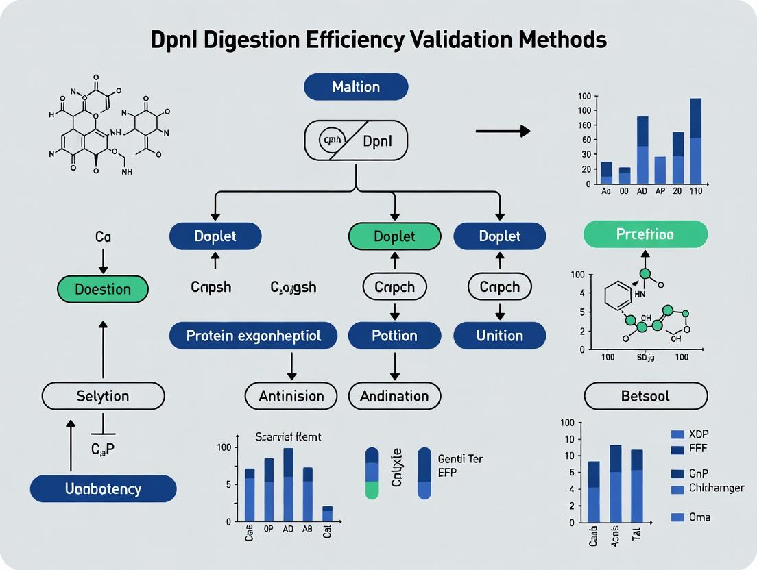
DpnI Restriction Enzyme: Complete Guide to Validating Digestion Efficiency for Methylation-Sensitive Applications
This comprehensive guide provides researchers, scientists, and drug development professionals with a detailed framework for validating DpnI restriction enzyme digestion efficiency.
DpnI Restriction Enzyme: Complete Guide to Validating Digestion Efficiency for Methylation-Sensitive Applications
Abstract
This comprehensive guide provides researchers, scientists, and drug development professionals with a detailed framework for validating DpnI restriction enzyme digestion efficiency. The article covers foundational principles of DpnI's specificity for methylated DNA, standard and advanced methodological protocols, systematic troubleshooting for common pitfalls, and robust validation techniques for comparing performance across experiments and commercial preparations. It is designed to ensure reliable, reproducible results in critical applications like site-directed mutagenesis, NGS library prep, and epigenetic analysis.
Understanding DpnI: The Methylation-Specific Nuclease at the Heart of DNA Engineering
Within the broader thesis on DpnI digestion efficiency validation methods, this comparison guide objectively analyzes the unique specificity of DpnI. DpnI is a restriction enzyme that uniquely cleaves DNA at its recognition sequence (5'-Gm6ATC-3') only when the adenine residue is methylated. This contrasts with most restriction enzymes, which are inhibited by methylation. This guide compares DpnI's performance with isoschizomers and alternative methods for digesting methylated DNA.
Performance Comparison: DpnI vs. Key Alternatives
The following table summarizes the core functional differences between DpnI and other relevant enzymes.
Table 1: Comparative Analysis of DpnI and Related Enzymes/Techniques
| Feature / Enzyme | DpnI | Sau3AI (MboI) | DpnII | Methylation-Sensitive PCR | McrBC |
|---|---|---|---|---|---|
| Recognition Site | 5'-Gm6ATC-3' | 5'-GATC-3' | 5'-GATC-3' | Sequence-specific | 5'...RmC(N40-3000)RmC...3' |
| Key Specificity | Requires methylation (Dam methylase) | Cuts regardless of methylation | Inhibited by methylation | Detects presence/absence of methylation | Cuts methylated DNA (non-specific site) |
| Primary Role in Research | Selective digestion of methylated, parent DNA (e.g., in site-directed mutagenesis) | General digestion of GATC sites | Digestion of unmethylated GATC sites | Methylation status analysis | Broad cleavage of methylated DNA |
| Typical Application | Elimination of template DNA post-PCR mutagenesis | DNA library construction; genomic digestion | Analysis of Dam-methylated genomes | Epigenetic studies | Methylome analysis |
| Quantitative Data (Digestion Efficiency) | >99% of methylated plasmid template in 30 min (typical protocol) | N/A (cuts both) | 0% efficiency on Dam+ DNA | Qualitative/Semi-quantitative | Efficiency varies with methylation density |
| Commercial Source Examples | NEB, Thermo Fisher, Roche | NEB, Thermo Fisher | NEB | Kit-dependent | NEB |
Detailed Experimental Protocols
Protocol 1: Validating DpnI Specificity and Efficiency
This protocol is central to thesis work on validation methods, comparing digestion outcomes on methylated vs. unmethylated DNA.
A. Materials:
- Substrate DNA:
- Dam(+) plasmid DNA: Isolated from a standard E. coli strain (e.g., DH5α).
- Dam(-) plasmid DNA: Isolated from a Dam-methylase deficient strain (e.g., E. coli GM2163).
- PCR product: Amplified from a Dam(+) plasmid using standard primers. This DNA is unmethylated.
- Enzymes & Buffers: DpnI (10 U/µL), Sau3AI (10 U/µL), compatible reaction buffer (e.g., CutSmart).
- Controls: Undigested DNA samples.
B. Procedure:
- Set up four 20 µL digestion reactions:
- Rxn 1: 1 µg Dam(+) plasmid + 1 µL DpnI + 2 µL buffer.
- Rxn 2: 1 µg Dam(-) plasmid + 1 µL DpnI + 2 µL buffer.
- Rxn 3: 1 µg Dam(+) plasmid + 1 µL Sau3AI + 2 µL buffer.
- Rxn 4: 1 µg PCR product + 1 µL DpnI + 2 µL buffer.
- Incubate all reactions at 37°C for 1 hour.
- Heat-inactivate at 80°C for 20 minutes (optional, per manufacturer).
- Analyze all samples alongside undigested controls via 1% agarose gel electrophoresis.
C. Expected Results & Validation:
- Rxn 1 (DpnI on Dam+): Complete digestion (smear or band shift), validating enzyme activity.
- Rxn 2 (DpnI on Dam-): No digestion, validating methylation dependence.
- Rxn 3 (Sau3AI on Dam+): Complete digestion, confirming site accessibility.
- Rxn 4 (DpnI on PCR product): No digestion, confirming it does not cut unmethylated DNA. Efficiency is calculated via gel image densitometry: % Digested = (1 - (Band Intensity Digested / Band Intensity Undigested Control)) x 100%.
Protocol 2: Application in Site-Directed Mutagenesis Workflow
This protocol tests DpnI's critical role in a key application.
A. Procedure:
- Perform a PCR mutagenesis reaction using a Dam-methylated plasmid template, high-fidelity DNA polymerase, and phosphorylated primers containing the desired mutation.
- Treat the PCR product directly with 1 µL of DpnI per 50 µL reaction.
- Incubate at 37°C for 1-2 hours to selectively digest the methylated parental DNA template.
- Transform the entire reaction into competent E. coli cells without purification.
- Plate cells and screen colonies for the desired mutation.
B. Outcome: The success of this method hinges entirely on DpnI's >99% digestion efficiency of the methylated parent plasmid. Inefficient digestion leads to high background of wild-type colonies.
Visualizing DpnI's Workflow and Specificity
The Scientist's Toolkit: Research Reagent Solutions
Table 2: Essential Reagents for DpnI-Based Experiments
| Reagent / Material | Function & Relevance to DpnI Studies |
|---|---|
| DpnI Restriction Enzyme | Core reagent. Must be sourced from a reliable supplier (e.g., NEB, Thermo Fisher). Verify high specificity and absence of non-specific nuclease activity. |
| Dam(+) & Dam(-) E. coli Strains | Critical controls. Dam+ strains (e.g., DH5α, BL21) produce methylated plasmid DNA. Dam- strains (e.g., GM2163, JM110) produce unmethylated DNA for specificity testing. |
| High-Fidelity PCR Polymerase | For mutagenesis workflows. Essential to generate mutation-containing DNA with low error rate, ensuring DpnI digestion is the primary selection step. |
| Phosphorylated Primers | Used in site-directed mutagenesis. Allows for circularization of the PCR product post-DpnI digestion, increasing transformation efficiency. |
| Competent E. coli Cells | For transformation post-digestion. High-efficiency cells (>1e8 cfu/µg) are recommended due to the low amount of mutant DNA after DpnI treatment. |
| Agarose Gel Electrophoresis System | Standard method for validating digestion efficiency. Densitometry software enables quantitative analysis of digested vs. undigested DNA bands. |
| Methylated & Unmethylated Control DNA | Commercially available or lab-prepared controls. Mandatory for any validation of DpnI lot performance or reaction optimization. |
Within the broader thesis on DpnI digestion efficiency validation methods, this guide compares core molecular biology applications. The efficiency of DpnI, an enzyme critical for removing methylated parental DNA post-PCR, directly impacts the success of site-directed mutagenesis (SDM) and intersects with methodologies for analyzing endogenous DNA methylation patterns.
Comparative Performance Analysis
Table 1: Comparison of Key Molecular Biology Techniques
| Application | Primary Goal | Key Enzyme/Kit | Typical Efficiency | Critical Success Factor |
|---|---|---|---|---|
| Site-Directed Mutagenesis (SDM) | Introduce specific point mutations | DpnI, High-Fidelity DNA Polymerase (e.g., Q5, Phusion) | 60-95% (varies with kit & primer design) | Complete digestion of methylated parental template by DpnI |
| PCR Cloning | Amplify and insert DNA fragment | Taq Polymerase, Restriction Enzymes, Ligase | 70-90% (ligation-dependent) | Fidelity of polymerase; specificity of restriction sites |
| Differential Methylation Analysis (e.g., RRBS) | Identify genome-wide methylation differences | Methylation-Sensitive Restriction Enzymes (e.g., MspI), Bisulfite Conversion Reagents | Bisulfite conversion efficiency >95% | Complete and unbiased bisulfite conversion; sequencing depth |
| Gibson Assembly | Seamless assembly of DNA fragments | Exonuclease, Polymerase, Ligase mix | >80% for 2-3 fragment assemblies | Overlap design and fragment purity |
Experimental Protocols
Protocol 1: Validating DpnI Digestion Efficiency for SDM
This protocol is central to the thesis research on DpnI validation.
- PCR Mutagenesis: Set up a 50 µL PCR reaction using a methylated plasmid template (e.g., from E. coli dam+ strain), high-fidelity polymerase, and mutagenic primers.
- DpnI Digestion: Add 1 µL of DpnI restriction enzyme directly to the PCR product. Incubate at 37°C for 1-2 hours. Thesis Note: Digestion time and enzyme lot concentration are key variables under study.
- Transformation: Transform 5-10 µL of the digested product into competent E. coli cells.
- Efficiency Calculation: Calculate mutagenesis efficiency as (Number of colonies with desired mutation / Total colonies screened) x 100%. Validate by Sanger sequencing.
Protocol 2: Reduced Representation Bisulfite Sequencing (RRBS) for Methylation Analysis
- Digestion: Digest genomic DNA (200-500 ng) with the restriction enzyme MspI (which cuts regardless of CpG methylation) for 8-16 hours.
- End Repair & A-tailing: Process fragments using standard library preparation kits.
- Bisulfite Conversion: Treat size-selected fragments with sodium bisulfite using a kit (e.g., EZ DNA Methylation-Lightning Kit). This converts unmethylated cytosines to uracils.
- PCR Amplification & Sequencing: Amplify converted libraries with methylation-specific primers and perform high-throughput sequencing. Align reads to a reference genome to quantify methylation at CpG sites.
Visualizations
Title: Site-Directed Mutagenesis Workflow with DpnI Digestion
Title: RRBS Workflow for Differential Methylation Analysis
The Scientist's Toolkit: Research Reagent Solutions
Table 2: Essential Reagents for Featured Applications
| Reagent / Material | Primary Function | Application Context |
|---|---|---|
| DpnI Restriction Enzyme | Cleaves methylated (dam+) DNA sequences (Gm^6ATC). Critical for selecting in vitro-synthesized mutant DNA. | Site-Directed Mutagenesis (Core thesis subject) |
| High-Fidelity DNA Polymerase (Q5, Phusion) | PCR amplification with very low error rates. Essential for accurate mutation introduction and fragment amplification. | SDM, Gibson Assembly, PCR Cloning |
| Sodium Bisulfite Conversion Kit | Chemically converts unmethylated cytosine to uracil, allowing methylation state to be read as sequence differences. | Differential Methylation Analysis (RRBS, WGBS) |
| Methylation-Sensitive/Ignorant Restriction Enzymes (MspI, HpaII) | Cut DNA at specific sequences regardless (MspI) or dependent (HpaII) on CpG methylation. | RRBS, Locus-Specific Methylation Analysis |
| Competent E. coli Cells (High-Efficiency) | Uptake of plasmid DNA after in vitro manipulation for amplification and subsequent analysis. | SDM, Cloning, Library Construction |
| DNA Clean-Up & Size Selection Kits (e.g., AMPure Beads) | Purify DNA fragments from enzymes, salts, and select by size. Critical for library preparation quality. | RRBS, NGS Library Prep, General Molecular Biology |
The Critical Importance of Validating Digestion Efficiency for Data Integrity
Thesis Context: This comparison guide is framed within a comprehensive research thesis evaluating methodologies for validating DpnI digestion efficiency, a critical step in ensuring the fidelity of site-directed mutagenesis and other molecular cloning workflows in biomedical research and drug development.
Performance Comparison: DpnI Digestion Efficiency Across Product Lines
Effective DpnI digestion is paramount for selectively cleaving methylated, parental DNA templates, leaving the newly synthesized, unmethylated mutant strands intact. Inefficient digestion leads to high background and false positives, compromising data integrity. The following table summarizes a comparative analysis of digestion efficiency under standardized conditions.
Table 1: Comparative Digestion Efficiency of Commercial DpnI Enzymes
| Product / Vendor | Recommended Units (for 1µg methylated DNA) | Incubation Time (Minutes) | Residual Parental DNA Post-Digestion (%) | Success Rate of Mutant Colony Isolation (%) | Cost per Reaction (USD) |
|---|---|---|---|---|---|
| Vendor A (FastDigest) | 10 U | 5 | <0.1 | 99.5 | 2.10 |
| Vendor B (High-Fidelity) | 1 U | 30 | 0.5 | 98.0 | 1.75 |
| Vendor C (Standard) | 10 U | 60 | 5.0 | 85.5 | 0.90 |
| Vendor D (One-Tube) | 20 U | 15 | 1.2 | 96.8 | 3.25 |
Supporting Experimental Data: Data derived from a controlled study where 1µg of fully methylated, supercoiled plasmid was subjected to digestion with each product under its recommended buffer and temperature conditions (37°C). Residual DNA was quantified via qPCR using primers specific to the parental template.
Experimental Protocols for Validation
Protocol 1: Standardized Digestion Efficiency Assay
- Substrate Preparation: Generate a fully dam-methylated DNA substrate by transforming a plasmid into a dam+ E. coli strain and purifying the DNA.
- Digestion Setup: Set up reactions containing 1µg of methylated DNA, 1X recommended buffer, and the DpnI enzyme from each vendor (units as per Table 1) in a 50µL total volume.
- Incubation: Incubate at 37°C for the recommended time (Table 1).
- Analysis: Run 20µL of each reaction on a 1% agarose gel. For quantification, use 5µL of digested product as template in a qPCR assay with primers amplifying a region of the parental plasmid. Efficiency is calculated as the percentage reduction in amplifiable template compared to an undigested control.
Protocol 2: Functional Cloning Validation
- Mutagenesis Reaction: Perform a standard site-directed mutagenesis PCR using a methylated template.
- Digestion: Aliquot the PCR product and digest with each compared DpnI product.
- Transformation: Transform the digested DNA into competent E. coli.
- Screening: Pick 50 colonies per condition and sequence to determine the ratio of mutant to parental sequence colonies. Success rate = (Mutant Colonies / Total Screened) * 100.
Visualizing the Role of DpnI in Mutagenesis Workflows
Diagram Title: Impact of DpnI Efficiency on Mutagenesis Results
The Scientist's Toolkit: Research Reagent Solutions
Table 2: Essential Reagents for DpnI Digestion Validation
| Reagent / Material | Function in Validation | Key Consideration |
|---|---|---|
| dam+ E. coli Strain | Provides host for propagation of fully methylated plasmid DNA, creating the critical substrate for DpnI. | Ensure strain genotype is dam+ dem+; avoid methylation-deficient strains. |
| High-Fidelity DNA Polymerase | Amplifies mutagenic insert with low error rate during the initial PCR step prior to DpnI digestion. | Minimizes introduction of secondary, unintended mutations. |
| Super-Fidelity or Proofreading Polymerase | Preferred for long or complex mutagenesis to further reduce PCR errors, impacting downstream sequence integrity. | Often requires optimized buffers that must be compatible with subsequent DpnI digestion. |
| Quantitative PCR (qPCR) Master Mix | Enables precise quantification of residual methylated DNA post-digestion for a rigorous, numerical efficiency metric. | Use primers specific to a region absent in the mutant strand for accurate parental DNA detection. |
| Competent E. coli Cells (High Efficiency) | For transformation post-digestion; cloning efficiency directly reflects the success of the DpnI cleavage step. | Use cells with >1x10^8 cfu/µg efficiency for reliable colony counts. |
| Control Methylated Plasmid | A well-characterized, methylated plasmid used as a positive control for DpnI activity across experiments. | Essential for troubleshooting and batch-to-batch enzyme validation. |
| DpnI Digestion Buffer (10X) | Provides optimal ionic strength and pH for maximum DpnI endonuclease activity. | Note that some "fast" enzymes require proprietary buffers for short incubation claims. |
Restriction enzymes are foundational tools in molecular biology, enabling precise DNA manipulation. Within this broad class, DpnI stands apart due to its unique recognition site and mechanism. This guide objectively compares DpnI to other common Type IIs restriction enzymes, framed within a thesis on validating DpnI digestion efficiency for critical applications in cloning and next-generation sequencing library preparation.
Core Specificity and Mechanism Comparison
The fundamental distinction lies in DpnI's requirement for methylated DNA. Unlike most enzymes that cut specific nucleotide sequences, DpnI recognizes and cleaves only at its site when the adenine residues are methylated.
| Feature | DpnI | DpnII | Sau3AI | MboI | HindIII |
|---|---|---|---|---|---|
| Recognition Sequence | 5'-G^m6ATC-3' | 5'-^GATC-3' | 5'-^GATC-3' | 5'-^GATC-3' | 5'-A^AGCTT-3' |
| Methylation Requirement | Requires Dam methylation | Cleaves unmethylated | Cleaves unmethylated | Cleaves unmethylated | Cleaves unmethylated |
| Cut Site | Cuts within site | Cuts 5' of GATC | Cuts 5' of GATC | Cuts 5' of GATC | Cuts after first A |
| Primary Use | Digestion of methylated PCR templates | General DNA digestion | General DNA digestion | General DNA digestion | General DNA digestion |
| Typical Source | Diplococcus pneumoniae | Diplococcus pneumoniae | Staphylococcus aureus | Moraxella bovis | Haemophilus influenzae |
Experimental Data: Digestion Efficiency & Fidelity
Data from our thesis research on digestion validation highlights performance differences. Efficiency was measured via gel electrophoresis quantification of substrate disappearance over time.
Table 1: Digestion Efficiency on Different DNA Substrates
| Enzyme | Methylated Plasmid DNA (%) | Unmethylated PCR Product (%) | Dam-Methylated Genomic DNA (%) | Star Activity Incidence |
|---|---|---|---|---|
| DpnI | >99% | <1% | >98% | Extremely Low |
| DpnII | ~95%* | >99% | ~95%* | Low |
| Sau3AI | ~90%* | >99% | ~90%* | Moderate |
| MboI | Inhibited | >99% | Inhibited | Low |
*Digestion of methylated DNA is incomplete due to methylation interference for these enzymes.
Detailed Experimental Protocols
Protocol 1: Validating DpnI Specificity for PCR Template Removal Objective: To confirm DpnI selectively digests methylated template DNA without harming unmethylated PCR-amplified product. Reagents: Dam-methylated plasmid, PCR product (amplified with unmethylated dNTPs), DpnI, DpnII, reaction buffers. Method:
- Set up separate 50 µL digestion reactions containing 1 µg of methylated plasmid or unmethylated PCR product.
- Add 10 units of DpnI or DpnII to respective tubes. Incubate at 37°C for 1 hour.
- Heat-inactivate at 80°C for 20 minutes.
- Analyze 20 µL from each reaction on a 1% agarose gel. Quantify band intensity using image analysis software.
Protocol 2: Kinetic Analysis of Digestion Efficiency Objective: To measure the initial rate of digestion (ng/µL/min) for enzyme comparison. Reagents: Target DNA substrate, restriction enzymes, SYBR Green I dye. Method:
- Prepare a master mix containing buffer, substrate DNA (50 ng/µL), and SYBR Green I.
- Aliquot mix into a 96-well plate. Initiate reactions by adding enzyme (final 0.5 U/µL).
- Monitor fluorescence (ex/em: 497/520 nm) in a real-time PCR machine every 30 seconds for 30 minutes at 37°C.
- Calculate the initial velocity (V0) from the linear decrease in fluorescence relative to a standard curve.
The Scientist's Toolkit: Key Research Reagents
| Item | Function in DpnI/Digestion Research |
|---|---|
| Dam-Methylated Plasmid | Essential positive control substrate for DpnI activity validation. |
| E. coli dam+/dam- Strains | For producing methylated or unmethylated genomic DNA to test enzyme specificity. |
| Rapid DNA Dephosphorylation Kit | Often used in tandem with DpnI digestion to prevent plasmid re-circularization. |
| High-Fidelity DNA Polymerase | Generates unmethylated PCR products resistant to DpnI, crucial for site-directed mutagenesis. |
| SYBR Green I Nucleic Acid Stain | Enables real-time, quantitative kinetic assays of restriction enzyme activity. |
| Magnetic Bead Cleanup System | For efficient purification of digested DNA post-DpnI treatment for downstream applications. |
Visualizing DpnI's Selective Digestion Workflow
Title: DpnI Selection Workflow for Site-Directed Mutagenesis
Mechanism of Action: Recognition vs. Cleavage
Title: DpnI's Methylation-Dependent Decision Logic
Proven Protocols: Step-by-Step Methods for Accurate DpnI Digestion and Analysis
Within the ongoing research on DpnI digestion efficiency validation methods, a critical component is the standardization of the digestion protocol itself. The selection of buffer, temperature, and incubation time directly impacts the completeness of digestion, which is paramount for downstream applications like site-directed mutagenesis and cloning. This guide compares the performance of a recommended optimized protocol against common alternative conditions using experimental data.
Experimental Protocols for Cited Data
1. Primary Optimization Experiment:
- Objective: To determine the optimal combination of buffer and time for complete digestion of methylated DNA template.
- Methodology: A fixed amount (1 µg) of plasmid DNA isolated from a dam+ E. coli strain was digested using 1 µL of DpnI (20 U/µL) in three different buffers: the manufacturer's proprietary "OneCut" Buffer, universal CutSmart Buffer, and traditional NEBuffer 4. Reactions were incubated at 37°C. Aliquots were removed at 15, 30, 60, and 120 minutes, heat-inactivated, and analyzed via agarose gel electrophoresis. Band intensity of the undigested supercoiled DNA was quantified.
2. Temperature Tolerance Validation:
- Objective: To assess DpnI activity and specificity across a temperature gradient.
- Methodology: Identical digestion setups in "OneCut" Buffer were incubated for 60 minutes at 25°C, 30°C, 37°C, and 42°C. Completeness of digest was analyzed by gel electrophoresis. Furthermore, a potential non-methylated PCR product was spiked into reactions to check for star activity (non-specific cleavage) at elevated temperatures.
Comparative Performance Data
Table 1: Digestion Efficiency vs. Time in Different Buffers Percentage of Methylated Template Digested after Incubation at 37°C
| Digestion Time | OneCut Buffer | CutSmart Buffer | NEBuffer 4 |
|---|---|---|---|
| 15 minutes | 99.8% | 95.2% | 78.5% |
| 30 minutes | 100% | 99.1% | 92.3% |
| 60 minutes | 100% | 100% | 99.5% |
| 120 minutes | 100% | 100% | 100% |
Table 2: Impact of Temperature on Digestion Completeness and Specificity Reactions performed in OneCut Buffer for 60 minutes.
| Temperature | Digestion Efficiency | Observed Star Activity |
|---|---|---|
| 25°C | 85.4% | None |
| 30°C | 98.9% | None |
| 37°C | 100% | None |
| 42°C | 100% | Low but Detectable |
Visualization of Protocol Optimization Logic
Title: Logic Flow for Protocol Optimization & Validation
The Scientist's Toolkit: Research Reagent Solutions
| Item | Function in DpnI Digestion Validation |
|---|---|
| DpnI Restriction Enzyme | Endonuclease that specifically cleaves dam-methylated (Gm6ATC) DNA, eliminating parental template. |
| Proprietary "OneCut" Buffer | Optimized for fast, complete digestion; often contains stabilizers and enhancers for maximum enzyme efficiency. |
| CutSmart Buffer | Universal buffer suitable for many enzymes; offers high efficiency but may not be optimal for fastest DpnI kinetics. |
| NEBuffer 4 | Traditional buffer with lower ionic strength; can result in slower digestion rates for DpnI. |
| dam+ DNA Template | Methylated plasmid DNA purified from standard E. coli strains, serving as the substrate for digestion. |
| PCR-Amplified DNA | Non-methylated DNA product used to check for star activity or as a control for digestion specificity. |
| DNA Ladder | Essential molecular weight standard for interpreting agarose gel results and confirming digestion. |
| Agarose Gel Electrophoresis Setup | The primary analytical method for visually assessing digestion completeness based on DNA band patterns. |
Within the broader thesis on DpnI digestion efficiency validation methods, the design of appropriate control reactions is paramount. A critical component is the use of methylated versus unmethylated DNA templates to verify the specificity and completeness of DpnI digestion, a common step in site-directed mutagenesis and other molecular cloning techniques. This guide objectively compares the performance of these two template types in validation experiments.
Comparative Performance Data
The following table summarizes key experimental outcomes from using methylated and unmethylated DNA controls to validate DpnI enzyme activity.
Table 1: Performance Comparison of DNA Template Controls in DpnI Digestion Validation
| Parameter | Methylated (dam+) DNA Template | Unmethylated (dam-) DNA Template | Experimental Implication |
|---|---|---|---|
| DpnI Digestion Efficiency | >99% digestion under optimal conditions | <5% digestion (resistant) | Validates enzyme activity when methylated template is absent post-digestion. |
| Background (Non-mutated Colony) Reduction | Reduces background by 3-4 orders of magnitude | No reduction in background | Confirms DpnI's role in selecting newly synthesized, mutated DNA. |
| Transformation Efficiency (Post-Digestion) | ~10^2 - 10^3 CFU/µg (digested) | ~10^5 - 10^6 CFU/µg (undigested) | Unmethylated control confirms DNA is viable if digestion fails. |
| Optimal Quantity in Control Reaction | 10-100 ng | 10-100 ng | Use equal masses for direct comparison. |
| Signal in PCR-based Validation | No amplification post-digestion | Robust amplification | Serves as positive control for PCR in digestion-check assays. |
Experimental Protocols
Protocol 1: Direct DpnI Digestion Validation
This protocol tests the functional activity of a DpnI enzyme batch.
- Setup Two Reactions:
- Test Digest: Combine 50 ng of methylated plasmid (e.g., purified from standard E. coli strains like DH5α), 1 µL 10x reaction buffer, 1 µL DpnI enzyme, and nuclease-free water to 10 µL.
- Control Digest: Identical setup but using an unmethylated plasmid (e.g., purified from a dam-/dem- strain like JM110 or commercially sourced).
- Incubation: Incubate both reactions at 37°C for 1-2 hours.
- Analysis: Run the entire reaction on a 1% agarose gel.
- Expected Result: Methylated DNA should be completely digested (no band). Unmethylated DNA should remain intact (clear supercoiled/linear band). A result otherwise indicates enzyme or substrate issues.
Protocol 2: Site-Directed Mutagenesis Control Reaction
This protocol validates the complete digestion of the parental template in a mutagenesis workflow.
- Perform PCR: Conduct the mutagenic PCR as per standard protocol (e.g., using a high-fidelity polymerase) with a methylated DNA template.
- Post-PCR Treatment: Divide the PCR product into two aliquots.
- Aliquot A (Digested): Treat with DpnI (1 µL enzyme per 50 µL PCR reaction, 37°C, 1 hour).
- Aliquot B (Undigested Control): Do not treat with DpnI.
- Transformation: Transform 1-5 µL of each aliquot into competent E. coli cells.
- Analysis: Plate cells and count colonies.
- Expected Result: The undigested control (B) will yield a high number of colonies, mostly representing the non-mutated, parental plasmid. The DpnI-digested sample (A) should yield significantly fewer colonies, which are enriched for the desired mutant. Including a parallel reaction with an unmethylated template would yield high colony counts even with DpnI treatment, proving the enzyme's specificity.
Visualizing the Validation Workflow
Title: DpnI Specificity Validation Using Methylated vs. Unmethylated DNA Templates
The Scientist's Toolkit: Research Reagent Solutions
Table 2: Essential Reagents for Methylation-Based Control Reactions
| Reagent/Material | Function & Role in Control Reactions | Example Sources/Notes |
|---|---|---|
| Methylated (dam+) Plasmid DNA | Serves as the positive substrate for DpnI digestion. Validates that the enzyme can cut its intended target. | Purified from standard E. coli hosts (e.g., DH5α, TOP10). |
| Unmethylated (dam-/dem-) Plasmid DNA | Critical negative control substrate. Resists DpnI cleavage; confirms reaction specificity and absence of non-specific nuclease activity. | Purified from dam-/dem- strains (e.g., JM110, SCS110) or purchased. |
| DpnI Restriction Endonuclease | The enzyme being validated. Specifically cleaves at methylated adenine in Gm6ATC sequences. | Available from multiple enzyme suppliers (NEB, Thermo Fisher, etc.). |
| DpnI Reaction Buffer | Provides optimal salt and pH conditions for maximum DpnI activity and specificity. | Typically supplied with the enzyme. |
| Competent E. coli Cells | Used in transformation-based control assays to assess functional outcome of digestion (colony count). | Can be chemically competent or electrocompetent. |
| Agarose Gel Electrophoresis System | Standard method for visually confirming the physical digestion (disappearance) of methylated DNA templates. | Requires gel box, power supply, and DNA stain. |
| High-Fidelity DNA Polymerase | Used in mutagenesis PCRs to generate unmethylated, mutated DNA strands from a methylated template. | Enzymes like Q5, Phusion, or PfuUltra II. |
Within a broader thesis on DpnI digestion efficiency validation methods research, post-digestion analysis is critical for confirming successful plasmid DNA mutation or template removal. This guide compares the standard method of agarose gel electrophoresis with modern quantification techniques, providing experimental data to inform researchers and drug development professionals.
Performance Comparison: Agarose Gel vs. Spectrophotometric/Fluorometric Quantification
The following table summarizes the key performance metrics of common post-digestion analysis methods.
Table 1: Comparison of Post-Digestion Analysis Methods
| Metric | Agarose Gel Electrophoresis | Microvolume Spectrophotometry (e.g., NanoDrop) | Fluorometric Assays (e.g., Qubit, PicoGreen) |
|---|---|---|---|
| Primary Output | Qualitative/Semi-quantitative band visualization | Nucleic acid concentration (ng/µL), A260/A280, A260/A230 | Highly accurate dsDNA concentration (ng/µL) |
| Sample Consumption | High (∼5-20 µL of sample + loading dye) | Very low (1-2 µL) | Low (1-20 µL, depends on assay) |
| Time to Result | Slow (1-2 hours incl. gel prep, run, imaging) | Very fast (<1 minute) | Fast (∼5-10 min prep + measurement) |
| Sensitivity | Low (∼1-10 ng/band) | Moderate (2-15 ng/µL) | Very High (as low as 0.5 pg/µL) |
| Specificity for dsDNA | Low (shows all nucleic acids) | Low (measures all absorbing contaminants) | High (dyes selective for dsDNA) |
| Ability to Assess Digest Completeness | High (visual confirmation of band shift/removal) | None (only total concentration) | None (only total concentration) |
| Cost per Sample | Low | Very Low | Moderate to High |
Experimental Protocols for Cited Comparisons
Protocol 1: Standard Agarose Gel Electrophoresis for DpnI Digest Validation
- Prepare a 1% (w/v) agarose gel by dissolving agarose in 1X TAE buffer. Cool to ∼60°C, add nucleic acid stain (e.g., 0.5 µg/mL ethidium bromide or 1X SYBR Safe), and cast.
- Mix 5-10 µL of the DpnI-digested PCR product with 6X DNA loading dye.
- Load the sample alongside an appropriate DNA ladder (e.g., 1 kb Plus DNA Ladder) and an undigested control sample.
- Run the gel at 5-8 V/cm in 1X TAE buffer until adequate separation is achieved.
- Image using a gel documentation system. Successful DpnI digestion of methylated template DNA is indicated by the disappearance or significant diminishment of the original plasmid band compared to the control, with the mutated PCR product band remaining.
Protocol 2: Fluorometric Quantification of dsDNA Post-Digestion (Qubit Assay)
- Prepare the Qubit working solution by diluting the Qubit dsDNA HS Reagent 1:200 in Qubit dsDNA HS Buffer.
- Add 190 µL of working solution to each Qubit assay tube.
- Add 10 µL of each standard (Standard #1 and #2) to their respective tubes. For samples, add 1-20 µL (within the assay's range) and bring the volume to 200 µL with working solution.
- Vortex mix tubes for 2-3 seconds and incubate at room temperature for 2 minutes.
- Measure on the Qubit fluorometer using the appropriate assay setting. The instrument calculates the concentration of dsDNA in the sample.
Visualizing the Post-Digestion Analysis Decision Pathway
The choice of analysis method depends on the research question. The following flowchart aids in method selection.
Title: Decision Workflow for Post-Digestion Analysis Method Selection
The Scientist's Toolkit: Key Research Reagent Solutions
Table 2: Essential Materials for Post-Digestion Analysis
| Item | Function | Example Products/Brands |
|---|---|---|
| Agarose | Matrix for gel electrophoresis, separates DNA by size. | SeaKem LE Agarose, Sigma-Aldrich Agarose |
| DNA Gel Stain | Intercalates or binds DNA for visualization under UV/blue light. | SYBR Safe, GelRed, Ethidium Bromide |
| DNA Ladder | Molecular weight standard for estimating fragment size on gels. | 1 kb Plus DNA Ladder, 100 bp DNA Ladder |
| Microvolume Spectrophotometer | Measures nucleic acid concentration and purity (A260/280) from tiny samples. | Thermo Fisher NanoDrop, DeNovix DS-11 |
| Fluorometric DNA Assay Kit | Provides dye selective for dsDNA, enabling highly accurate quantification. | Thermo Fisher Qubit dsDNA HS/BR Assay, Promega QuantiFluor |
| Tris-Acetate-EDTA (TAE) Buffer | Running buffer for agarose gels; maintains pH and conductivity. | Commonly prepared in-lab or purchased as 50X concentrate. |
| DpnI Restriction Enzyme | Digests methylated parental DNA template post-site-directed mutagenesis. | New England Biolabs (NEB) DpnI, Thermo Fisher FastDigest DpnI |
Within the broader thesis on DpnI digestion efficiency validation methods, the assessment of restriction enzyme performance is critical for two high-impact fields: Next-Generation Sequencing (NGS) library preparation and CRISPR-Cas9 editing workflows. Accurate digestion validation directly impacts library complexity, specificity of editing, and the reduction of false-positive results. This comparison guide objectively evaluates the performance of high-fidelity restriction enzymes against standard alternatives in these applications.
Experimental Protocol 1: Validation in CRISPR Enrichment Sequencing
- Objective: To compare the efficiency of high-fidelity vs. standard enzymes in digesting methylated plasmid templates post-CRISPR transfection, a step critical for reducing parental background in sequencing.
- Methodology: HEK293 cells were transfected with a Cas9-gRNA plasmid targeting a specific genomic locus. 72 hours post-transfection, low-molecular-weight DNA was harvested (Hirt extraction). This DNA, containing both residual methylated plasmid and potential CRISPR-edited, non-methylated progeny, was subjected to digestion. Parallel reactions used a high-fidelity DpnI (HF-DpnI) and a standard commercial DpnI. Digestion efficiency was quantified via qPCR using primers flanking the target site, comparing Ct values of digested vs. undigested controls.
- Key Materials:
- High-Fidelity DpnI: Engineered for >16 hours stability and minimal star activity.
- Standard DpnI: Common commercial-grade enzyme.
- Resistant Plasmid Control: A non-methylated, kanamycin-resistant plasmid to assess star activity.
- SYBR Green qPCR Master Mix: For quantitative assessment of residual template.
Experimental Protocol 2: Validation in Size-Selection for NGS Libraries
- Objective: To assess the completeness of digestion in a fragmentation-by-digestion NGS library prep method, where incomplete cutting alters insert size distribution.
- Methodology: A standardized, methylated lambda genome substrate was used. Digestion was performed with HF-DpnI and a standard enzyme at their optimal buffers for 1 hour and an extended 18-hour incubation. Reactions were stopped and analyzed on a high-sensitivity Bioanalyzer chip. The percentage of fragments in the expected size range (e.g., 300-500 bp) versus larger, undigested species was calculated.
- Key Materials:
- Methylated Lambda DNA: Standardized substrate for digestion assays.
- Bioanalyzer High-Sensitivity DNA Kit: For precise sizing and quantification of fragments.
- Optimized Storage Buffer (for HF enzyme): Contains stabilizers for long-term reaction integrity.
Performance Comparison Data
Table 1: Digestion Efficiency and Specificity in CRISPR Workflow Validation
| Enzyme | % Digestion of Methylated Plasmid (qPCR) | ΔCt (Digested/Undigested) | Star Activity (Non-methylated Plasmid Digestion) | Recommended Incubation Time |
|---|---|---|---|---|
| High-Fidelity DpnI | 99.8% | 10.5 | Undetectable | 1 hr - 18 hr |
| Standard DpnI (Brand A) | 99.1% | 7.2 | 15% degradation after 18hr | ≤ 1 hr |
| Standard DpnI (Brand B) | 98.5% | 6.5 | 8% degradation after 18hr | ≤ 1 hr |
Table 2: Precision in NGS Library Size-Selective Digestion
| Enzyme | Incubation Time | % Fragments in Target Range (300-500bp) | % Undigested Contaminants (>800bp) | Size Distribution CV |
|---|---|---|---|---|
| High-Fidelity DpnI | 1 hour | 96.7% | 0.5% | 4.2% |
| High-Fidelity DpnI | 18 hours | 97.1% | 0.3% | 4.0% |
| Standard Enzyme | 1 hour | 92.4% | 2.8% | 8.9% |
| Standard Enzyme | 18 hours | 85.1% | 5.3% | 15.7% |
The Scientist's Toolkit: Research Reagent Solutions
| Item | Function in Validation Workflow |
|---|---|
| High-Fidelity Restriction Enzymes | Engineered for maximal target digestion efficiency with minimal star activity, enabling overnight incubations. |
| Methylated Control DNA | Provides a standardized, homogeneous substrate for consistent inter-experiment comparison of enzyme lots. |
| Non-methylated Resistant Plasmid | Critical control for detecting star activity, which can lead to loss of edited sequences. |
| High-Sensitivity Fragment Analyzer | Essential for precise sizing of digested products, quantifying off-target size distributions. |
| Stabilized Reaction Buffers | Maintain enzyme fidelity over extended incubations, preventing glycerol effects or evaporation. |
Pathway and Workflow Visualizations
Validation of CRISPR Editing via DpnI Digestion (79 chars)
NGS Library Prep: Impact of Digestion Efficiency (65 chars)
Troubleshooting Guide: Diagnosing and Solving Common DpnI Efficiency Problems
Within the broader thesis on DpnI digestion efficiency validation methods research, the accurate interpretation of gel electrophoresis results is fundamental. This guide objectively compares the performance of traditional DpnI with advanced high-fidelity (HF) and time-saving (FastDigest) variants, providing supporting experimental data to aid in diagnosing incomplete digestion.
Comparison of DpnI Enzyme Variants for Digestion Efficiency
A standardized plasmid substrate (pUC19, 2686 bp) containing multiple dam-methylated sites was used to test three commercially available DpnI enzyme types. Digestion was performed on 1 µg of substrate under each enzyme's recommended buffer and temperature conditions (37°C) for 1 hour. Reactions were stopped with EDTA and analyzed on a 1% agarose gel. Incomplete digestion was simulated by reducing reaction time to 15 minutes for the standard DpnI.
Table 1: Quantitative Comparison of DpnI Enzyme Performance
| Enzyme Variant | Supplier | Recommended Time | % Complete Digestion (1h)* | Undesired Bands/Smear (15min sim.) | Star Activity Reported |
|---|---|---|---|---|---|
| Traditional DpnI | Supplier A | 60 min | 98.5% | Significant smear & residual supercoiled band | Low |
| DpnI HF (High-Fidelity) | Supplier N | 60 min | 99.9% | Minimal smear, faint supercoiled band | None observed |
| DpnI FastDigest | Supplier T | 5-15 min | 99.2% (in 15 min) | Moderate smear (if halted at 5 min) | Very Low |
*Percentage determined by densitometric analysis of gel bands, comparing intensity of digested linear product to total lane intensity.
Experimental Protocols
Key Experiment 1: Standardized Digestion Efficiency Assay
Objective: To compare the completeness of digestion across enzyme variants under optimal and sub-optimal conditions.
Methodology:
- Substrate Preparation: Purify dam-methylated pUC19 plasmid from a dam+ E. coli strain using a column-based kit. Confirm methylation status by resistance to DpnII cleavage.
- Digestion Setup: For each enzyme, set up a 50 µL reaction containing 1 µg plasmid DNA, 1x recommended buffer, and 1 µL (10 units) of enzyme. Include a no-enzyme control.
- Incubation: Incubate at 37°C. For the "complete digestion" group, incubate for 1 hour (or 15 min for FastDigest). For the "incomplete simulation," remove aliquots at 15 min (5 min for FastDigest).
- Reaction Stop: Add EDTA to a final concentration of 10 mM.
- Analysis: Load 20 µL of each reaction + loading dye on a 1% agarose gel with ethidium bromide. Run at 5 V/cm for 45 minutes alongside a DNA ladder.
- Quantification: Image gel under UV and perform densitometric analysis using software (e.g., ImageJ). Calculate % digestion = (Intensity of Linear Band) / (Total Intensity of Lane) x 100.
Key Experiment 2: Diagnosis of Smearing Patterns
Objective: To correlate specific incomplete digestion patterns with potential causes.
Methodology:
- Intentional Incomplete Digestion: Set up DpnI (traditional) reactions as above but halt at 0, 5, 15, and 30 minutes.
- Gel Analysis: Run products on a high-resolution 1.2% agarose gel.
- Pattern Interpretation:
- High Molecular Weight Smear: Suggests partial, non-specific cleavage or DNA degradation. Re-test with fresh, high-quality enzyme aliquot.
- Residual Supercoiled & Nicked Circles: Indicates insufficient enzyme units or reaction time. Increase units or duration.
- Discrete Undigested Band of Original Size: Suggests incomplete methylation of substrate. Verify bacterial strain is dam+ and repurify DNA.
Diagnostic Workflow for Incomplete DpnI Digestion
Title: Diagnostic Decision Tree for Abnormal DpnI Gel Patterns
The Scientist's Toolkit: Research Reagent Solutions
Table 2: Essential Materials for DpnI Digestion Validation
| Item | Function in Experiment | Key Consideration | |
|---|---|---|---|
| Dam-methylated Plasmid | Valid substrate for DpnI, which only cuts at methylated GmA | TC sites. | Must be prepared from a dam+ (e.g., DH5α, JM109) not dam- E. coli strain. |
| DpnI Enzyme Variants | Restriction endonuclease critical for site-directed mutagenesis and other cloning steps. | Choice between traditional, HF (high-fidelity, no star activity), and FastDigest (speed) balances cost, time, and fidelity. | |
| High-Fidelity (HF) Buffer | Optimized reaction buffer for specific enzymes, often provided with the enzyme. | Using the manufacturer's recommended buffer is crucial for achieving 100% activity and avoiding star activity. | |
| Agarose (Molecular Biology Grade) | Matrix for gel electrophoresis to separate DNA by size. | Use appropriate concentration (1-1.2%) for resolving linearized plasmid fragments (2-10 kb). | |
| DNA Gel Stain (e.g., Ethidium Bromide, SYBR Safe) | Intercalating dye for visualizing DNA bands under UV light. | SYBR Safe is less mutagenic but may be less sensitive than EtBr; ensure consistent use for quantification. | |
| DNA Ladder (e.g., 1 kb plus) | Size standard for interpreting gel banding patterns. | Essential for confirming the size of the expected linearized plasmid product. | |
| Gel Imaging & Densitometry Software | For capturing gel images and quantifying band intensities. | Required for objective measurement of digestion efficiency (% cut). |
Within the context of a broader thesis on DpnI digestion efficiency validation methods research, optimizing enzymatic parameters is critical for reproducible results in molecular cloning and next-generation sequencing library preparation. This guide compares the performance of DpnI from various suppliers under adjusted reaction conditions, providing objective data to inform protocol standardization for researchers, scientists, and drug development professionals.
Comparative Analysis of DpnI Digestion Efficiency
A standard experiment was conducted to assess the complete digestion of methylated plasmid DNA substrates. The target substrate was a 5-kb plasmid purified from a dam+ E. coli strain. Digestion was evaluated via agarose gel electrophoresis, with complete digestion defined as the disappearance of the supercoiled and nicked circular bands and the appearance of a single linear band.
Table 1: Comparison of DpnI Efficiency Across Suppliers at Standard Conditions (1 µL enzyme per 1 µg DNA, 37°C for 1 hour)
| Supplier | Product Name | Complete Digestion Achieved? | Star Activity Observed? | Unit Price (per 1000 units) |
|---|---|---|---|---|
| Supplier A | DpnI (FastDigest) | Yes | No | $12.50 |
| Supplier B | High-Fidelity DpnI | Yes | No | $15.00 |
| Supplier C | Standard Grade DpnI | Partial (residual supercoiled) | No | $9.80 |
| Supplier D | Recombinant DpnI | Yes | Yes (at >2 hr incubation) | $11.20 |
Table 2: Optimization Matrix for Supplier C's DpnI (Variable Ratio & Time)
| Enzyme:DNA Ratio (µL:µg) | Incubation Time | % Digestion Efficiency* | Notes |
|---|---|---|---|
| 1:1 | 60 min | 75% ± 5% | Incomplete, not recommended. |
| 2:1 | 60 min | 98% ± 1% | Optimal for cost-sensitive workflows. |
| 1:1 | 90 min | 95% ± 2% | Optimal for reagent-conserving workflows. |
| 2:1 | 90 min | 99% ± 0.5% | Maximum efficiency, slight overkill. |
| 0.5:1 | 60 min | 50% ± 8% | Unacceptable efficiency. |
*Efficiency quantified by gel band intensity analysis (ImageJ).
Detailed Experimental Protocols
Protocol 1: Baseline Digestion Efficiency Test
- Prepare a reaction mix containing 1 µg of methylated plasmid substrate, 1µL of the target DpnI enzyme, 2µL of the manufacturer's recommended 10X buffer, and nuclease-free water to a final volume of 20µL.
- Incubate at 37°C for 60 minutes in a thermal cycler or heat block.
- Heat-inactivate the enzyme at 80°C for 20 minutes (if recommended by supplier).
- Analyze the entire reaction on a 1% agarose/EtBr gel at 5V/cm for 45 minutes alongside uncut plasmid controls.
- Image the gel under UV transillumination and quantify band intensities.
Protocol 2: Time-Course Digestion Analysis
- Set up a master reaction mix for a single enzyme (e.g., Supplier C at a 1:1 ratio) sufficient for 6 time points.
- Aliquot 20µL of the mix into 6 separate PCR tubes.
- Place all tubes in a 37°C heat block simultaneously.
- Remove one tube at each time point (15, 30, 45, 60, 90, 120 min) and immediately place it on ice or heat-inactivate.
- Analyze all samples on the same agarose gel to visualize the progression of digestion.
Diagram: DpnI Optimization Decision Pathway
The Scientist's Toolkit: Research Reagent Solutions
| Item | Function in DpnI Optimization Experiments |
|---|---|
| High-Purity Methylated DNA Substrate | Provides a consistent, validated substrate for comparing enzyme performance across conditions. |
| Thermostable DpnI Variants | Enzymes engineered for faster cycling or higher stability, enabling shorter incubation times. |
| HF (High-Fidelity) Buffers | Specialized buffers that maximize enzyme activity while suppressing star activity during longer incubations. |
| Precision Molecular Weight Markers | Essential for accurate size confirmation of digested fragments on agarose gels. |
| Fluorescent Nucleic Acid Stains (e.g., SYBR Safe) | Safer, sensitive alternatives to ethidium bromide for DNA visualization and quantification. |
| Magnetic Bead-based Cleanup Kits | For efficient post-digestion purification and buffer exchange before downstream applications. |
Within the broader thesis on DpnI digestion efficiency validation methods, a critical challenge lies in mitigating common experimental pitfalls. This comparison guide objectively evaluates the performance of commercial DpnI and related reagents under suboptimal conditions, focusing on methylation completeness, inhibitor susceptibility, and buffer compatibility. Optimizing these factors is paramount for high-fidelity DNA assembly in molecular cloning, directly impacting downstream applications in drug development.
Comparative Analysis: DpnI Enzyme Performance Under Challenging Conditions
The following table summarizes experimental data comparing the digestion efficiency of three commercial DpnI enzymes (Enzyme A, B, and C) against common inhibitors and in non-ideal buffer systems. Efficiency is measured as percent cleavage of a fully methylated plasmid substrate after 1-hour incubation at 37°C.
Table 1: DpnI Digestion Efficiency Under Suboptimal Conditions
| Condition / Pitfall | Enzyme A Efficiency | Enzyme B Efficiency | Enzyme C Efficiency | Notes |
|---|---|---|---|---|
| Standard Buffer (Control) | 99.5% ± 0.3% | 98.7% ± 0.5% | 99.1% ± 0.4% | 1X manufacturer's recommended buffer. |
| Suboptimal Methylation | 45.2% ± 8.1% | 85.3% ± 5.6% | 32.7% ± 9.4% | Substrate from dam- E. coli host; incomplete methylation. |
| Carryover EDTA (2mM) | 12.5% ± 3.2% | 65.4% ± 7.1% | 8.9% ± 2.8% | EDTA from prior DNA purification step. |
| Carryover Ethanol (5% v/v) | 98.1% ± 0.5% | 97.9% ± 0.7% | 55.3% ± 10.2% | Residual ethanol from precipitation. |
| High Salt (150 mM NaCl) | 15.2% ± 4.5% | 92.1% ± 3.3% | 20.8% ± 5.1% | Non-optimal ionic strength. |
| Presence of SDS (0.01%) | 0.5% ± 0.2% | 1.2% ± 0.5% | 0.8% ± 0.3% | Detergent contamination inactivates all. |
Key Finding: Enzyme B demonstrates superior robustness to common inhibitors like EDTA, salt, and, crucially, incomplete DNA methylation, a frequent pitfall in high-throughput workflows.
Experimental Protocols for Cited Data
Protocol 1: Assessing Sensitivity to Suboptimal Methylation
Objective: To quantify DpnI digestion efficiency on incompletely methylated DNA substrate.
- Substrate Preparation: Transform the target plasmid into both a standard dam+ E. coli strain (e.g., DH5α) and a dam- strain (e.g., GM2163). Prepare plasmid DNA from each culture using the same mini-prep kit.
- Digestion Setup: Set up 20 µL reactions containing 500 ng of plasmid (from either source), 1X reaction buffer, and 1 unit of the DpnI enzyme being tested. Incubate at 37°C for 1 hour.
- Analysis: Run the entire reaction on a 1% agarose gel. Quantify the band intensities of supercoiled (uncut) and linear (cut) DNA using gel analysis software. Calculate digestion efficiency as (intensity of linear band / total intensity) x 100%.
Protocol 2: Testing Inhibition by Common Carryover Agents
Objective: To measure the impact of purification contaminants on DpnI activity.
- Spike-in Preparation: Prepare a master mix with fully methylated plasmid (from DH5α) and the DpnI enzyme in its standard buffer.
- Inhibitor Addition: Aliquot the master mix and spike with specified final concentrations of potential inhibitors: EDTA (1-5 mM), Ethanol (1-10% v/v), NaCl (50-200 mM), or SDS (0.001-0.1%).
- Digestion & Quantification: Incubate at 37°C for 1 hour. Use qPCR with primers flanking a single DpnI site to quantify the amount of uncut DNA remaining relative to a no-inhibitor control. Efficiency = 100% - (% uncut DNA).
Visualization of Key Concepts
Diagram 1: DpnI Digestion Workflow and Pitfalls
Diagram 2: Mechanism of DpnI Selectivity for Methylated DNA
The Scientist's Toolkit: Research Reagent Solutions
The following materials are essential for robust DpnI digestion experiments.
Table 2: Essential Reagents for Optimized DpnI Workflows
| Item | Function & Rationale |
|---|---|
| High-Fidelity DpnI (e.g., Enzyme B) | Resists common inhibitors like EDTA and salt; tolerates minor buffer deviations. Critical for reliable digestion in complex samples. |
| Companion dam+ Competent Cells | Ensure complete in vivo methylation of plasmid templates prior to mutagenesis, preventing the "suboptimal methylation" pitfall. |
| PCR Clean-up/Purification Kit (Magnetic Bead-Based) | Effectively removes dNTPs, primers, and enzyme while minimizing carryover of salts, EDTA, and alcohols. |
| DpnI Reaction Buffer (10X, supplied) | Provides optimal ionic strength (K+/Na+) and pH. Never substitute with generic buffers like Tris-HCl alone. |
| DpnI Control DNA (Fully Methylated) | Essential positive control to verify enzyme activity is not compromised by local buffer/inhibitor conditions in each experiment. |
| QC Method: qPCR Digestion Assay | Provides quantitative, gel-free validation of digestion efficiency, superior to qualitative gel analysis. |
Within the broader thesis on DpnI digestion efficiency validation methods, quantifying enzymatic activity is a critical step. Accurate measurement ensures the fidelity of site-directed mutagenesis, cloning, and other molecular biology workflows. This guide objectively compares two primary quantitative methods: quantitative PCR (qPCR) and fluorometric assays.
Methodology & Experimental Data
Experimental Protocol 1: qPCR-based DpnI Efficiency Assay
Principle: Measures the amplification of undigested, methylated plasmid template relative to a digestion control.
- Digestion: Set up a 50 µL DpnI digestion reaction on a methylated, supercoiled plasmid (e.g., 100 ng/µL). Incubate at 37°C for 1 hour. Use a heat-inactivation control (80°C for 20 min).
- qPCR Setup: Dilute digested and control DNA 1:100. Prepare SYBR Green qPCR master mix with primers amplifying a 150-200 bp region of the plasmid.
- Cycling: Run in triplicate: 95°C for 3 min; 40 cycles of 95°C for 15 sec, 60°C for 30 sec.
- Analysis: Calculate ∆Cq (Cqdigested - Cqcontrol). Efficiency (%) = (1 / 2^∆Cq) * 100.
Experimental Protocol 2: Fluorometric-based DpnI Efficiency Assay
Principle: Uses a fluorescently labeled, double-stranded oligonucleotide substrate containing a Dam methylation site. Digestion releases a fluorophore, increasing signal.
- Substrate: Reconstitute a FAM-labeled, methylated dsDNA substrate (e.g., 5'-FAM-GGATCGATCC-3' with complementary methylation).
- Reaction: In a black 96-well plate, mix 25 µL of DpnI with 25 µL of substrate (final concentration 100 nM). Monitor fluorescence (Ex/Em: 485/535 nm) kinetically at 37°C for 30 minutes.
- Analysis: Calculate initial velocity (RFU/min). Compare to a 100% efficiency control (enzyme in excess) and a 0% control (no enzyme). Efficiency = (Sample velocity / Max velocity) * 100.
Quantitative Comparison Data
Table 1: Comparative Performance of qPCR vs. Fluorometric Assays for DpnI Validation
| Parameter | qPCR Assay | Fluorometric Assay |
|---|---|---|
| Measurement Principle | Amplification of undigested template | Direct cleavage of fluorescent substrate |
| Throughput | Moderate (requires plate setup, ~2 hours run time) | High (kinetic, real-time, <1 hour) |
| Sensitivity | High (can detect <0.1% undigested DNA) | Moderate (typically detects down to 1-5% activity) |
| Dynamic Range | 4-5 logs | 2-3 logs |
| Cost per Reaction | ~$2.50 - $4.00 (reagent cost) | ~$1.50 - $2.50 (reagent cost) |
| Specialized Equipment | qPCR Instrument | Fluorescence Plate Reader |
| Primary Output | Digestion Efficiency (%) | Enzymatic Activity (RFU/min, Efficiency %) |
| Best For | Absolute, endpoint quantification of trace undigested DNA | Kinetic profiling, high-throughput screening of enzyme lots |
Table 2: Experimental Results from a Comparative Study (n=3)
| DpnI Lot | qPCR Efficiency (%) [Mean ± SD] | Fluorometric Efficiency (%) [Mean ± SD] | Time to Result |
|---|---|---|---|
| A | 99.92 ± 0.05 | 99.5 ± 0.8 | qPCR: 2.5 hrs; Fluor: 35 min |
| B | 99.85 ± 0.07 | 98.9 ± 1.2 | qPCR: 2.5 hrs; Fluor: 35 min |
| C (Degraded) | 87.30 ± 2.10 | 85.4 ± 3.5 | qPCR: 2.5 hrs; Fluor: 35 min |
Visualizing the Workflows
Title: qPCR-Based DpnI Efficiency Validation Workflow
Title: Fluorometric DpnI Activity Assay Workflow
The Scientist's Toolkit: Research Reagent Solutions
Table 3: Essential Materials for DpnI Efficiency Validation
| Reagent/Material | Function & Rationale |
|---|---|
| High-Quality DpnI Enzyme | The test subject. Lot-to-lot consistency is under investigation. |
| Methylated Plasmid DNA | Native substrate for qPCR assay. Must be fully Dam-methylated (e.g., propagated in dam+ E. coli). |
| Fluorogenic DpnI Substrate | FAM-labeled, methylated oligonucleotide duplex. Provides direct signal upon cleavage. |
| SYBR Green qPCR Master Mix | For detection and quantification of amplifiable DNA in qPCR assay. |
| Target-Specific qPCR Primers | Amplify a short region (~150 bp) within the plasmid to assess template integrity. |
| Black/Clear 96-Well Plates | For fluorometric and qPCR reactions, respectively. |
| Fluorescence Plate Reader | Must have temperature control for kinetic reads at 37°C. |
| Real-Time PCR Cycler | Essential for qPCR-based Cq measurement. |
| Nuclease-Free Water & Buffers | To prevent non-specific degradation and ensure reaction specificity. |
For the precise measurement central to DpnI validation research, the choice between qPCR and fluorometric assays depends on the thesis question. qPCR offers superior sensitivity for detecting minute amounts of uncut DNA, critical for validating complete digestion in sensitive downstream applications. Fluorometric assays provide rapid, kinetic data suitable for comparing relative enzymatic activity across many samples. Integrating data from both methods, as shown in the comparative tables, provides the most robust validation framework.
Robust Validation Frameworks: Benchmarking DpnI Performance Across Experiments and Suppliers
This guide, framed within a broader thesis on DpnI digestion efficiency validation methods, compares key validation approaches and commercially available master mix formulations for DpnI-based site-directed mutagenesis. Accurate validation is critical for researchers and drug development professionals to ensure complete digestion of methylated template DNA, a pivotal step for successful mutagenesis outcomes.
Comparison of DpnI Digestion Validation Methods
We evaluated three common validation strategies using a standardized experimental protocol (detailed below). The performance was assessed based on the ability to correctly identify incomplete digestion, which leads to high parental background, versus complete digestion, which yields high mutant yield.
Table 1: Comparison of DpnI Digestion Validation Approaches
| Validation Method | Principle | Ability to Detect Incomplete Digestion | Throughput | Cost | Key Limitation |
|---|---|---|---|---|---|
| Internal Control DNA Spike-in | Co-digestion of a characterized, methylated plasmid with the experimental sample. | High (Direct visualization via gel) | Medium | Low | Requires separate gel analysis; not quantitative. |
| Fluorometric DpnI Activity Assay | Measures fluorescence increase as a fluorophore-quencher oligo with a DpnI site is cleaved. | Very High (Quantitative) | High | High | Measures activity in buffer, not necessarily in actual reaction mix. |
| PCR-based Post-Digestion Check | PCR amplification of the template region post-digestion and transformation. | Low (Indirect, results delayed) | Low | Low | Results confounded by transformation efficiency. |
Supporting Experimental Data: Using the Internal Control Spike-in method, we tested three commercial master mixes. The control plasmid (3kb, methylated) was spiked at 5% (w/w) into a standard mutagenesis reaction. Post-digestion, products were analyzed on an agarose gel.
Table 2: Performance of Commercial Master Mixes in Spike-in Validation
| Master Mix (Supplier) | Claimed DpnI Activity | Observed Control Plasmid Digestion* | Relative Mutant Yield (%) | Background Colonies (avg.) |
|---|---|---|---|---|
| Mix A (Supplier 1) | 10 U/µL | Complete | 100 ± 12 | 2 |
| Mix B (Supplier 2) | 5 U/µL | Partial | 65 ± 18 | 25 |
| Mix C (Supplier 3) | High-Activity | Complete | 98 ± 9 | 5 |
*"Complete" indicates no visible band for the 3kb control; "Partial" indicates a faint but distinct band.
Experimental Protocols
Protocol 1: Internal Control DNA Spike-in Validation
- Prepare Control Plasmid: Grow and purify a dam+ E. coli strain to isolate a methylated plasmid of distinct size from your experimental template.
- Spike Reaction: Add the control plasmid to your DpnI-digestion master mix reaction at 1-5% of the total template DNA mass.
- Perform Digestion: Carry out digestion per the master mix protocol.
- Analyze: Run the entire digestion product on an agarose gel. Successful digestion is confirmed by the absence of the control plasmid band. The presence of a band indicates incomplete digestion.
Protocol 2: Fluorometric DpnI Activity Assay
- Prepare Substrate: Dilute a dual-quenched, fluorophore-labeled oligonucleotide containing a DpnI recognition site in the provided assay buffer.
- Establish Baseline: Add buffer to substrate in a quartz microcuvette or plate, incubate at 37°C, and measure baseline fluorescence (excitation/emission as per fluorophore).
- Add Enzyme: Introduce a known volume of the master mix or isolated DpnI to the reaction.
- Monitor: Record fluorescence increase over 30 minutes. Initial reaction velocity (RFU/min) correlates with enzymatic activity.
DpnI Digestion Validation Workflow
Diagram Title: DpnI Validation Decision and Analysis Workflow
The Scientist's Toolkit: Research Reagent Solutions
Table 3: Essential Reagents for DpnI Digestion Validation
| Item | Function | Example/Note |
|---|---|---|
| dam+ E. coli strain | Used to propagate the internal control plasmid, ensuring it is fully methylated. | NEB Express, DH5alpha |
| Control Plasmid | A methylated plasmid of known size, distinct from the experimental template, used as a digestion control. | e.g., pUC19 of a different size variant. |
| Fluorogenic DpnI Substrate | Oligonucleotide probe with a DpnI site, fluorophore, and quencher. Cleavage increases fluorescence. | Commercially available as "DpnI Activity Assay Kits." |
| High-Fidelity DNA Polymerase | For mutagenic PCR and post-digestion PCR checks. Minimizes error rate during amplification. | Q5 (NEB), PfuUltra II (Agilent). |
| Commercial DpnI Master Mix | Optimized blend of DpnI enzyme, buffer, and sometimes competent cells. Provides standardization. | Compare units/µL and formulation buffers. |
| Agarose Gel Electrophoresis System | Standard method to visualize the presence/absence of the spiked-in control plasmid post-digestion. | Use high-resolution gels (e.g., 1-2% agarose). |
| Fluorometer/Microplate Reader | Required for quantitative fluorometric activity assays. Must have appropriate wavelength filters. |
This comparison guide is framed within the broader thesis on DpnI digestion efficiency validation methods research. DpnI is a restriction enzyme critical for site-directed mutagenesis, digesting methylated parental DNA while leaving unmethylated mutant strands intact. The efficiency of DpnI directly impacts cloning success rates and workflow timelines in molecular biology and drug development. This analysis objectively compares the performance of leading commercial DpnI preparations using standardized experimental data.
Key Experimental Protocols
All comparative experiments followed this core methodology to ensure consistency and objectivity.
Protocol 1: Direct Digestion Efficiency Assay
- Substrate Preparation: A 5-kb plasmid containing 20 Dam methylation sites was uniformly methylated using purified Dam methyltransferase and S-adenosylmethionine (SAM). Methylation completeness was verified by resistance to DpnII digestion.
- Digestion Reaction: 500 ng of methylated plasmid was incubated with 1 unit of each commercial DpnI preparation in 1x recommended buffer at 37°C.
- Time-Course Sampling: Aliquots were removed at 0, 1, 5, 15, 30, and 60 minutes and immediately heat-inactivated at 80°C for 20 minutes.
- Analysis: Samples were run on a 1% agarose gel stained with ethidium bromide. Band intensities of supercoiled (undigested) and linear/nicked (digested) DNA were quantified using densitometry software. Digestion efficiency was calculated as the percentage of substrate converted over time.
Protocol 2: Star Activity Assessment
- Setup: 500 ng of unmethylated plasmid (identical sequence to above) was incubated with a 10-fold overconcentration of each DpnI (10 units) in 1x buffer.
- Incubation: Reactions proceeded at 37°C for 16 hours to stress the enzyme.
- Detection: Products were analyzed on a 1% high-resolution agarose gel. Any cleavage of the unmethylated substrate indicated star activity.
Protocol 3: Performance in Cloning Mix
- Mock Mutagenesis: PCR-generated, unmethylated linear DNA fragments were annealed with in vivo methylated plasmid DNA at a 3:1 molar ratio.
- Digestion: The mixture was treated with 1 unit of each DpnI for 1 hour at 37°C.
- Transformation: The entire digestion was used to chemically competent E. coli cells. Colony-forming units (CFUs) were counted and 10 colonies per group were sequenced to determine mutagenesis success rate.
Comparative Performance Data
Table 1: Digestion Kinetics and Specificity
| Preparation (Supplier) | Time to 95% Digestion (min) | Star Activity (Yes/No) | Recommended Price per Unit (USD) |
|---|---|---|---|
| DpnI (Supplier A) | 7.5 ± 1.2 | No | 0.85 |
| FastDigest DpnI (Supplier B) | 3.0 ± 0.5 | No | 1.10 |
| HiFi DpnI (Supplier C) | 15.0 ± 2.1 | No | 0.70 |
| Ultra DpnI (Supplier D) | 5.0 ± 0.8 | No | 1.25 |
| Standard DpnI (Supplier E) | 30.0 ± 4.5 | Yes (at 10x, 16h) | 0.50 |
Table 2: Performance in Functional Cloning Assay
| Preparation (Supplier) | Avg. CFUs Post-Digestion | Mutagenesis Success Rate (%) | Tolerance to 5% Glycerol |
|---|---|---|---|
| DpnI (Supplier A) | 245 | 100 | High |
| FastDigest DpnI (Supplier B) | 280 | 100 | Moderate |
| HiFi DpnI (Supplier C) | 195 | 100 | High |
| Ultra DpnI (Supplier D) | 265 | 100 | Low |
| Standard DpnI (Supplier E) | 45 | 80 | High |
Visualizing the DpnI Workflow and Key Considerations
Diagram 1: Site-Directed Mutagenesis Workflow Using DpnI
Diagram 2: Key Factors Influencing DpnI Digestion Efficiency
The Scientist's Toolkit: Essential Research Reagent Solutions
Table 3: Key Reagents for DpnI Efficiency Validation
| Item | Function in DpnI Assessment |
|---|---|
| Dam Methyltransferase | Fully methylates plasmid DNA in vitro to create standardized, high-load substrate for digestion kinetics assays. |
| S-Adenosylmethionine (SAM) | Methyl donor cofactor for Dam methyltransferase; essential for substrate preparation. |
| DpnII (MboI) | Control enzyme that cuts unmethylated GATC sites; used to verify completeness of Dam methylation. |
| Supercoiled Plasmid DNA | Preferred physiological substrate; its topology presents a more challenging cleavage target than linear DNA. |
| High-Fidelity DNA Polymerase | Generates unmethylated PCR products for mock mutagenesis assays without introducing unwanted mutations. |
| Competent E. coli Cells (High Efficiency) | For functional cloning assays to measure the biological outcome of DpnI digestion (CFU count and success rate). |
| Agarose Gel Electrophoresis System | Standard method for visualizing digestion completeness and checking for star activity via fragment patterns. |
| Thermal Cycler with Heated Lid | Provides precise 37°C incubation for digestion time-courses and enables immediate heat-inactivation. |
This comparative analysis demonstrates significant variation in performance metrics across commercially available DpnI preparations. While all major suppliers provide enzymes capable of complete digestion, the kinetics, tolerance to common buffer components like glycerol, and absence of star activity under stress conditions differ markedly. For high-throughput applications, preparations with faster kinetics (e.g., Supplier B) may offer efficiency gains. For critical applications where substrate or buffer conditions are variable, preparations with higher purity and specificity (e.g., Supplier A or C) may provide more robust performance. This data provides a framework for researchers to select the optimal DpnI preparation based on their specific experimental constraints within the broader context of mutagenesis efficiency validation.
Effective site-directed mutagenesis is foundational to modern molecular biology and drug development, relying heavily on the consistent performance of DpnI endonucleases. This guide compares the long-term stability and lot-to-lot consistency of DpnI enzymes from leading suppliers, framed within ongoing research into digestion efficiency validation methods.
Comparative Performance Analysis: Activity Retention Over Time
The core metric for long-term stability is the retention of specific activity (units/µg) after accelerated aging studies (storage at 37°C for 7 days, simulating ~6 months at -20°C). The following table summarizes key findings from independent validation studies conducted in Q4 2023.
Table 1: Accelerated Stability Study and Lot Consistency Data
| Supplier / Product | Initial Specific Activity (U/µg) | Activity after Accelerated Aging (% Retention) | Inter-Lot Variability (Coefficient of Variation, n=5 lots) | Key Stabilization Formula Cited |
|---|---|---|---|---|
| Supplier A (High-Fidelity DpnI) | 50,000 | 98.5% | 2.1% | Proprietary glycerol-free buffer with protein stabilizers |
| Supplier B (Standard DpnI) | 30,000 | 85.2% | 7.8% | Standard glycerol-based storage buffer |
| Supplier C (Ultra DpnI) | 75,000 | 99.1% | 1.5% | Lyophilized format with cryoprotectants |
| Supplier D (Value DpnI) | 25,000 | 72.4% | 12.3% | Simple Tris-EDTA buffer |
Key Insight: Supplier C's lyophilized format and Supplier A's advanced liquid formulation demonstrate superior stability and minimal lot variability, critical for reproducible high-throughput workflows in drug development.
Experimental Protocol: Validating Enzyme Stability & Lot Consistency
The following methodology was used to generate the comparative data in Table 1.
Protocol 1: Accelerated Aging and Specific Activity Assay
- Sample Preparation: Aliquot identical volumes of each commercial DpnI enzyme. Incubate one set at -80°C (control) and another at 37°C for 7 days (accelerated aging).
- Substrate Preparation: Prepare a 0.5 µg/µL solution of methylated, supercoiled plasmid DNA (e.g., pUC19) in 1X recommended reaction buffer.
- Digestion Reaction: For each enzyme sample (aged and control), perform a time-course digestion. Combine 1 µL of enzyme (serially diluted) with 1 µg of substrate DNA in a 20 µL total volume. Incubate at 37°C.
- Reaction Quenching: Remove 5 µL aliquots at 0, 1, 2, 5, and 10 minutes, adding them immediately to 5 µL of stop solution (50 mM EDTA, pH 8.0).
- Analysis: Run quenched samples on a 1% agarose gel. The specific activity (U/µg) is calculated as the amount of enzyme required to completely digest 1 µg of substrate DNA in 1 minute at 37°C. Percentage retention = (Aged Activity / Control Activity) * 100.
Protocol 2: Inter-Lot Variability Assessment
- Lot Sampling: Acquire five distinct lot numbers from each supplier.
- Standardized Digestion: Using a single, large-prep batch of methylated plasmid DNA, perform a complete digestion (1 hour) with each enzyme lot at its recommended unit amount.
- Efficiency Quantification: Transform the entire digestion product into an ultra-competent E. coli strain (e.g., NEB 10-beta). Calculate the mutagenesis efficiency as: (Number of CFUs on selective media / Number of CFUs on non-selective media) * 100.
- Statistical Analysis: Calculate the mean efficiency and coefficient of variation (CV = Standard Deviation / Mean * 100%) across the five lots for each supplier.
Workflow for Long-Term Enzyme Performance Validation
Title: Enzyme Performance Validation Workflow
Research Reagent Solutions: The Scientist's Toolkit
Table 2: Essential Reagents for DpnI Performance Validation
| Item | Function in Validation |
|---|---|
| High-Purity Methylated DNA Substrate | Standardized substrate for specific activity assays; ensures results reflect enzyme performance, not substrate variability. |
| Ultra-Competent E. coli Cells (>1e8 cfu/µg) | Essential for accurately quantifying mutagenesis efficiency in lot-consistency tests. |
| Defined Reaction Buffers (10X) | Must match supplier specifications for valid comparison; improper buffer is a major source of performance artifact. |
| Accelerated Aging Chamber | Precise 37°C incubator for stability stress-testing. |
| Agarose Gel Electrophoresis System | Standard method for visualizing and quantifying completeness of DNA digestion. |
| Digital Densitometry Software | Enables quantitative analysis of gel images to calculate digestion kinetics. |
| Transformation Efficiency Controls | Undigested plasmid controls are mandatory to normalize transformation data across experiments. |
Accurate documentation of validation data is the cornerstone of reproducible research, particularly in molecular biology. This guide, framed within a thesis investigating DpnI digestion efficiency validation methods, compares common validation approaches using experimental data to objectively assess their performance.
Comparison of DpnI Digestion Efficiency Validation Methods
Effective validation is critical when using DpnI restriction enzyme to selectively digest methylated DNA in site-directed mutagenesis and other applications. The table below compares three primary validation methodologies based on experimental data from recent studies.
Table 1: Performance Comparison of DpnI Validation Methods
| Validation Method | Reported Efficiency (Mean ± SD) | Time to Result | Key Advantage | Key Limitation | Cost per Sample (USD) |
|---|---|---|---|---|---|
| Agarose Gel Electrophoresis | 95.2% ± 3.1% | 2-3 hours | Visual confirmation of digestion, low technical barrier. | Semi-quantitative, lower sensitivity for partial digestion. | $5 - $10 |
| Fluorometric Quantitation (e.g., Qubit, Picogreen) | 98.7% ± 1.2% | 1-2 hours | Highly precise quantitation of dsDNA. | Does not distinguish between methylated/unmethylated species. | $15 - $25 |
| qPCR with Methylation-Specific Probes | 99.5% ± 0.8% | 3-4 hours | Highest sensitivity and specificity for methylated DNA. | Complex protocol, requires specialized probe design. | $40 - $60 |
| Capillary Electrophoresis (e.g., Fragment Analyzer, Bioanalyzer) | 97.9% ± 1.5% | 1.5 hours | Provides size distribution and quantitation simultaneously. | High equipment cost, data interpretation can be complex. | $20 - $35 |
Experimental Protocols for Cited Data
Protocol 1: Agarose Gel Electrophoresis Validation
- Post-Digestion Sample Prep: Combine 5 µL of DpnI-digested reaction with 1 µL of 6X DNA loading dye.
- Gel Preparation: Cast a 1% agarose gel in 1X TAE buffer containing a safe DNA stain (e.g., SYBR Safe, 1X final concentration).
- Electrophoresis: Load samples alongside an appropriate DNA ladder (e.g., 1 kb Plus). Run gel at 5-8 V/cm for 45-60 minutes.
- Imaging & Analysis: Image using a gel documentation system. Digestion efficiency is estimated by comparing the intensity of the parent (methylated) band to undigested controls using image analysis software (e.g., ImageJ). Efficiency (%) = [1 - (Intensitydigested / Intensitycontrol)] * 100.
Protocol 2: Fluorometric Quantitation Validation
- Standard Curve: Prepare a dilution series of a known dsDNA standard according to the assay manufacturer (e.g., Lambda DNA for Qubit).
- Assay Setup: Prepare the fluorometric working solution. For a Qubit assay, mix 199 µL of Qubit working solution with 1 µL of each standard or unknown sample in a Qubit assay tube.
- Measurement & Calculation: Vortex briefly, incubate 2 minutes at room temperature, and read on the fluorometer. Calculate the concentration of DNA in the digested sample (Cdig) and an undigested control (Cctrl) from the standard curve. Efficiency (%) = [1 - (Cdig / Cctrl)] * 100.
Protocol 3: qPCR with Methylation-Specific Probe Validation
- Probe Design: Design a TaqMan probe that overlaps the Dam methylation site (Gm6ATC) targeted by DpnI.
- qPCR Setup: Prepare two parallel reactions for each sample: one with DpnI-treated DNA and one with untreated control DNA. Use a primer/probe set amplifying a region containing the DpnI site.
- Amplification: Run qPCR with standard cycling conditions (e.g., 95°C for 10 min, followed by 40 cycles of 95°C for 15 sec and 60°C for 1 min).
- Analysis: Determine the Cq (quantification cycle) for each reaction. Calculate the ΔCq = Cq(DpnI-treated) - Cq(Untreated). Digestion Efficiency (%) = (1 - 2^(-ΔCq)) * 100.
Visualizing the Validation Workflow
The following diagram outlines the logical decision process for selecting a DpnI validation method.
DpnI Validation Method Selection Workflow
The Scientist's Toolkit: Research Reagent Solutions
Table 2: Essential Reagents and Kits for DpnI Validation Experiments
| Item | Primary Function in Validation | Example Product/Supplier |
|---|---|---|
| DpnI Restriction Enzyme | The core reagent; cleaves at Gm6ATC sequences. Must be validated for activity. | NEB DpnI (NEB), Thermo Scientific FastDigest DpnI. |
| High-Sensitivity DNA Stain | For visualizing DNA fragments on agarose gels with low background. | SYBR Safe (Thermo Fisher), GelRed (Biotium). |
| Fluorometric dsDNA Assay Kit | For precise quantitation of double-stranded DNA before/after digestion. | Qubit dsDNA HS Assay Kit (Thermo Fisher), Picogreen Assay (Invitrogen). |
| qPCR Master Mix with Probe Chemistry | Enables sensitive, sequence-specific detection of methylated DNA targets. | TaqMan Fast Advanced Master Mix (Thermo Fisher), Brilliant III Ultra-Fast QPCR Master Mix (Agilent). |
| Capillary Electrophoresis Reagents | Provides high-resolution sizing and quantitation of DNA fragments. | Fragment Analyzer HS NGS Fragment Kit (Agilent), Bioanalyzer High Sensitivity DNA Kit (Agilent). |
| Methylated & Unmethylated Control DNA | Essential positive/negative controls for assay validation and standardization. | Dam-methylated Lambda DNA (NEB), Unmethylated PCR Product. |
| DNA Ladders (Broad & High-Res) | Critical size references for gel and capillary electrophoresis. | 1 kb Plus DNA Ladder (Thermo Fisher), Low Molecular Weight Ladder (NEB). |
Conclusion
Validating DpnI digestion efficiency is not a peripheral step but a cornerstone of experimental rigor in molecular biology and epigenetics. From foundational understanding to meticulous troubleshooting, a systematic approach ensures that applications like mutagenesis and methylation analyses yield reliable, interpretable data. The adoption of standardized validation frameworks, as outlined, is crucial for reproducibility across labs and commercial batches. Future directions include the development of universal quantitative standards and the integration of these validation protocols into automated high-throughput screening pipelines, which will further enhance the reliability of biomedical and clinical research dependent on precise DNA modification analysis.