Directed Evolution Enzyme Engineering: Protocols, Machine Learning Advances, and Biomedical Applications
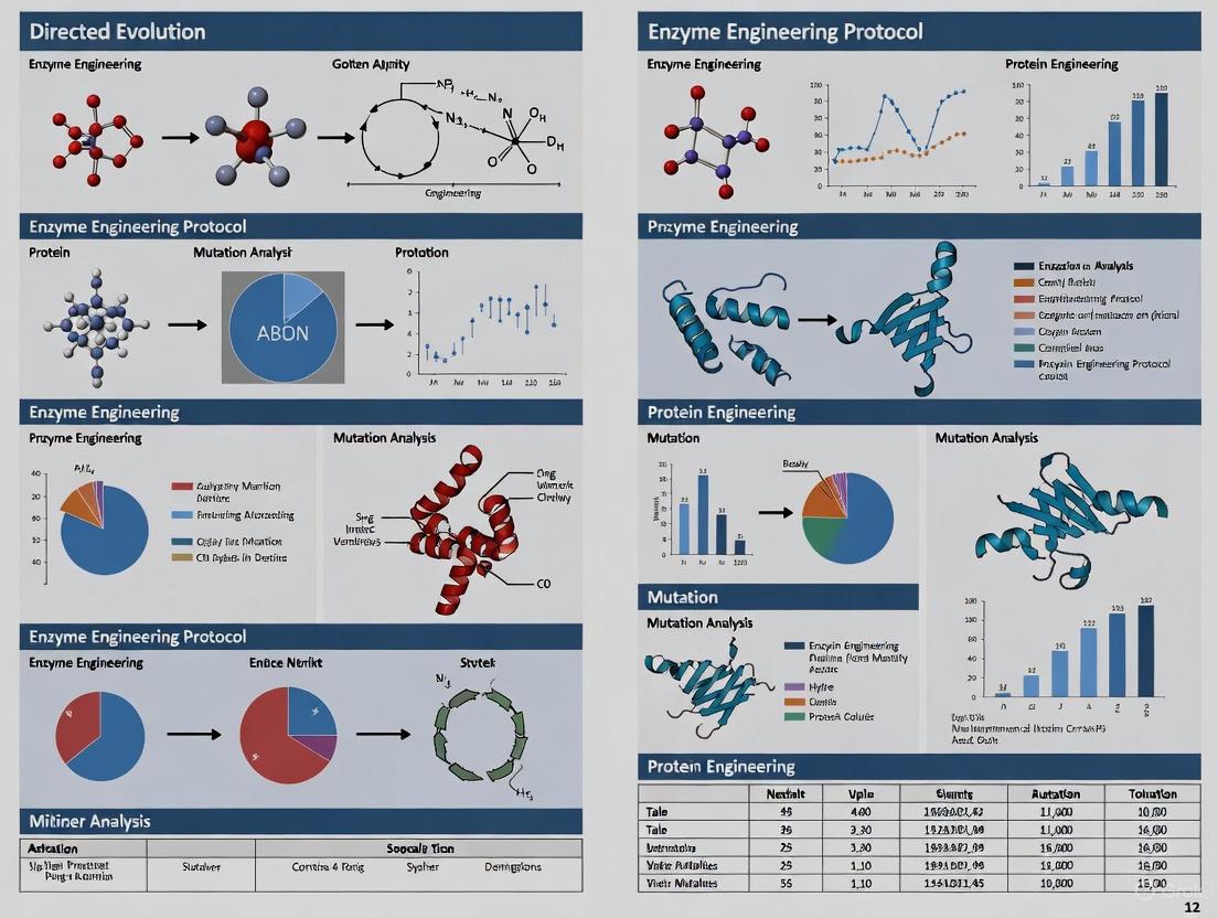
This article provides a comprehensive guide to directed evolution for enzyme engineering, tailored for researchers, scientists, and drug development professionals.
Directed Evolution Enzyme Engineering: Protocols, Machine Learning Advances, and Biomedical Applications
Abstract
This article provides a comprehensive guide to directed evolution for enzyme engineering, tailored for researchers, scientists, and drug development professionals. It covers foundational principles, from generating genetic diversity to high-throughput screening methodologies. The scope extends to modern protocols, including continuous in vivo evolution and machine learning-assisted frameworks like Active Learning-assisted Directed Evolution (ALDE) and Bayesian optimization. It also addresses common troubleshooting scenarios and offers a comparative analysis of different directed evolution systems, highlighting their validation in biomedical research for developing advanced tools such as improved degron technologies and therapeutic enzymes.
The Principles and Power of Directed Evolution: Mimicking Nature in the Lab
Directed evolution is a powerful protein engineering method that mimics the process of natural selection in a laboratory setting to steer proteins or nucleic acids toward a user-defined goal [1]. This approach has become one of the most useful and widespread tools in basic and applied biology, revolutionizing how scientists engineer enzymes for therapeutic, industrial, and research applications [2]. The profound impact of this approach was formally recognized with the 2018 Nobel Prize in Chemistry, awarded to Frances H. Arnold for her pioneering work that established directed evolution as a cornerstone of modern biotechnology and industrial biocatalysis [3].
The core premise of directed evolution is an iterative, recursive process that compresses geological timescales of natural evolution into weeks or months through intentional acceleration of mutation rates and application of unambiguous, user-defined selection pressures [3]. Unlike rational design approaches that require extensive knowledge of protein structure and function, directed evolution can deliver robust solutions without requiring detailed a priori knowledge of a protein's three-dimensional structure or its catalytic mechanism [3]. This capability allows it to bypass the inherent limitations of rational design, which relies on a predictive understanding of sequence-structure-function relationships that is often incomplete [3].
The Three-Phase Iterative Cycle
The directed evolution workflow functions as a three-part iterative engine, relentlessly driving a protein population toward a desired functional goal through repeated cycles of diversification, selection, and amplification [1] [3]. This process is represented in the following workflow:
Phase 1: Diversification - Generating Genetic Diversity
Diversification represents the first critical step in directed evolution, where genetic variation is introduced to create a library of protein variants [1] [3]. The quality, size, and nature of this diversity directly constrain the potential outcomes of the entire evolutionary campaign [3]. The creation of a diverse library of gene variants defines the boundaries of the explorable sequence space [3].
Quantitative Parameters for Diversification Methods
Table 1: Comparison of Genetic Diversification Methods in Directed Evolution
| Method | Mechanism | Mutation Rate | Library Size | Key Applications |
|---|---|---|---|---|
| Error-Prone PCR (epPCR) | Random mutagenesis via low-fidelity PCR | 1-5 base mutations/kb [3] | 10^4-10^6 variants [4] | Initial rounds of evolution, no structural data available [3] |
| DNA Shuffling | Recombination of DNA fragments | N/A (recombination-based) | 10^6-10^8 variants [2] | Combining beneficial mutations from multiple parents [2] |
| Site-Saturation Mutagenesis | Targeted randomization of specific codons | All 19 amino acids at targeted positions [4] | 10^2-10^4 variants per position [4] | Hotspot optimization, active site engineering [4] |
| In vivo Methods (e.g., EvolvR) | CRISPR-based targeted mutagenesis in living cells | 10^7-fold increase over wildtype [4] | Limited by transformation efficiency [1] | Continuous evolution, genomic integration required [4] |
Protocol: Error-Prone PCR (epPCR) for Random Mutagenesis
Principle: Error-prone PCR is a modified PCR that intentionally reduces the fidelity of the DNA polymerase, thereby introducing errors during gene amplification [3]. This technique is particularly valuable during initial evolution rounds when no structural information is available.
Reagents and Equipment:
- Target gene in appropriate vector
- Taq polymerase (lacks 3' to 5' proofreading exonuclease activity)
- dNTP mix
- MgCl₂ and MnCl₂
- PCR primers flanking target gene
- Thermocycler
- Agarose gel electrophoresis equipment
Procedure:
- Prepare Reaction Mix:
- 1X PCR buffer
- 0.2 mM each dATP and dGTP
- 1 mM each dCTP and dTTP (creates nucleotide imbalance)
- 0.5 mM MnCl₂ (concentration can be adjusted to tune mutation rate)
- 5-10 ng DNA template
- 0.2 μM forward and reverse primers
- 5 U Taq polymerase
- Nuclease-free water to 50 μL
Amplification Conditions:
- Initial denaturation: 95°C for 2 minutes
- 25-30 cycles of:
- Denaturation: 95°C for 30 seconds
- Annealing: 50-60°C (primer-specific) for 30 seconds
- Extension: 72°C for 1 minute per kb
- Final extension: 72°C for 5 minutes
Purification and Cloning:
- Purify PCR product using gel extraction or PCR cleanup kit
- Digest with appropriate restriction enzymes
- Ligate into expression vector
- Transform into competent host cells (E. coli typically used)
Critical Parameters:
- Mn²⁺ concentration directly controls mutation rate (typically 0.1-0.5 mM)
- dNTP imbalance enhances error rate
- Cycle number should be minimized to prevent accumulation of deleterious mutations
- Target 1-2 amino acid substitutions per variant for optimal results [3]
Phase 2: Selection - Identifying Improved Variants
The selection phase represents the critical bottleneck in directed evolution where rare improved variants are identified from large mutant libraries [1] [3]. This step, which links the genetic code of a variant (genotype) to its functional performance (phenotype), is widely recognized as the primary bottleneck in the process [3]. The success of a campaign is dictated by the axiom, "you get what you screen for" [3].
Key Consideration: A crucial distinction exists between screening and selection. Screening involves the individual evaluation of every member of the library for the desired property, while selection establishes a system where the desired function is directly coupled to the survival or replication of the host organism, automatically eliminating non-functional variants [1] [3].
Quantitative Metrics for Selection Methods
Table 2: High-Throughput Selection and Screening Platforms in Directed Evolution
| Method | Throughput | Quantitative Output | Key Advantage | Implementation Complexity |
|---|---|---|---|---|
| Microtiter Plate Screening | 10^2-10^3 variants/day [3] | Yes (colorimetric/fluorometric) [3] | Quantitative data on each variant [3] | Low [3] |
| Flow Cytometry | 10^7-10^8 variants/day [4] | Limited | Extreme throughput for binding assays [4] | Medium [4] |
| Droplet Microfluidics | >10^6 variants/day [4] | Yes | Compartmentalization enables ultrasensitive detection [4] | High [4] |
| Phage Display | 10^9-10^11 variants [1] | No (enrichment-based) | Direct genotype-phenotype linkage [1] | Medium [1] |
| In vivo Selection | Limited by transformation efficiency [1] | No | Couples enzyme activity to survival [1] | High (design) [1] |
Protocol: Growth-Coupled In Vivo Selection for Metabolic Engineering
Principle: This selection strategy directly couples the desired enzyme activity to host cell survival, enabling automatic selection of improved variants without individual screening [1]. This approach is particularly valuable for engineering enzymes in biosynthetic pathways.
Reagents and Equipment:
- Knockout host strain (auxotrophic for target metabolite)
- Expression vector library encoding enzyme variants
- Selective media lacking essential metabolite
- Non-selective media for control
- Sterile culture tubes/flasks
- Spectrophotometer for OD measurements
Procedure:
- Strain Preparation:
- Use a host strain with a knockout in the gene encoding the enzyme that produces an essential metabolite
- Alternatively, use a toxin-antitoxin system where enzyme activity degrades a toxin
Library Transformation:
- Transform the mutant library into the selection host strain
- Plate an aliquot on non-selective media to determine total library size
- Plate the remainder on selective media lacking the essential metabolite
Selection Process:
- Incubate plates at appropriate temperature for 24-72 hours
- Only clones expressing functional enzyme variants will survive on selective media
- Pick surviving colonies for further analysis
Validation and Amplification:
- Isolate plasmids from surviving colonies
- Restreak on selective media to confirm phenotype
- Sequence variants to identify beneficial mutations
Critical Parameters:
- Selection stringency must be carefully tuned - too stringent may yield no survivors, too lenient may allow false positives
- Include appropriate controls (empty vector, wildtype enzyme)
- Monitor for compensatory mutations in host genome that bypass the selection
- Use counterselection methods when possible to eliminate false positives
Phase 3: Amplification - Preparing for Subsequent Cycles
Amplification completes the iterative cycle by regenerating genetic material from selected variants to serve as templates for subsequent evolution rounds [1]. When functional proteins have been isolated, it is necessary that their genes are recovered too, therefore a genotype–phenotype link is required [1].
Key Functions of Amplification:
- Genetic Expansion: Increase the copy number of selected variant genes
- Template Preparation: Generate clean genetic material for the next diversification step
- Mutation Combination: Enable recombination of beneficial mutations from different variants
- Population Expansion: Grow sufficient biomass for downstream analysis and storage
Protocol: Gene Recovery and Template Preparation
Principle: This protocol describes the recovery of genetic material from selected variants and preparation for subsequent evolution rounds, including optional recombination of beneficial mutations.
Reagents and Equipment:
- Selected clones from screening/selection
- Plasmid miniprep kit
- PCR reagents and equipment
- Restriction enzymes and ligase
- Competent E. coli cells
- Agar plates with appropriate antibiotics
- Gel electrophoresis equipment
Procedure:
- Gene Recovery:
- Isolate plasmids from selected variants using plasmid miniprep protocol
- Alternatively, amplify gene directly from colonies using colony PCR
- Verify gene sequence by Sanger sequencing
Template Preparation Options: Option A: Sequential Evolution
- Use the best single variant as template for next diversification round
- Simple approach but may lead to evolutionary dead ends
Option B: Recombination Pool
- Mix genes from top 5-10 performers
- Use DNA shuffling to recombine beneficial mutations
- Creates chimeric genes with combined improvements
Quality Control:
- Verify sequence integrity of template genes
- Confirm expression and activity of template variants
- Store glycerol stocks of template strains
Critical Parameters:
- Include back-crossing steps to remove neutral or deleterious mutations
- Maintain population diversity when possible rather than focusing on single best variant
- Archive intermediate variants to prevent loss of beneficial traits
- Monitor for accumulation of neutral mutations that don't contribute to function
Research Reagent Solutions for Directed Evolution
Successful implementation of directed evolution requires carefully selected reagents and materials that enable efficient diversification, selection, and amplification. The following toolkit represents essential components for establishing a robust directed evolution pipeline.
Table 3: Essential Research Reagent Solutions for Directed Evolution
| Reagent/Material | Function | Key Considerations | Example Applications |
|---|---|---|---|
| Error-Prone PCR Kit | Introduces random mutations throughout gene | Adjustable mutation rate; Mn²⁺ concentration critical [3] | Initial diversification; exploring unknown sequence spaces [3] |
| DNA Shuffling Reagents | Recombines beneficial mutations from multiple parents | Requires >70% sequence identity for efficiency [3] | Combining hits from different evolution lineages [2] |
| Site-Directed Mutagenesis Kit | Targets specific residues for saturation mutagenesis | NNK codons reduce library size while maintaining diversity [4] | Active site optimization; hotspot engineering [4] |
- High-Throughput Screening Platform: Enables rapid assessment of variant libraries; Selection of platform depends on throughput needs and assay compatibility [3] [4]
- Specialized Expression Vectors: Maintains genotype-phenotype linkage; Vectors with inducible promoters allow controlled expression during selection [1]
- Chemical Mutagens: Alternative to epPCR for in vivo mutagenesis; Agents like EMS or NTG can provide different mutational spectra [2]
Advanced Applications and Integrated Approaches
OrthoRep: A Continuous Evolution System
The OrthoRep system developed by Chang Liu's laboratory represents a revolutionary approach to directed evolution that enables continuous evolution in yeast [5]. This system utilizes a cytoplasmic linear plasmid that replicates with an error-prone DNA polymerase, achieving mutation rates 100,000-fold higher than natural evolution while maintaining host genome stability [5].
Key Features:
- Targeted Mutagenesis: Mutations are confined to the target gene on the plasmid
- Continuous Operation: No need for iterative cloning between rounds
- High Mutation Rate: Enables exploration of vast sequence spaces
- Automation Compatibility: Suitable for extended evolution campaigns
Machine Learning-Enhanced Directed Evolution
Recent advances have integrated machine learning with directed evolution to create more efficient and predictive engineering pipelines [5] [6]. AI tools can now accurately propose beneficial mutations and predict function from sequence, dramatically shortening experimental cycles [6].
Implementation Framework:
- Initial Evolution Rounds: Generate training data through traditional directed evolution
- Model Training: Use sequence-function data to train predictive algorithms
- Virtual Screening: Prioritize promising variants in silico before experimental testing
- Active Learning: Iteratively improve models with new experimental data
Troubleshooting and Optimization Strategies
Successful directed evolution campaigns require careful optimization and problem-solving throughout the iterative cycle. The following strategies address common challenges:
Low Library Diversity:
- Increase mutation rate by adjusting Mn²⁺ concentration in epPCR
- Incorporate multiple diversification methods (epPCR + DNA shuffling)
- Use family shuffling with homologous genes from different species
Poor Selection Efficiency:
- Validate screening assay with known positive and negative controls
- Optimize selection stringency to balance identification of improved variants with maintaining sufficient survivors
- Implement counter-selection methods to eliminate false positives
Premature Convergence:
- Maintain population diversity rather than focusing only on best performer
- Incorporate neutral drifts to explore sequence space without strong selection pressure
- Use recombination methods to escape local fitness maxima
The iterative cycle of diversification, selection, and amplification represents the foundational engine of directed evolution, enabling researchers to engineer proteins with novel functions and optimized properties. By systematically applying and optimizing these core concepts, scientists can harness the power of evolution to create biological solutions to challenging problems across biotechnology, medicine, and industrial manufacturing.
The field of directed evolution, which enables researchers to engineer biomolecules with enhanced or entirely new functions, traces its origins to a pioneering experiment in the 1960s. Sol Spiegelman's groundbreaking work demonstrated that biomolecules could be evolved in a test tube, establishing the core principles that would later be refined and expanded into modern protein engineering methodologies [7]. This in vitro evolution approach mimicked natural selection by applying selective pressure for rapid replication, leading to the emergence of optimized RNA molecules that paved the way for contemporary enzyme engineering protocols [8] [9].
These foundational experiments established the fundamental cycle of directed evolution: diversification, selection, and amplification [1]. Over subsequent decades, this framework has been sophisticated through technological advances, including the integration of artificial intelligence and high-throughput screening methods, transforming directed evolution into a powerful tool for drug development, biotechnology, and basic research [10] [11]. This article traces this methodological evolution and provides detailed protocols for implementing these techniques in modern research settings.
Historical Breakthrough: Spiegelman's Monster Experiment
Original Experimental Protocol
In 1965, Sol Spiegelman and colleagues conducted what became known as the "Spiegelman's Monster" experiment, which achieved the first synthesis of a biologically competent, infectious nucleic acid in a test tube [7]. The protocol created an autonomous evolutionary system for RNA molecules.
Materials:
- Qβ bacteriophage RNA (4500 nucleotide bases)
- Qβ RNA replication enzyme (Qβ replicase)
- Free nucleotide triphosphates (ATP, GTP, CTP, UTP)
- Salts and appropriate reaction buffers
Methodology:
- Initial Mixture: Combine Qβ RNA template, Qβ replicase, free nucleotides, and salts in a test tube to create a replicating system [7].
- Serial Transfer: After allowing time for replication, transfer a small aliquot of the resulting RNA mixture to a fresh tube containing only buffer, nucleotides, and enzymes—no additional RNA template [8] [7].
- Iterative Evolution: Repeat this transfer process multiple times (74 generations in the original experiment), each time selecting the fastest-replicating molecules to seed the next generation [7].
Observations and Results: The original 4500-nucleotide RNA strand evolved into a dramatically shortened "dwarf genome" of only 220 bases after 74 generations [7]. This minimized RNA replicator, dubbed "Spiegelman's Monster," had shed any genetic information not essential for replication under the experimental conditions, demonstrating that molecules under selection pressure for rapid multiplication eliminate functional ballast [8] [7].
Modern Reevaluation (1997 Protocol)
Thirty years later, researchers revisited Spiegelman's work with advanced molecular biology techniques, employing a modified self-sustained sequence replication (3SR) method that mimics part of the HIV-1 replication cycle in vitro [8].
Materials:
- RNA template (220b RNA representing a 220-base segment of the HIV-1 genome)
- HIV-1 reverse transcriptase
- T7 RNA polymerase
- Nucleotide triphosphates
- Reaction buffers
- Thiazole orange intercalating dye for fluorescence monitoring
- Serial Transfer Apparatus (STA) for automated monitoring and transfer
Methodology:
- 3SR Reaction Setup: The RNA template undergoes reverse transcription by HIV-1 reverse transcriptase, followed by second-strand synthesis and transcription of the resulting dsDNA by T7 RNA polymerase [8].
- Automated Serial Transfer: The STA monitors nucleic acid concentration online via laser-induced fluorescence from thiazole orange intercalation, automatically transferring aliquots to fresh reaction mixtures during exponential growth phase [8].
- Selection Pressure: Transfers during exponential growth phase select for faster-replicating variants, as all enzymes and nucleotides are present in excess [8].
Results: After just two serial transfers, two shorter RNA species (EP1 [48b] and EP2 [54b]) emerged through deletion mutations, displacing the original 220b RNA template within thirty transfers due to superior replication rates [8]. Sequence analysis suggested these variants formed via HIV-1 reverse transcriptase strand-transfer reactions [8].
The Evolution of Methodologies: Key Transitions
The transition from early evolutionary experiments to modern protein engineering has been marked by significant methodological advances, particularly in library generation and screening techniques. The table below summarizes the quantitative evolution of these methods.
Table 1: Evolution of Directed Evolution Methodologies
| Era | Library Generation Methods | Library Size | Screening Throughput | Key Advantages |
|---|---|---|---|---|
| 1960s-70s | Serial transfer of natural molecules [7] | Limited | Low | Simple setup, foundational principles |
| 1980s-90s | Error-prone PCR, DNA shuffling [12] [1] | 10⁴-10⁶ variants | Moderate (10³-10⁴) | Whole-gene randomization, recombination benefits [12] |
| 2000s-2010s | Site-saturation mutagenesis, targeted libraries [12] | 10⁶-10⁸ variants | High (10⁷ via FACS) | Focused diversity, better coverage of mutational space [12] |
| 2020s-Present | AI-informed constraints, inverse folding models [10] [11] | 10⁸-10¹⁵ variants | Very high (10⁷-10⁹) | Reduced useless variants, predictive design [11] |
Modern Library Generation Techniques
Contemporary directed evolution employs sophisticated library generation methods that maximize functional diversity while minimizing library size:
Site-Saturation Mutagenesis:
AI-Informed Constraints for Protein Engineering (AiCE):
- Principle: Uses protein inverse folding models to predict high-fitness mutations guided by structural and evolutionary constraints [11]
- Protocol: Sample sequences from inverse folding models, integrate structural constraints, identify beneficial single and multi-mutations [11]
- Performance: Success rates of 11%-88% across eight protein engineering tasks, spanning proteins from tens to thousands of residues [11]
Contemporary AI-Driven Protein Engineering
The integration of artificial intelligence has transformed protein engineering from a largely empirical process to a systematic engineering discipline. A 2025 review established the first comprehensive roadmap for AI-driven protein design, organizing tools into a coherent seven-toolkit workflow [10].
Table 2: AI-Driven Protein Design Toolkit (Adapted from Noivirt et al., 2025 [10])
| Toolkit | Purpose | Key Tools | Research Application |
|---|---|---|---|
| T1: Protein Database Search | Find sequence/structural homologs | BLAST, Foldseek | Identify starting scaffolds and templates |
| T2: Protein Structure Prediction | Predict 3D structures from sequences | AlphaFold2, RoseTTAFold | Determine structures for wild-type and mutant proteins |
| T3: Protein Function Prediction | Annotate function, predict binding sites | DeepFRI, protein language models | Predict functional consequences of mutations |
| T4: Protein Sequence Generation | Generate novel sequences | ProteinMPNN, ESM | Design sequences for desired structures/functions |
| T5: Protein Structure Generation | Create novel protein backbones | RFDiffusion, RoseTTAFold | De novo design of protein scaffolds |
| T6: Virtual Screening | Computationally assess candidates | Molecular docking, MD simulations | Prioritize variants for experimental testing |
| T7: DNA Synthesis & Cloning | Translate designs to DNA sequences | Custom gene synthesis, codon optimization | Prepare designed proteins for experimental validation |
Case Study: AI-Driven Directed Evolution Protocol
The AiCE (AI-informed constraints for protein engineering) approach represents the cutting edge of directed evolution methodology, combining computational predictions with experimental validation [11].
Materials:
- Protein inverse folding models (e.g., ProteinMPNN)
- Structural prediction software (e.g., AlphaFold2)
- Target protein gene sequence
- Appropriate expression system (E. coli, yeast, etc.)
- High-throughput screening assay components
Methodology:
- Initial Sequence Analysis:
- Input wild-type sequence into inverse folding model
- Generate structural predictions using AlphaFold2
- Identify evolutionarily conserved regions from multiple sequence alignments
Mutation Design:
- Sample potential mutations using inverse folding models
- Apply structural constraints to maintain folding stability
- Apply evolutionary constraints to preserve functional regions
- Generate library of single and multi-mutations with predicted high fitness
Experimental Validation:
- Synthesize and clone designed variants
- Express protein variants in appropriate host system
- Screen for desired activity using high-throughput methods
- Iterate design process with experimental feedback
Applications:
- Development of enhanced base editors (enABE8e, enSdd6-CBE, enDdd1-DdCBE) [11]
- Engineering proteins with varying sizes, structures, and functions [11]
- Success rates of 11%-88% across diverse protein engineering tasks [11]
Advanced Screening and Selection Methods
Modern directed evolution relies on sophisticated screening methodologies that enable researchers to efficiently identify rare beneficial variants from large libraries.
High-Throughput Screening Protocols
Fluorescence-Activated Droplet Sorting (FADS) Protocol:
Materials:
- Microfluidic droplet generation device
- Fluorogenic enzyme substrate
- Water-in-oil emulsion components
- FACS instrument capable of sorting droplets
- Library of cells expressing protein variants
Methodology:
- Droplet Generation:
Incubation and Reaction:
- Incubate droplets to allow enzyme expression and substrate turnover
- Fluorescent product accumulates within each droplet
Sorting and Recovery:
- Analyze droplets using fluorescence-activated cell sorter
- Sort droplets based on fluorescence intensity corresponding to enzyme activity
- Recover cells from sorted droplets for gene sequencing and further rounds of evolution
Performance Metrics:
- Throughput: >10⁷ variants per screening round [12]
- Application examples: Evolution of β-galactosidase, horseradish peroxidase, serum paraoxonase (100× improved activity) [12]
Research Reagent Solutions
Table 3: Essential Research Reagents for Directed Evolution
| Reagent Category | Specific Examples | Function/Application | Key Features |
|---|---|---|---|
| Library Generation | Error-prone PCR kits, Trimer phosphoramidites [12] | Create genetic diversity in target genes | Controlled mutation rates, reduced codon bias |
| Expression Systems | E. coli, yeast, in vitro transcription/translation systems [12] [1] | Produce protein variants from DNA libraries | High transformation efficiency, proper folding |
| Screening Reagents | Fluorogenic substrates, cell surface display scaffolds [12] | Link genotype to phenotype for screening | Sensitivity, compatibility with high-throughput formats |
| AI/Computational Tools | ProteinMPNN, RFDiffusion, AlphaFold2 [10] [11] | Predict structures, generate novel sequences | Integration capabilities, high accuracy |
| Selection Materials | Immobilized ligands, antibiotics, essential metabolites [1] | Apply selective pressure for desired function | Specificity, tunable stringency |
The journey from Spiegelman's RNA evolution experiments to contemporary AI-driven protein engineering represents a remarkable transformation in biological engineering capabilities. What began as a fundamental demonstration of evolutionary principles in a test tube has evolved into a systematic discipline capable of designing biomolecules with precision [8] [7] [11].
Current research frontiers include closing the gap between in silico predictions and in vivo performance, reducing computational costs, and establishing robust biosecurity governance frameworks [10]. The integration of deep learning methods with high-throughput experimental validation continues to expand the scope of protein engineering, enabling researchers to address challenges in medicine, sustainability, and biotechnology that were previously inaccessible [13].
As these methodologies become increasingly sophisticated and accessible, they empower researchers to not only modify existing proteins but to design entirely novel biomolecules from first principles, opening new frontiers in synthetic biology and personalized medicine [10] [13]. The continued refinement of these protocols promises to accelerate the development of next-generation biocatalysts, therapeutic proteins, and functional biomaterials.
Directed evolution is a powerful protein engineering methodology that mimics the process of natural selection in a laboratory setting to optimize biomolecules for human-defined applications. Unlike rational design, which requires detailed prior knowledge of protein structure and function to make specific, targeted mutations, directed evolution explores vast sequence spaces through iterative cycles of diversification and selection without needing structural blueprints [9]. This approach is particularly valuable because the relationship between a protein's amino acid sequence, its three-dimensional structure, and its ultimate function remains remarkably difficult to predict, even with advanced computational models [14]. Since the first in vitro evolution experiments in the 1960s, directed evolution has diversified into numerous techniques that can tackle increasingly complex protein engineering challenges, from altering enzyme substrate specificity to creating entirely novel protein switches [9] [15].
The fundamental advantage of directed evolution lies in its ability to navigate protein sequence spaces of astronomical size. For a modest protein of just 100 amino acids, the theoretical sequence space encompasses approximately 20100 (∼1.3 × 10130) possible variants – a number far exceeding the atoms in the known universe [14]. Where rational design struggles to predict which mutations will yield improvements, especially when epistasis (where the effect of one mutation depends on others) is prevalent, directed evolution uses empirical screening to efficiently discover beneficial combinations of mutations that would be impossible to foresee through structure-based design alone [16] [14].
Key Advantages Over Rational Design
Handling of Epistasis and Non-Additive Effects
Epistasis presents a fundamental challenge for rational design approaches, as the optimal amino acid at one position often depends on the identity of other residues in the sequence. This non-additive behavior makes it difficult to predict the effects of multiple mutations, even when single mutations are well-characterized. Directed evolution excels in navigating such rugged fitness landscapes by experimentally testing variant combinations and selecting those with synergistic effects.
A compelling example comes from the optimization of five epistatic residues in the active site of a Pyrobaculum arsenaticum protoglobin (ParPgb) for a non-native cyclopropanation reaction. Single-site saturation mutagenesis at these positions failed to identify significantly improved variants, and simple recombination of the best single mutants did not yield improved function, demonstrating strong negative epistasis [16]. Through directed evolution, researchers successfully optimized these epistatic residues, improving the yield of the desired product from 12% to 93% in just three rounds of experimentation [16]. This case highlights how directed evolution can identify optimal mutational combinations in highly epistatic regions where rational design would likely fail.
Table 1: Comparison of Rational Design and Directed Evolution Approaches
| Feature | Rational Design | Directed Evolution |
|---|---|---|
| Structural Information Required | High (detailed 3D structure essential) | Minimal to none |
| Handling of Epistasis | Poor (difficult to predict non-additive effects) | Excellent (empirically selects beneficial combinations) |
| Exploration of Sequence Space | Limited, focused on predetermined mutations | Broad, can discover unexpected solutions |
| Suitability for Novel Functions | Limited to functions relatable to known mechanisms | High (can optimize for any screenable function) |
| Automation Potential | Lower (requires expert analysis for each decision) | High (can be fully automated in screening workflows) |
Exploration of Vast Sequence Spaces
The sequence space of even small proteins is astronomically large, creating what is known as the "needle in a haystack" problem for protein engineering. Where rational design attempts to intellectually navigate this space using physical principles and structural knowledge, directed evolution employs experimental sampling guided by functional selection to efficiently locate optimal sequences.
This advantage is particularly evident when engineering proteins for non-native functions or substrates. For instance, when engineering the LaccID enzyme for proximity labeling in mammalian cells, researchers performed 11 rounds of directed evolution starting from an ancestral fungal laccase template [17]. The resulting enzyme gained the ability to function effectively in mammalian cellular environments – a feat that would have been extraordinarily difficult through rational design alone due to the complex interplay of factors including pH sensitivity, halide inhibition, glycosylation requirements, and copper coordination [17]. Through iterative diversification and selection, directed evolution discovered a sequence solution that optimally balanced these constraints without requiring explicit understanding of each contributing factor.
Machine Learning Enhancement Without Structural Dependency
Modern directed evolution increasingly incorporates machine learning (ML) to further enhance its efficiency in navigating sequence space. ML-assisted approaches can predict promising variants based on experimental data, dramatically reducing the number of variants that need to be experimentally tested.
Active Learning-assisted Directed Evolution (ALDE) represents a cutting-edge example of this integration. ALDE leverages uncertainty quantification in machine learning models to balance exploration of new sequence regions with exploitation of known promising variants [16]. In the ParPgb optimization case study, ALDE used batch Bayesian optimization to suggest which variants to test in each round based on previous experimental results [16]. Importantly, these ML methods typically use sequence-based representations rather than structure-based features, maintaining the advantage of not requiring structural information. The models learn the sequence-function relationship directly from experimental data, capturing epistatic effects and guiding the exploration toward sequences with higher fitness.
Diagram 1: Active Learning-assisted Directed Evolution (ALDE) Workflow. This iterative process combines wet-lab experimentation with machine learning to efficiently navigate protein sequence space without structural information.
Experimental Protocols and Methodologies
Library Creation Methods
The initial step in any directed evolution campaign involves creating genetic diversity in the target protein sequence. Various methodologies have been developed to generate libraries of variants, each with distinct advantages and applications.
Table 2: Key Library Generation Methods in Directed Evolution
| Method | Principle | Advantages | Disadvantages | Typical Library Size |
|---|---|---|---|---|
| Error-prone PCR | Random point mutations through low-fidelity PCR | Easy to perform; no prior knowledge needed | Mutational bias; limited sampling | 104–106 variants |
| DNA Shuffling | Random recombination of homologous sequences | Recombines beneficial mutations | Requires sequence homology | 106–108 variants |
| Site-Saturation Mutagenesis | All amino acids tested at specific positions | Comprehensive exploration of key positions | Limited to predefined positions | 20n (n=number of positions) |
| Nonhomologous Recombination | Recombination of unrelated genes | Creates novel protein folds and functions | Often disrupts protein folding | 105–107 variants |
Protocol: Site-Saturation Mutagenesis at Epistatic Residues
Application: Focused exploration of specific positions suspected to exhibit epistatic interactions, as demonstrated in the ParPgb active site engineering [16].
Materials:
- Template plasmid containing gene of interest
- NNK degenerate codon primers (NNK = A/T/G/C + A/T/G/C + G/T)
- High-fidelity DNA polymerase
- DpnI restriction enzyme
- Competent E. coli cells
Procedure:
- Design forward and reverse primers containing NNK codons at the target positions, with 15-20 bp flanking sequences complementary to the template.
- Set up PCR reaction: 10 ng template plasmid, 0.5 μM each primer, 200 μM dNTPs, 1× polymerase buffer, 1 U polymerase in 50 μL total volume.
- Run thermocycling program: 95°C for 2 min; 18 cycles of 95°C for 30 s, 55°C for 30 s, 68°C for 1 min/kb; 68°C for 5 min.
- Digest template plasmid with DpnI (10 U, 37°C for 1 h) to eliminate methylated parental DNA.
- Purify PCR product and transform into competent E. coli cells.
- Plate transformed cells on selective media and incubate overnight at 37°C.
- Harvest library for screening, typically achieving 105-106 transformants.
Critical Notes: The NNK degeneracy encodes all 20 amino acids while reducing stop codons to only one (TAG). For five simultaneous positions (as in the ParPgb example), the theoretical diversity is 3.2 × 10⁶ variants, requiring careful library size management [16].
Screening and Selection Methods
Identifying improved variants from libraries represents the second critical phase of directed evolution. The choice of screening method depends on the desired property and available assay throughput.
Protocol: Yeast Surface Display for Enzyme Engineering
Application: Evolution of LaccID from an ancestral fungal laccase for improved activity in mammalian cells [17].
Materials:
- Yeast surface display vector (e.g., pCTCON2)
- Saccharomyces cerevisiae EBY100 strain
- Fluorescent substrate (e.g., biotin-phenol for LaccID)
- Streptavidin-fluorophore conjugate
- Fluorescence-activated cell sorter (FACS)
- SD-CAA and SG-CAA media
Procedure:
- Clone laccase library into display vector as Aga2p fusion, transform into EBY100 yeast.
- Induce expression in SG-CAA medium at 20°C for 24-48 h.
- Incubate 107 cells with biotin-phenol substrate (250-500 μM) in appropriate buffer for 1-2 h.
- Wash cells and incubate with streptavidin-fluorophore conjugate (1:100 dilution) on ice for 15 min.
- Wash and resuspend in ice-cold PBS for FACS analysis.
- Sort top 1-5% of fluorescent population, collect into recovery media.
- Plate sorted cells on selective media and grow at 30°C for 2-3 days.
- Harvest plasmid DNA for subsequent rounds or analysis.
Critical Notes: In the LaccID evolution, selection stringency was progressively increased over 11 rounds by reducing labeling time and adding radical quenchers to minimize trans-labeling between cells [17]. This protocol enabled a 4-fold improvement in biotinylation efficiency compared to the starting template.
Table 3: Essential Research Reagent Solutions for Directed Evolution
| Reagent/Category | Function in Protocol | Example Application |
|---|---|---|
| NNK Degenerate Codon Primers | Encodes all 20 amino acids at target positions | Site-saturation mutagenesis at epistatic sites [16] |
| Biotin-Phenol Probes | Radical-based labeling for detection | Screening laccase activity in yeast display [17] |
| Fluorescent-Activated Cell Sorter (FACS) | High-throughput screening based on fluorescence | Enriching active enzyme variants from libraries [17] |
| Error-Prone PCR Kit | Introduces random mutations throughout gene | Creating diverse variant libraries [9] |
| Specialized Media (SD/SG-CAA) | Controls expression of surface-displayed proteins | Yeast surface display system [17] |
Case Studies and Applications
Optimization of Non-Natural Enzyme Function
The ParPgb engineering case exemplifies how directed evolution excels at optimizing enzymes for functions not found in nature. The goal was to improve cyclopropanation yield and stereoselectivity – a non-biological carbene transfer reaction [16]. After defining a combinatorial space of five epistatic active site residues, researchers applied ALDE through three rounds of experimentation:
Round 1: Initial library generation and screening provided the first sequence-fitness data points. Round 2: ML model trained on round 1 data proposed variants likely to have improved fitness. Round 3: Additional data collection further refined the model and identified optimal variants.
The final optimized variant achieved 99% total yield and 14:1 diastereoselectivity for the desired cyclopropane product – a dramatic improvement from the starting 12% yield [16]. Notably, the mutations in the final variant were not predictable from initial single-mutation scans, underscoring how directed evolution navigates epistatic interactions without requiring structural understanding of the underlying mechanism.
Creating Protein Switches Through Nonhomologous Recombination
Directed evolution enables the creation of entirely novel protein functions not observed in nature, such as allosteric switches. In one pioneering example, researchers recombined genes for maltose-binding protein (MBP) and TEM1 β-lactamase (BLA) to create MBP-BLA hybrids where maltose binding regulates β-lactamase activity [15].
The process involved iterative library construction and screening:
- Creating libraries of circularly permuted BLA genes inserted at various positions in MBP
- Selecting for ampicillin resistance in the presence of maltose
- Screening for maltose-dependent nitrocefin hydrolysis activity
This approach yielded effective "on-off" switches with maltose altering catalytic activity by up to 600-fold [15]. The success of this nonhomologous recombination strategy demonstrates directed evolution's power to discover functional sequences in vast combinatorial spaces where rational design would struggle to identify viable connections between unrelated protein domains.
Diagram 2: Creating Protein Switches through Nonhomologous Recombination. This workflow demonstrates how directed evolution can create novel allosteric regulation not found in nature.
Directed evolution represents a powerful paradigm for protein engineering that leverages empirical screening rather than structural prediction to navigate vast sequence spaces. Its key advantages over rational design include the ability to handle epistatic interactions, explore unprecedented sequence territory, and optimize proteins without requiring structural information. The integration of machine learning approaches like ALDE further enhances the efficiency of this exploration by leveraging uncertainty quantification to guide experimental efforts [16].
As synthetic biology capabilities advance, enabling more sophisticated library construction and screening methodologies, directed evolution continues to grow as an essential tool for biocatalyst development. Its application to increasingly complex challenges – from engineering non-natural enzyme functions to creating novel protein switches – demonstrates its versatility and power. For researchers seeking to optimize enzyme properties, especially when structural information is limited or epistatic effects are significant, directed evolution provides a robust methodology for discovering dramatically improved variants that would remain inaccessible to purely rational approaches.
In both natural evolution and laboratory-directed evolution, fitness is the paramount concept defining an enzyme's success. It is a quantifiable measure of an enzyme's ability to perform its function in a specific environment, directly driving its selection or amplification. In nature, fitness is determined by an enzyme's contribution to organismal survival and reproduction [18]. In directed evolution, fitness is an engineered parameter designed by researchers to select for desired enzymatic properties, such as activity, stability, or specificity [19]. Understanding the paradigms of natural enzyme evolution provides a foundational framework for designing effective directed evolution protocols. Natural evolution primarily operates through mechanisms such as gene duplication and divergence, which allows one gene copy to acquire new functions while the other maintains essential ancestral activities [18]. Furthermore, the Innovation-Amplification-Divergence (IAD) model highlights the critical role of promiscuous activities—a latent pool of low-level side activities—as a starting point for the evolution of new functions [18]. This report bridges these evolutionary principles with cutting-edge laboratory protocols, detailing how modern directed evolution harnesses and refines these natural processes to solve complex challenges in biocatalysis and therapeutic development.
Theoretical Foundations of Enzyme Evolution
The evolution of enzyme function is governed by well-established evolutionary paradigms that explain how new activities emerge and are optimized. These models provide the conceptual toolkit for designing effective directed evolution campaigns.
Key Evolutionary Models
- Gene Duplication and Divergence: This classic model, proposed by Susumo Ohno, posits that after a gene duplication event, one copy is freed from selective pressure to maintain the original function. This copy can accumulate mutations, potentially leading to the neofunctionalization and emergence of a new enzymatic activity [18].
- The Innovation-Amplification-Divergence (IAD) Model: This model addresses limitations of the neofunctionalization model by incorporating the role of promiscuity [18]. It outlines a three-step process:
- Innovation: A mutation grants an enzyme a novel, typically low-level, promiscuous activity that has no immediate fitness consequence.
- Amplification: The gene undergoes duplication or amplification, increasing the dosage and thus the total cellular level of the promiscuous activity. This provides a immediate fitness advantage if the new activity becomes beneficial.
- Divergence: The amplified gene copies then independently mutate and specialize, with some copies refining the new activity and others being lost, eventually resulting in a specialized new enzyme [18].
- Evolution by Domain Recombination: Beyond the duplication of whole genes, the recombination of protein domains—functional and structural units—allows for larger evolutionary jumps and more significant changes of function by mixing and matching functional modules [18].
Quantifying Evolutionary Constraints
The influence of an enzyme's active site extends beyond its immediate vicinity, imposing varying degrees of evolutionary constraint. The range of this evolutionary coupling varies significantly among different enzymes.
Table 1: Variation in Evolutionary Coupling Range Among Enzymes
| Characteristic | Reported Range | Implication for Directed Evolution |
|---|---|---|
| Evolutionary Coupling Range | 2 to 20 Å [20] | Mutations far from the active site can impact function; the "mutable landscape" is enzyme-specific. |
| Underlying Physical Coupling | Short-range for all enzymes [20] | The root cause of long-range evolutionary effects is functional selection pressure, not physical energy transfer. |
| Determining Factor | Functional selection pressure [20] | The strength and nature of the selection pressure defined in a screen dictate which mutations are beneficial. |
Application Note: Growth-Coupled Continuous Directed Evolution
Experimental Principle and Workflow
Growth-coupled continuous directed evolution (GCCDE) represents a paradigm shift in enzyme engineering. It seamlessly integrates in vivo mutagenesis with a powerful selection system that directly links desired enzyme activity to host cell survival and growth [19]. This protocol automates the evolutionary process, allowing for the rapid and high-throughput exploration of vast sequence spaces exceeding 10⁹ variants in a single continuous culture [19]. The core logic of the experimental workflow is as follows:
Detailed Experimental Protocol
Objective: To enhance a specific enzymatic activity (e.g., low-temperature β-galactosidase activity of CelB) while maintaining other key properties (e.g., thermostability) through growth-coupled continuous directed evolution.
Background: The thermostable enzyme CelB from Pyrococcus furiosus has low activity at moderate temperatures. Its activity is coupled to E. coli growth by making the bacterium dependent on CelB's ability to hydrolyze lactose for survival in a minimal medium [19].
Protocol Steps:
Strain and Plasmid Construction
- Clone the gene encoding the target enzyme (e.g.,
celB) into an appropriate expression vector. - Integrate this construct into an E. coli host strain that is deficient in endogenous lactose metabolism (e.g., ∆lacZ).
- Co-transform or integrate the MutaT7 mutagenesis system, which typically consists of a T7 RNA polymerase mutant and a mutagenic plasmid, to enable in vivo hypermutation of the target gene [19].
- Clone the gene encoding the target enzyme (e.g.,
Establishment of Growth Coupling
- Prepare a minimal medium where lactose is the sole carbon source.
- Inoculate the engineered E. coli strain into this medium. Only cells expressing a functional CelB enzyme can hydrolyze lactose and grow.
- Validate the coupling by demonstrating that growth rate is proportional to enzyme activity in a controlled experiment.
Continuous Cultivation and Evolution
- Initiate a continuous culture in a bioreactor (e.g., a turbidostat or chemostat) containing the lactose minimal medium.
- The MutaT7 system will continuously generate random mutations in the
celBgene during cell division, creating a diverse library of variants (>10⁹ variants) [19]. - Allow the culture to grow under constant selection pressure. Variants with improved β-galactosidase activity will hydrolyze lactose more efficiently, leading to a faster growth rate. These fitter variants will outcompete others and dominate the population over time.
- Maintain the continuous culture for multiple generations (e.g., for weeks), allowing for the accumulation of beneficial mutations.
Variant Isolation and Characterization
- Sample the culture at intervals and plate cells on solid medium to isolate individual clones.
- Screen isolated clones for the desired improved activity (e.g., higher β-galactosidase activity at 37°C) using a colorimetric assay (e.g., ONPG hydrolysis).
- Sequence the gene from the best-performing clones to identify the causative mutations.
- Purify the evolved enzyme and perform biochemical assays to confirm that enhanced low-temperature activity was achieved without compromising thermostability.
Research Reagent Solutions
The following table details the key reagents and their critical functions in the GCCDE protocol.
Table 2: Essential Research Reagents for Growth-Coupled Continuous Directed Evolution
| Reagent / Material | Function and Importance in the Protocol |
|---|---|
| MutaT7 Mutagenesis System | An in vivo mutagenesis tool that uses a mutated T7 RNA polymerase to introduce random mutations into the target gene during transcription, enabling continuous library generation without manual intervention [19]. |
| Specialized E. coli Strain (e.g., ∆lacZ) | A genetically engineered host cell that lacks the native ability to metabolize lactose. This is essential for creating a tight growth coupling where survival depends solely on the function of the evolved target enzyme [19]. |
| Minimal Medium with Lactose | A culture medium containing only essential salts and lactose as the sole carbon source. It creates the essential selection pressure that forces the host cell to rely on improved enzyme function for growth and survival [19]. |
| Continuous Bioreactor (Turbidostat/Chemostat) | A cultivation system that maintains a constant cell density and continuously supplies fresh medium. It allows for prolonged evolution over hundreds of generations and enables real-time, automated selection of superior variants [19]. |
Data Analysis and Interpretation
The success of a directed evolution campaign is validated through a combination of phenotypic analysis and genotypic characterization.
- Phenotypic Analysis: Monitor the culture's growth rate (doubling time) and optical density over the course of the experiment. A steady increase in growth rate under the selective conditions is a primary indicator of successful evolution [19].
- Genotypic Analysis: DNA sequencing of evolved variants identifies the specific mutations responsible for improved function. In the case of CelB evolution, mutations likely affecting substrate binding and catalytic turnover were identified [19]. Mapping these mutations onto a 3D protein structure can provide mechanistic insights.
- Biochemical Characterization: Compare the kinetic parameters (e.g., kcat, KM) and stability profiles (e.g., thermal denaturation temperature, Tm) of the evolved enzyme to the wild-type. Successful outcomes, as demonstrated with CelB, show significantly enhanced activity under the desired conditions (e.g., lower temperature) while preserving ancestral stability [19].
The Growth-Coupled Continuous Directed Evolution (GCCDE) protocol, powered by systems like MutaT7, effectively mirrors and accelerates natural evolutionary principles in the laboratory. By establishing a direct link between enzyme function and cellular fitness, it automates the selection of optimal variants from incredibly diverse libraries. This approach bypasses the traditional, labor-intensive cycles of error-prone PCR and screening, dramatically accelerating the enzyme engineering pipeline [19]. The principles outlined here—harnessing evolutionary models like IAD, defining fitness through clever selection pressures, and leveraging continuous evolution—provide a robust framework for optimizing enzymes for industrial biocatalysis, therapeutic development, and fundamental research. As these tools become more sophisticated and widely adopted, they will undoubtedly unlock new frontiers in our ability to tailor biological catalysts to meet the world's evolving chemical and medical needs.
A Practical Toolkit: Library Generation, Screening, and Selection Protocols
Within the framework of directed evolution for enzyme engineering, the strategic generation of genetic diversity is a critical first step. The choice of library construction method profoundly influences the efficiency and outcome of the entire engineering campaign. Researchers and drug development professionals are often faced with a strategic decision: whether to use random mutagenesis techniques, such as error-prone PCR (epPCR), which introduce mutations throughout the gene, or targeted approaches like saturation mutagenesis, which focus on specific amino acid positions. This application note provides a detailed comparison of these two fundamental strategies, supported by quantitative data and explicit protocols, to guide researchers in selecting and implementing the optimal methodology for their specific protein engineering goals.
Comparative Analysis: epPCR vs. Saturation Mutagenesis
The table below summarizes the core characteristics, advantages, and limitations of error-prone PCR and saturation mutagenesis to facilitate methodological selection.
Table 1: Comparison of Random and Targeted Mutagenesis Approaches
| Feature | Error-Prone PCR (epPCR) | Saturation Mutagenesis |
|---|---|---|
| Core Principle | Uses low-fidelity PCR conditions to introduce random mutations throughout the gene sequence [21] [3]. | Systematically replaces amino acid(s) at one or more predefined positions using degenerate primers [22] [23]. |
| Mutation Spectrum | Predominantly point mutations (base substitutions); inefficient for insertions/deletions [22] [3]. | Focused amino acid substitutions at targeted sites. |
| Prior Knowledge Required | Minimal; does not require structural or mechanistic data [22]. | Essential; relies on structural data or hotspot identification to choose target residues [3]. |
| Library Quality & Bias | Inherent bias towards transition mutations; accesses ~5-6 of 19 possible amino acids per position on average [3]. | Can achieve comprehensive coverage of all 20 amino acids at a single site; bias depends on degenerate codon used (e.g., NNK) [22]. |
| Primary Application | Initial exploration of sequence space, improving general stability, or when structural data is unavailable [3]. | Optimizing specific regions like active sites, substrate-binding pockets, or combinatorial active sites (CASTing) [23]. |
| Key Limitation | Mutation bias limits accessible sequence space; high frequency of neutral/deleterious mutations [22] [3]. | Restricted to known or presumed important regions; can miss beneficial distal mutations [3]. |
Experimental Protocols
Protocol 1: Library Generation via Error-Prone PCR
This protocol is adapted from established methodologies for creating random mutant libraries using epPCR, followed by a highly efficient cloning step [21].
Research Reagent Solutions & Materials
- Template DNA: Plasmid containing the wild-type gene of interest (e.g., pDsRed2, ~20-50 ng).
- Primers: Forward and reverse primers flanking the multiple cloning site of your expression vector.
- Error-Prone PCR Mix:
- DNA Polymerase: Low-fidelity polymerase without proofreading (e.g., from GeneMorph II Random Mutagenesis kit).
- dNTPs: Often used at unequal concentrations to promote misincorporation.
- MgCl₂: Elevated concentration (e.g., 7 mM) to reduce polymerase fidelity.
- MnCl₂: A critical additive (e.g., 0.5 mM) to further increase error rate [3].
- Cloning Reagents: Restriction enzymes and T4 DNA Ligase for Ligation-Dependent Cloning Process (LDCP), or reagents for Circular Polymerase Extension Cloning (CPEC) [21].
- E. coli Strain: Electrocompetent cells (e.g., TOP 10) for transformation.
Step-by-Step Procedure
- epPCR Setup:
- Set up a 50 µL PCR reaction as follows:
- 1X proprietary error-prone polymerase buffer
- dNTP mix (concentrations as per kit protocol)
- 0.3-0.5 µM each of forward and reverse primer
- 20 ng template DNA
- 5 U error-prone DNA polymerase
- Add MgCl₂ and/or MnCl₂ as required by the specific kit or protocol.
- Set up a 50 µL PCR reaction as follows:
- Thermocycling:
- Perform PCR using conditions such as: 1 cycle of 94 °C for 2 min; 30 cycles of 94 °C for 15 s, 60-68 °C for 30 s, 72 °C for 1-2 min/kb; and 1 final cycle of 72 °C for 5 min [21].
- Product Analysis and Purification:
- Verify the PCR product by 1% agarose gel electrophoresis.
- Purify the amplified product using a commercial PCR purification kit.
- Cloning (CPEC Method Recommended):
- Linearize Vector: Amplify your expression vector using primers that overlap with the ends of your purified epPCR product.
- CPEC Reaction: Mix the purified epPCR product (insert) and the linearized vector in a 1:1 molar ratio. Use a high-fidelity DNA polymerase (e.g., TAKARA LA Taq) in a PCR reaction without primers. The thermocycling conditions are: 94 °C for 2 min; 30 cycles of 94 °C for 15 s, 63 °C for 30 s, 68 °C for 4 min; and 72 °C for 5 min. The polymerase extends the overlapping regions, seamlessly circularizing the plasmid [21].
- Transformation and Library Analysis:
- Transform the CPEC reaction product into electrocompetent E. coli cells.
- Plate on selective media and incubate to obtain colonies.
- Assess library diversity by colony PCR and sequencing a representative number of clones.
Protocol 2: Library Generation by Saturation Mutagenesis
This protocol describes an improved two-stage, whole-plasmid PCR method for creating high-quality saturation mutagenesis libraries, even for difficult-to-amplify templates [23].
Research Reagent Solutions & Materials
- Template DNA: Plasmid containing the wild-type gene (e.g., pETM11-P450-BM3, ~50 ng).
- Primers: A pair of mutagenic primers containing the degenerate codon (e.g., NNK, where N=A/C/G/T, K=G/T) at the target position, designed with complementary ends. An optional "antiprimer" (a non-mutagenic primer) can be used in place of one mutagenic primer.
- High-Fidelity PCR Mix:
- DNA Polymerase: High-fidelity, thermostable polymerase (e.g., KOD Hot Start).
- dNTPs: Standard equilibrium concentration.
- MgSO₄: Concentration as optimized for the polymerase.
- Restriction Enzyme: DpnI (for digesting the methylated template plasmid post-PCR).
- E. coli Strain: Chemocompetent cells (e.g., DH5α) for transformation.
Step-by-Step Procedure
- Primary PCR (Megaprimer Synthesis):
- Set up a 50 µL PCR reaction containing:
- 1X polymerase buffer
- 0.2 mM dNTPs
- 0.3 µM mutagenic primer and antiprimer (or 0.3 µM of each mutagenic primer)
- 50 ng plasmid template
- 1 U high-fidelity DNA polymerase
- Thermocycling for megaprimer generation: 5-10 cycles of 94 °C for 30 s, 50-55 °C for 1 min, 70 °C for 1-2 min/kb [23].
- Set up a 50 µL PCR reaction containing:
- Secondary PCR (Whole-Plasmid Amplification):
- Without purifying the primary PCR product, directly initiate the second stage by increasing the annealing temperature to 65-75 °C to prevent primer annealing. Run an additional 20 cycles to amplify the plasmid using the megaprimers.
- Template Digestion:
- Treat the PCR product with DpnI (10 U, 37 °C for 1-2 hours) to digest the methylated parental template DNA.
- Transformation and Library Validation:
- Transform the DpnI-treated DNA directly into chemocompetent E. coli cells.
- Plate on selective media and incubate.
- The transformation step effectively closes nicks in the amplified plasmid.
- Sequence individual clones to confirm the introduction of the desired diversity and assess library quality.
Workflow Visualization
The following diagram illustrates the key procedural and logical differences between the two mutagenesis approaches within a directed evolution cycle.
Discussion and Strategic Outlook
The integration of machine learning (ML) is revolutionizing both random and targeted approaches. ML models can predict fitness landscapes from sequence-function data, guiding the design of smarter libraries and reducing experimental burden [24] [25]. For instance, ML-guided platforms integrating cell-free expression have been used to map fitness landscapes for amide synthetases, leading to the identification of variants with 1.6- to 42-fold improved activity [24].
Furthermore, advanced high-throughput methods using chip-based oligonucleotide synthesis are emerging. These allow for the precise construction of comprehensive mutagenesis libraries, such as full-length amber codon scanning libraries, with very high coverage (e.g., 93.75%) [22]. The choice between epPCR and saturation mutagenesis is not mutually exclusive. A powerful strategy involves an initial round of epPCR to identify beneficial "hotspots," followed by iterative saturation mutagenesis (ISM) to deeply explore those key positions, efficiently accumulating beneficial mutations [3] [23].
Selecting the appropriate library design methodology is a critical determinant of success in directed evolution. Error-prone PCR is a versatile, knowledge-independent tool ideal for broad exploration of sequence space and initial improvements. In contrast, saturation mutagenesis is a highly efficient, targeted strategy for rational optimization of specific protein regions once key residues have been identified. The decision should be guided by the availability of structural information, the specific protein property being engineered, and the available screening capacity. As the field advances, the convergence of classical methods with machine learning and synthetic biology promises to further accelerate the engineering of robust biocatalysts for therapeutic and industrial applications.
Directed evolution is a powerful protein engineering technique that mimics natural selection in laboratory settings to generate biomolecules with improved or novel functions, such as enhanced catalytic efficiency, altered substrate specificity, or increased stability [9]. Since its conceptual origins in Spiegelman's in vitro evolution experiments in the 1960s, the field has diversified considerably, with modern applications spanning industrial biocatalysis, therapeutic development, and biosensor engineering [9] [26]. A critical bottleneck in any directed evolution campaign remains the identification of improved variants from vast genetic libraries, which necessitates robust high-throughput screening (HTS) and selection methodologies [26].
This Application Note details three principal high-throughput screening platforms—optical methods, fluorescence-activated cell sorting (FACS), and emulsion microdroplet technologies—within the context of directed enzyme evolution. We provide experimental protocols, comparative performance metrics, and implementation guidelines to assist researchers in selecting and optimizing appropriate screening strategies for their specific engineering objectives.
Optical Screening Methods
Optical screening methods utilize colorimetric or fluorimetric changes to report enzymatic activity. These assays are typically performed in microtiter plates (MTPs), which miniaturize reactions to volumes of 100-200 µL in 96-well formats, or even lower volumes in 384-well and 1536-well formats [26]. The primary advantage of optical methods is their direct compatibility with traditional enzyme assays, requiring only that substrate consumption or product formation generates a measurable change in absorbance or fluorescence.
Recent advancements have integrated automation and online monitoring systems. For instance, the Biolector system enables online monitoring of light scatter and NADH fluorescence signals during cultivation, providing real-time data on cell growth and enzyme activity without manual sampling [26].
Protocol: Microtiter Plate-Based Screening for Hydrolase Activity
Purpose: To identify enzyme variants with enhanced hydrolase activity from a library of mutants expressed in E. coli.
Materials:
- Reagents: LB medium with appropriate antibiotics, isopropyl β-D-1-thiogalactopyranoside (IPTG), assay buffer (e.g., phosphate-buffered saline, pH 7.4), fluorogenic or chromogenic substrate (e.g., p-nitrophenyl acetate for esterases), stop solution (e.g., 1 M sodium carbonate).
- Equipment: 96-well deep-well plates, 96-well clear optical bottom assay plates, microplate shaker/incubator, multi-channel pipettes, plate reader capable of measuring absorbance and/or fluorescence.
Procedure:
- Library Expression:
- Inoculate individual enzyme variant clones into 96-well deep-well plates containing 500 µL LB medium with antibiotics.
- Grow cultures at 37°C with shaking (800 rpm) until OD600 reaches ~0.6.
- Induce protein expression by adding IPTG to a final concentration of 0.1-1.0 mM.
- Continue incubation for 16-24 hours at appropriate temperature (e.g., 20-30°C for improved soluble expression).
Cell Harvesting and Lysis:
- Centrifuge deep-well plates at 3,000 × g for 10 minutes to pellet cells.
- Discard supernatant and resuspend cell pellets in 200 µL assay buffer.
- Lyse cells by adding lysozyme (0.2 mg/mL final concentration) and incubating for 30 minutes on a shaker, or by repeated freeze-thaw cycles.
Enzymatic Assay:
- Transfer 50 µL of cell lysate (or clarified supernatant for secreted enzymes) to a 96-well assay plate.
- Initiate the reaction by adding 50 µL of substrate solution prepared in assay buffer. For p-nitrophenyl acetate, use a final concentration of 0.1-1.0 mM.
- Incubate at assay temperature (e.g., 30°C) for 10-60 minutes.
- Stop the reaction by adding 50 µL of 1 M sodium carbonate (for chromogenic substrates where alkaline conditions stabilize the signal).
Detection and Analysis:
- Measure absorbance at 405 nm for p-nitrophenol release using a plate reader.
- Normalize enzyme activity to cell density (OD600) or total protein concentration.
- Select clones exhibiting significantly higher specific activity than the wild-type control for further characterization.
Considerations: Ensure substrate saturation and linear reaction kinetics by optimizing substrate concentration and reaction time. Include positive (wild-type enzyme) and negative (empty vector or inactive mutant) controls on each plate.
Digital Imaging for Solid-Phase Screening
Digital imaging (DI) extends optical screening to solid-phase assays, enabling direct colorimetric analysis of microbial colonies on agar plates [26]. This approach is particularly valuable for screening enzymes acting on problematic or insoluble substrates. A key application involves screening transglycosidases, where colonies expressing desired transferase activity develop intense coloration in the presence of appropriate acceptor molecules [26]. This method achieved a 70-fold improvement in the transglycosidase-to-hydrolysis activity ratio in one application [26].
Fluorescence-Activated Cell Sorting (FACS)
FACS is a powerful high-throughput screening method capable of analyzing and sorting individual cells at rates up to 30,000 cells per second based on their fluorescent properties [26]. The technique requires establishing a linkage between the desired enzymatic function and a fluorescent output, typically achieved through intracellular product entrapment, surface display systems, or genetic reporter constructs.
Product entrapment relies on differential transport properties of substrates and products across the cell membrane. A fluorescent substrate that can freely diffuse into and out of the cell is converted into a charged or bulky product that becomes trapped intracellularly, directly linking fluorescence intensity to enzymatic activity [26]. This approach enabled identification of a glycosyltransferase variant with 400-fold enhanced activity for fluorescent selection substrates [26].
Cell surface display fuses enzyme libraries to anchoring motifs on the outer membrane of bacteria, yeast, or mammalian cells, making the enzyme accessible to externally added substrates [26]. When combined with FACS, this system enabled a 6,000-fold enrichment of active bond-forming enzyme clones after a single sorting round [26].
Protocol: FACS-Based Screening via Intracellular Product Entrapment
Purpose: To isolate enzyme variants with enhanced activity from a large library using intracellular product accumulation.
Materials:
- Reagents: Fluorogenic substrate (membrane-permeant, e.g., fluorescein di-β-D-galactopyranoside for β-galactosidase), assay buffer, growth medium.
- Equipment: Flow cytometer with cell sorter (e.g., BD FACS Aria), microcentrifuge, incubator shaker.
Procedure:
- Library Preparation and Expression:
- Transform the enzyme variant library into an appropriate expression host (e.g., E. coli or yeast).
- Grow transformed cells under selective conditions to mid-log phase (OD600 ~0.5-0.8).
- Induce enzyme expression if using an inducible promoter.
Substrate Loading:
- Harvest cells by centrifugation at 2,000 × g for 5 minutes.
- Wash cells once with assay buffer to remove residual medium.
- Resuspend cells to OD600 ~1.0 in assay buffer containing the fluorogenic substrate.
- Incubate for 10-30 minutes at room temperature to allow substrate uptake and enzymatic conversion.
Product Entrapment and Washing:
- Dilute the cell suspension 10-fold with ice-cold assay buffer to stop the reaction.
- Pellet cells by centrifugation and wash twice with ice-cold buffer to remove extracellular substrate and product.
- Resuspend cells in ice-cold buffer at a density of ~1×10^7 cells/mL for sorting.
FACS Analysis and Sorting:
- Configure the flow cytometer with appropriate excitation lasers and emission filters for the fluorescent product.
- Set sorting gates based on fluorescence intensity of control cells (negative control: empty vector; positive control: known active variant if available).
- Sort the top 0.1-1% of highly fluorescent cells into collection tubes containing recovery medium.
- Plate sorted cells on selective agar plates for expansion and further analysis.
Considerations: Optimize substrate concentration and incubation time to maximize signal-to-noise ratio. Include control populations to establish appropriate gating strategies. Verify sorted clone activities through secondary validation assays.
Advanced FACS Applications: Double Emulsion Droplets
Traditional FACS is limited to intracellular or surface-associated products. Double emulsion (DE) droplets address this limitation by encapsulating individual cells in picoliter-scale aqueous compartments surrounded by a fluorinated oil phase and an outer aqueous phase, making them compatible with standard flow cytometers [27]. This water-in-oil-in-water structure retains secreted extracellular products in proximity to the producing cell, maintaining genotype-phenotype linkage [27].
Table 1: Comparison of High-Throughput Screening Platforms
| Screening Method | Throughput (variants/day) | Key Requirement | Typical Volume | Primary Application |
|---|---|---|---|---|
| Microtiter Plates | 10^2 - 10^4 | Spectral or fluorescent changes in bulk culture | 50-200 µL | Low-complexity libraries; validation assays |
| Digital Imaging | 10^3 - 10^4 | Colorimetric change on solid phase | N/A (solid phase) | Colony-based assays; insoluble substrates |
| FACS | 10^7 - 10^9 | Fluorescence linked to enzyme activity at single-cell level | N/A (single cell) | Intracellular or surface-displayed enzymes |
| Microdroplets | 10^7 - 10^9 | Compartmentalization of single cells | 1 pL - 10 nL | Extracellular products; secreted enzymes |
Emulsion Microdroplet Technologies
Microfluidic droplet technologies compartmentalize single cells or genes in monodisperse aqueous droplets surrounded by an immiscible oil phase, creating picoliter-volume reactors ideal for ultrahigh-throughput screening [28] [29]. This approach maintains critical genotype-phenotype linkage while enabling screening rates exceeding 10^7 variants per day [29]. The extreme miniaturization reduces reagent consumption by several orders of magnitude compared to microtiter plate-based assays.
Droplet microfluidics offers significant advantages over bulk emulsification methods, producing droplets with size variations of less than 3% (coefficient of variation) compared to 20-50% for bulk methods [29]. This monodispersity is critical for accurate quantitative screening, as fluorescence intensity directly correlates with product concentration only when droplet volumes are uniform.
Protocol: Fluorescence-Activated Droplet Sorting (FADS)
Purpose: To screen a metagenomic library or enzyme variant library for activity using microfluidic droplets.
Materials:
- Reagents: Fluorinated oil (e.g., HFE-7500) with 1-2% (w/w) PEG-PFPE amphiphilic block copolymer surfactant, aqueous phase (cell suspension in growth medium), fluorogenic enzyme substrate, lysis agent (e.g., lysozyme or B-PER reagent).
- Equipment: PDMS or glass microfluidic droplet generation and sorting chips, pressure-based fluid handling system or syringe pumps, fluorescence microscope, incubation vessel for droplets.
Procedure:
- Droplet Generation:
- Prepare a cell suspension of the enzyme library at approximately 1×10^6 cells/mL in growth medium containing the fluorogenic substrate and lysis agent.
- Load the aqueous phase and oil phase into separate syringes.
- Prime the microfluidic droplet generation device according to manufacturer's instructions.
- Generate monodisperse droplets (10-30 µm diameter, 0.5-10 pL volume) by flowing the aqueous and oil phases through the device at optimized pressure or flow rates.
- Collect emitted droplets in a syringe or plastic tube.
Droplet Incubation:
- Incubate the collected emulsion at appropriate temperature (e.g., 30°C) for 2-24 hours to allow cell growth, enzyme expression, and substrate conversion.
- Gently agitate the emulsion during incubation to prevent droplet sedimentation or coalescence.
Droplet Sorting:
- Reinject the incubated emulsion into the sorting device at controlled pressure or flow rate.
- As droplets pass through the laser detection region, measure fluorescence of each droplet.
- Apply a dielectrophoretic sorting pulse to deflect droplets exceeding the fluorescence threshold into a collection channel.
- Collect sorted droplets in a separate tube containing breaking buffer (e.g., 1H,1H,2H,2H-perfluoro-1-octanol in oil) or directly plate on selective media.
Cell Recovery and Analysis:
- Break the collected emulsion by adding 20% (v/v) 1H,1H,2H,2H-perfluoro-1-octanol, vortexing, and centrifuging.
- Recover the aqueous phase containing the sorted cells.
- Plate on selective agar media for outgrowth and further characterization of individual clones.
Considerations: Optimize cell density to maximize single-cell encapsulation according to Poisson distribution (typically ~20% of droplets contain exactly one cell). Validate sorting efficiency using control populations before running valuable libraries.
Double Emulsion Droplets for FACS Compatibility
Standard water-in-oil droplets are incompatible with conventional flow cytometers due to their oil continuous phase. Double emulsion (DE) droplets address this limitation by encapsulating the aqueous reaction compartment within an outer aqueous phase, creating water-in-oil-in-water structures compatible with FACS instrumentation [27]. This approach combines the high-throughput screening capabilities of microfluidics with the widespread availability of flow cytometers, though technical challenges include droplet rupture and sorting efficiency optimization [27].
Table 2: Essential Research Reagent Solutions for Droplet-Based Screening
| Reagent | Function | Example Formulation | Application Notes |
|---|---|---|---|
| Fluorinated Oil | Continuous phase for emulsion stability | HFE-7500 with 1.5% (w/w) PEG-PFPE surfactant | Biocompatible; oxygen-permeable; minimal small molecule diffusion |
| Block Copolymer Surfactant | Stabilizes droplets against coalescence | PEG-PFPE amphiphilic block copolymer, 1-2% in fluorinated oil | Prevents fusion during incubation and sorting |
| Fluorogenic Substrate | Reports enzymatic activity | 0.1-1.0 mM in aqueous buffer | Must be membrane-permeant for whole-cell assays |
| Lysis Agent | Releases intracellular enzymes | 0.2 mg/mL lysozyme or commercial B-PER | Required for intracellular targets in lysate-based screens |
| Breaking Buffer | Recovers cells from droplets | 20% (v/v) 1H,1H,2H,2H-perfluoro-1-octanol in carrier oil | Demulsifies collected droplets for cell plating |
Workflow Integration and Comparative Analysis
The selection of an appropriate screening method depends on multiple factors, including library size, enzyme characteristics, available instrumentation, and throughput requirements. The following diagram illustrates a generalized decision workflow for selecting optimal screening strategies in directed evolution campaigns:
Figure 1: Decision workflow for selecting high-throughput screening methods in directed enzyme evolution.
Emerging methodologies are further enhancing these screening platforms. Machine learning-guided approaches now integrate cell-free expression systems with predictive modeling, enabling more efficient exploration of protein sequence space [24]. Additionally, in vivo continuous evolution systems couple targeted mutagenesis with ultrahigh-throughput screening, allowing rapid enzyme optimization without iterative cloning steps [30].
Optical methods, FACS, and emulsion microdroplets represent a complementary toolkit for addressing diverse screening challenges in directed enzyme evolution. While optical methods provide accessibility and compatibility with standard laboratory equipment, FACS and droplet microfluidics offer substantially higher throughput for surveying vast sequence spaces. The integration of these platforms with emerging technologies in machine learning, biosensor development, and automated strain engineering promises to further accelerate the creation of novel biocatalysts for industrial and therapeutic applications.
Researchers should select screening methodologies based on their specific library characteristics, instrumentation access, and engineering objectives, while remaining cognizant of the continuous technological innovations expanding the capabilities of each platform.
Application Notes
Phage-Assisted Continuous Evolution (PACE)
PACE is a powerful directed evolution platform that enables rapid protein improvement in a continuous, automated manner without the need for sequential rounds of library creation and screening. This system directly links the desired activity of a protein of interest (POI) to the propagation of a bacteriophage, creating a strong selection pressure for improved variants over hundreds of generations.
Key Applications:
- Altering Enzyme Specificity: PACE has been successfully used to completely reprogram protease specificity. In one landmark study, TEV protease was evolved over ~2500 generations to cleave the human IL-23 sequence HPLVGHM, which differs at six of seven positions from its native substrate ENLYFQS. The resulting protease contained 20 amino acid substitutions and effectively inhibited IL-23-mediated immune signaling in murine splenocytes [31].
- Developing Genome Editing Tools: PACE is instrumental for evolving novel prime editors. The PE6 series of editors were developed using phage-assisted evolution, resulting in enzymes 516–810 base pairs smaller than previous editors while achieving a 24-fold improvement in loxP insertion efficiency in the murine brain cortex [32].
- Evolving Recombinases: Bridge recombinases for gene replacement therapies are being improved using PACE, linking recombinase activity directly to phage propagation to evolve enzymes capable of inserting healthy gene copies into specific genomic loci for treating genetic diseases like Alpha-1 Antitrypsin Deficiency [33].
Auxin-Inducible Degrons (AID)
The AID system provides a robust method for rapid, conditional protein depletion in non-plant systems by leveraging the plant auxin signaling pathway. Recent advancements have addressed initial limitations, making this technology suitable for more sensitive applications, including in vivo models.
Key Applications:
- Sharp Degradation Control in Multiple Systems: The next-generation AID2 system, employing an OsTIR1(F74G) mutant and 5-Ph-IAA ligand, demonstrates no detectable leaky degradation and functions at 670-times lower ligand concentrations. This system achieves rapid target depletion in yeast, mammalian cells, and mice, enabling precise functional studies of essential proteins [34].
- Studying Dynamic Cellular Processes: The improved ARF-AID system preserves basal protein levels by co-expressing the PB1 domain of ARF, which suppresses constitutive degradation of AID-tagged proteins. This allows for more accurate study of transcription factor dynamics, as demonstrated in genome-wide analyses of ZNF143 depletion effects on RNA polymerase pausing [35].
- Functional Genomics: AID tagging of endogenous genes enables rapid perturbation of protein function to define primary molecular responses. This is particularly valuable for studying essential genes where chronic depletion would be lethal or cause compensatory adaptations [35].
Protocols
Protocol 1: Implementing PACE for Protease Engineering
This protocol adapts methodology from Dickinson et al. (2017) for evolving proteases with altered substrate specificity [31].
Stage 1: System Design and Setup
1.1. Selection Phage (SP) Construction:
- Replace gene III in an M13 phage genome with your protease gene of interest.
- Clone the protease gene with a ribosome binding site and appropriate regulatory elements.
1.2. Accessory Plasmid (AP) Design:
- Implement a protease-activated RNA polymerase (PA-RNAP) system.
- Construct a PA-RNAP by fusing T7 RNA polymerase to T7 lysozyme via a linker containing the target substrate sequence.
- Place gene III under control of a T7 promoter on the AP.
1.3. Host Cell Preparation:
- Use E. coli harboring the AP and an optional mutagenesis plasmid (MP).
- For accelerated evolution, use MP6 which increases mutation rates approximately 300,000-fold.
Stage 2: Continuous Evolution Process
2.1. Lagoon Operation:
- Maintain a fixed-volume lagoon (typically 10-15 mL) with continuous inflow of fresh host cells.
- Set dilution rate to allow phage replication while washing out non-infectious particles.
- For challenging specificity changes, implement evolutionary stepping stones with intermediate substrates.
2.2. Monitoring and Harvesting:
- Regularly titer phage from the lagoon to monitor population dynamics.
- Harvest phage population once desired activity is achieved or after predetermined generations.
- Sequence protease genes from harvested phage to identify mutations.
Table 1: Quantitative Parameters for TEV Protease PACE [31]
| Parameter | Value | Notes |
|---|---|---|
| Generations to evolve IL-23 cleavage | ~2500 | From wild-type TEV to IL-23 cleaver |
| Number of amino acid substitutions | 20 | In final evolved variant |
| Positions differing from wild-type substrate | 6/7 | ENLYFQS → HPLVGHM |
| Mutation rate increase with MP6 | ~300,000× | Compared to wild-type E. coli |
Protocol 2: AID2 System for Sharp Protein Degradation in Mammalian Cells and Mice
This protocol is based on the AID2 system described by Nishimura et al. (2020) [34].
Stage 1: System Component Engineering
1.1. Target Protein Tagging:
- Fuse the mini-AID (mAID) degron (approximately 7 kD from Arabidopsis IAA17) to the protein of interest.
- For endogenous tagging, use CRISPR/Cas9 to insert mAID at the C- or N-terminus.
1.2. E3 Ligase Expression:
- Express the OsTIR1(F74G) mutant in target cells.
- Generate stable cell lines using lentiviral transduction or targeted integration.
- For in vivo applications, use tissue-specific or inducible promoters as needed.
Stage 2: Degradation Induction and Monitoring
2.1. Ligand Preparation:
- Prepare 5-Ph-IAA stock solution in DMSO (e.g., 1-10 mM).
- For mammalian cells, use working concentrations of 1 nM to 1 μM.
- For mouse studies, optimize dosing based on administration route.
2.2. Degradation Kinetics Assessment:
- Treat cells or animals with 5-Ph-IAA and monitor protein levels over time.
- For quantitative analysis, use Western blotting with densitometry.
- Determine degradation half-life from time-course data.
Table 2: Quantitative Comparison of AID Systems [34] [35]
| Parameter | Original AID | ARF-AID | AID2 |
|---|---|---|---|
| Basal degradation (leakiness) | High | Suppressed | Undetectable |
| Typical IAA concentration | 100-500 μM | 100-500 μM | 0.1-1 μM 5-Ph-IAA |
| DC50 (ligand concentration) | 300 ± 30 nM | Not specified | 0.45 ± 0.01 nM |
| Degradation half-life (reporter) | 147.1 ± 12.5 min | Improved vs. original | 62.3 ± 2.0 min |
| Application in mice | Challenging due to toxicity | Not demonstrated | Successful |
Visualization of Systems
Diagram 1: PACE Selection Mechanism
Diagram 2: AID2 Degradation Pathway
The Scientist's Toolkit
Table 3: Essential Research Reagents for PACE and AID Systems
| Reagent / Material | Function / Application | Specifications / Notes |
|---|---|---|
| OsTIR1(F74G) mutant | E3 ligase component for AID2 | Critical for reduced basal degradation and enhanced sensitivity [34] |
| 5-Ph-IAA | Synthetic auxin for AID2 | Enables degradation at nanomolar concentrations (DC50 = 0.45 nM) [34] |
| mini-AID (mAID) tag | 7 kD degron from Arabidopsis IAA17 | Fused to protein of interest for targeted degradation [34] |
| Selection Phage (SP) | M13 phage with gene III replaced | Carries evolving protease gene in PACE [31] |
| Accessory Plasmid (AP) | Host cell plasmid for PACE | Contains PA-RNAP and T7 promoter-controlled gene III [31] |
| Mutagenesis Plasmid (MP6) | Accelerates evolution in PACE | Increases mutation rate ~300,000× [31] |
| Protease-Activated RNAP | Links protease activity to gene III expression | T7 RNAP fused to T7 lysozyme via cleavable linker [31] |
| Bridge RNA (bRNA) | Guides recombination in evolved systems | Binds both genomic target and donor DNA for precise integration [33] |
Directed evolution (DE) is a cornerstone of modern protein engineering, enabling the development of enzymes and biomolecules with novel or enhanced functions. However, its efficiency is often hampered by the vastness of protein sequence space and the prevalence of epistasis, where the effect of one mutation depends on the presence of others, creating rugged fitness landscapes that are difficult to navigate [16] [36]. Machine learning (ML) has emerged as a powerful tool to overcome these limitations. This article details the application of two advanced ML frameworks—Active Learning-assisted Directed Evolution (ALDE) and Bayesian Optimization (BO)—for efficient navigation of protein fitness landscapes. Aimed at researchers and drug development professionals, these protocols provide a structured approach to accelerate the engineering of biocatalysts, therapeutics, and other functional proteins.
## The ALDE Framework: An Active Learning Paradigm for Directed Evolution
Active Learning-assisted Directed Evolution (ALDE) is an iterative machine learning workflow designed to optimize protein fitness more efficiently than traditional DE, especially in scenarios involving significant epistasis [16]. It functions by strategically selecting the most informative variants to test in the lab, thereby reducing experimental burden.
### Workflow and Protocol
The ALDE cycle consists of several key stages, as illustrated in the diagram below:
Diagram 1: The ALDE iterative workflow. This process cycles between wet-lab experimentation and computational modeling to efficiently navigate the fitness landscape. The key differentiator from traditional DE is the use of a machine learning model to actively guide the design of each subsequent library [16].
Step 1: Define the Combinatorial Design Space
- Objective: Identify a set of
ktarget residues for optimization. These are typically active site residues or regions known to influence the function of interest. - Protocol: The choice of
kinvolves a trade-off; a largerkcan capture more complex epistatic interactions but exponentially increases the sequence space (20^k possibilities). The design space should be informed by structural data or prior knowledge [16].
Step 2: Initial Library Construction and Screening
- Objective: Generate an initial dataset of sequence-fitness pairs to train the first ML model.
- Protocol: Simultaneously mutate all
kresidues using methods like PCR-based mutagenesis with NNK degenerate codons. Screen hundreds of variants using a relevant functional assay (e.g., GC-MS for product yield, activity assays) to measure fitness [16].
Step 3: Machine Learning Model Training and Variant Ranking
- Objective: Use the collected data to learn a mapping from protein sequence to fitness and propose the next set of variants to test.
- Protocol:
- Encoding: Represent protein sequences numerically (e.g., one-hot encoding).
- Model Training: Train a supervised ML model. The ALDE study found that models providing frequentist uncertainty quantification can be more consistent than Bayesian approaches in this context [16].
- Variant Ranking: Apply an acquisition function (e.g., Upper Confidence Bound) to the model's predictions to rank all sequences in the design space. This balances exploration of uncertain regions with exploitation of predicted high-fitness regions [16].
Step 4: Iterative Library Refinement
- Objective: Experimentally validate the computationally selected variants and update the model.
- Protocol: The top
N(e.g., tens to hundreds) ranked variants are synthesized and screened in the wet lab. The new sequence-fitness data is pooled with the existing data, and the cycle returns to Step 3. The process repeats until a fitness goal is met.
### Case Study: Application Notes for Optimizing a Protoglobin
Background: ALDE was successfully applied to engineer the protoglobin ParPgb for a non-native cyclopropanation reaction, a challenge where standard DE failed due to negative epistasis among five active-site residues (W56, Y57, L59, Q60, F89) [16].
Key Application Notes:
- Initial SSM Failure: Single-site saturation mutagenesis (SSM) at the five positions failed to yield variants with a significant desirable shift in the objective (product yield and selectivity) [16].
- Failed Recombination: Simple recombination of the best single mutants did not produce a high-fitness variant, confirming strong epistasis and the ineffectiveness of a greedy DE approach [16].
- ALDE Success: In just three rounds of ALDE, exploring only ~0.01% of the possible sequence space (32,000 variants), an optimal variant was identified that achieved 99% total yield and 14:1 diastereoselectivity for the desired cyclopropane product [16]. The mutations in the final variant were not predictable from the initial single-mutant screens, underscoring the critical role of ML in modeling epistatic interactions.
Table 1: Summary of Key Experimental Findings from the ParPgb Case Study
| Method | Number of Rounds | Key Experimental Observation | Final Variant Performance |
|---|---|---|---|
| Single-Site Saturation Mutagenesis (SSM) | N/A | No significant improvement in yield or selectivity from single mutants. | Not achieved [16]. |
| Simple Recombination of SSM Hits | N/A | Failed to combine beneficial mutations; epistasis prevented improvement. | Not achieved [16]. |
| Active Learning-assisted DE (ALDE) | 3 | Optimal combination of epistatic mutations identified from ~0.01% of sequence space. | 99% yield, 14:1 selectivity [16]. |
## Bayesian Optimization as a Core Engine for ML-Guided Evolution
Bayesian Optimization (BO) is a powerful strategy for global optimization of expensive black-box functions, making it ideally suited for guiding directed evolution where each fitness measurement requires a laborious experiment [37] [38]. It can serve as the computational engine within frameworks like ALDE.
### The Bayesian Optimization Cycle
The BO process is a sequential, model-based optimization strategy. Its core cycle is depicted below:
Diagram 2: The Bayesian Optimization cycle. The surrogate model approximates the fitness landscape, and the acquisition function uses it to decide which sequence to test next, balancing exploration and exploitation [37] [38].
### Protocols for Implementing Bayesian Optimization
1. Selecting and Training a Surrogate Model
- Objective: Create a probabilistic model that predicts fitness for any sequence in the design space and quantifies its own uncertainty.
- Protocol:
- Gaussian Process (GP): A common choice, especially for smaller datasets. It uses a kernel function (e.g., Tanimoto kernel for molecular fingerprints) to model covariance [37] [38].
- Bayesian Neural Networks (BNNs): Offer flexibility for more complex, high-dimensional data but may require more data and computational resources [39] [38].
- Ranking Models: An emerging alternative. Instead of predicting absolute fitness values, ranking models learn the relative order of sequences. Rank-based Bayesian Optimization (RBO) has shown similar or improved performance compared to regression-based BO, particularly on rough fitness landscapes with "activity cliffs" [39].
2. Utilizing the Acquisition Function
- Objective: Determine the next sequence(s) to evaluate experimentally by trading off exploration and exploitation.
- Protocol:
- Upper Confidence Bound (UCB): A popular function defined as
UCB(x) = μ(x) + λ * σ(x), whereμ(x)is the predicted mean fitness,σ(x)is the uncertainty, andλis a parameter controlling the balance. A higherλfavors exploration [37] [38]. - Expected Improvement (EI): Selects the point with the highest expected improvement over the current best observation [38].
- Batch Selection: For ALDE, which tests batches of variants per round, use a batch variant of the acquisition function (e.g., batch UCB) to select multiple diverse sequences simultaneously [16].
- Upper Confidence Bound (UCB): A popular function defined as
### Advanced Application: Regularized Bayesian Optimization
To prevent the optimization from proposing functionally improved but structurally unstable or non-native-like proteins, regularization can be incorporated.
- Protocol: Reformulate the acquisition function to include a penalty term. For example:
Acquisition_Regularized(x) = UCB(x) + β * R(x), whereR(x)is a regularization term [38]. - Structure-Based Regularization:
R(x)can be the predicted change in folding free energy (ΔΔG) computed by tools like FoldX. This biases the search toward thermodynamically stable variants [38]. - Evolutionary-Based Regularization:
R(x)can be the log-likelihood of the sequence under a generative model of natural protein sequences (e.g., a protein language model). This biases the search toward "native-like" sequences [38]. Evidence suggests structure-based regularization is consistently beneficial, while evolutionary regularization has mixed results [38].
Table 2: Key Software and Modeling Choices for Bayesian Optimization
| Component | Options | Application Notes and Considerations |
|---|---|---|
| Software Packages | BoTorch, Ax, GPyOpt, Dragonfly | Provides pre-built implementations of GPs, acquisition functions, and optimization loops [37]. |
| Surrogate Models | Gaussian Process (GP), Bayesian Neural Network (BNN), Ranking Model | GP: Best for low-data regimes. BNN/Ranking: Scalable for complex, high-dimensional data; ranking is robust to activity cliffs [39] [37] [38]. |
| Acquisition Functions | Upper Confidence Bound (UCB), Expected Improvement (EI), Probability of Improvement (PI) | UCB: Simple, tunable with λ. EI: No hyperparameters, widely effective [37] [38]. |
| Regularization | Structure-based (e.g., FoldX ΔΔG), Evolutionary (e.g., sequence likelihood) | Structure-based: Highly recommended to maintain protein stability. Evolutionary: Use with caution, as it may constrain the search space excessively [38]. |
## The Scientist's Toolkit: Essential Research Reagents and Materials
The following table details key reagents and computational tools essential for implementing ML-guided directed evolution protocols.
Table 3: Research Reagent Solutions for ML-Guided Directed Evolution
| Reagent / Tool | Function / Description | Example Use in Protocol |
|---|---|---|
| NNK Degenerate Codon | Allows for saturation mutagenesis by encoding all 20 amino acids and one stop codon. | Used in the initial library construction (ALDE Step 2) to randomize target residues [16]. |
| High-Fidelity DNA Polymerase (e.g., Q5) | Accurate DNA amplification for library construction with low error rates. | Used in inverse PCR for site-directed mutagenesis during library generation [40]. |
| FoldX Suite | Protein design software for fast computational prediction of protein stability (ΔΔG). | Provides the structural regularization term in regularized Bayesian optimization [38]. |
| Gaussian Process Software (e.g., GPyTorch) | Library for building and training flexible Gaussian Process models. | Serves as the surrogate model within the Bayesian optimization cycle [39] [37]. |
| Protein Language Model (e.g., ESM) | Deep learning model trained on millions of protein sequences to infer evolutionary constraints. | Can be used for sequence encoding or to compute an evolutionary regularization score [38]. |
| ALDE Codebase | A dedicated computational package for running ALDE workflows. | Implements the full ALDE cycle, including model training, uncertainty quantification, and variant ranking (https://github.com/jsunn-y/ALDE) [16]. |
Overcoming Experimental Hurdles: Optimization and Pitfall Avoidance
In directed evolution, epistasis—the phenomenon where the functional effect of a mutation depends on the genetic background in which it appears—creates a rugged fitness landscape that can hinder the efficient engineering of improved enzymes [41] [42]. This ruggedness means that adaptive paths may not be monotonic, requiring researchers to navigate through fitness valleys or explore alternative mutational trajectories. Understanding and addressing epistasis is therefore critical for optimizing directed evolution campaigns, as it influences library design, screening strategies, and the interpretation of genetic interaction data.
The challenge is particularly pronounced when engineering complex enzyme properties such as thermostability, substrate specificity, and catalytic efficiency, which often require multiple mutations with non-additive effects [43]. As we move toward engineering more sophisticated enzyme functions, the traditional assumption of independence between mutations becomes increasingly inadequate. This application note provides a structured framework for identifying, quantifying, and navigating epistatic interactions to optimize directed evolution outcomes.
Quantitative Analysis of Genetic Interactions
Measuring Epistatic Effects
A multilinear regression framework provides a robust method for quantifying epistatic effects from quantitative trait measurements [42]. For two genes X and Y under a controlled signal S, the trait T can be modeled as:
T(s,x,y) = β₀ + βₛs + βₓx + βᵧy + βₛₓsx + βₛᵧsy + βₓᵧxy + βₛₓᵧsxy + ε
Where the regression parameters capture the individual effects of the signal (βₛ), gene deletions (βₓ, βᵧ), and their interaction terms (βₓᵧ, βₛₓᵧ). The interaction term βₓᵧ specifically captures the epistatic interaction between genes X and Y that cannot be explained by their individual effects.
Table 1: Regression Parameters for Epistasis Analysis
| Parameter | Biological Interpretation |
|---|---|
| β₀ | Baseline trait level without signal or mutations |
| βₛ | Effect of signal on wild-type background |
| βₓ | Effect of deleting gene X without signal |
| βᵧ | Effect of deleting gene Y without signal |
| βₓᵧ | Epistatic interaction between X and Y without signal |
| βₛₓ | Interaction between signal and gene X deletion |
| βₛᵧ | Interaction between signal and gene Y deletion |
| βₛₓᵧ | Three-way interaction between signal, X, and Y |
Functional Regression for High-Dimensional Data
When analyzing interactions between sets of genetic variants (e.g., between entire protein domains), functional regression models offer a powerful alternative to traditional pairwise approaches [41]. This method treats genetic variant profiles as functions of genomic position rather than discrete genotype values, effectively reducing dimensionality while preserving critical interaction information. The functional regression model for quantitative traits takes the form:
Yᵢ = μ + ∫₋ G₁ᵢ(s)α(s)ds + ∫₋ G₂ᵢ(t)β(t)dt + ∫₋∫₋ G₁ᵢ(s)G₂ᵢ(t)γ(s,t)dsdt + εᵢ
Where G₁ᵢ(s) and G₂ᵢ(t) are genotype functions for the two genomic regions, α(s) and β(t) are genetic additive effect functions, and γ(s,t) represents the interaction effect function between positions s and t.
Experimental Protocols for Epistasis Mapping
Library Design for Epistasis Analysis
Objective: Create comprehensive mutant libraries that enable detection of epistatic interactions.
Procedure:
- Targeted Library Construction: Identify critical regions or specific amino acid positions based on structural data or evolutionary conservation. Use site-directed mutagenesis with degenerate codons (NNK or NNS) at target positions [44].
- Trimer Codon Optimization: Employ trimer phosphoramidite mixes coding for optimal codons to avoid skewed representations or rare codons that could confound fitness measurements [44].
- Combinatorial Library Generation: For known beneficial single mutants, create combinatorial libraries by shuffling mutations to detect synergistic or antagonistic interactions [44].
- Error-Prone PCR: For global exploration, use error-prone PCR with optimized mutation rates to sample sequence space broadly, particularly when prior structural information is limited [44].
Quality Control: Sequence validate library diversity across targeted positions. Ensure coverage of at least 3× library size to adequately represent all variants.
High-Throughput Screening for Genetic Interactions
Objective: Quantitatively measure fitness effects of single and double mutants to identify epistatic interactions.
Procedure:
- Emulsion-Based Screening:
Double Emulsion FACS:
Matrix Capture Methods:
Data Collection:
- For each variant, measure fitness or activity under multiple conditions (e.g., with/without signal).
- Include sufficient replicates to ensure statistical power for detecting interactions.
- Normalize measurements using internal controls to account for experimental variability.
Table 2: Research Reagent Solutions for Epistasis Studies
| Reagent/Category | Specific Examples | Function in Experiment |
|---|---|---|
| Mutagenesis Kits | Trimer phosphoramidite mixes (IDT) | Creates balanced codon representation in synthetic libraries |
| Sorting Matrices | Streptavidin-coated beads, agarose with inducible gelling | Maintains genotype-phenotype linkage during multi-step assays |
| Detection Reagents | Fluorogenic enzyme substrates | Reports on enzyme activity within droplets or on solid support |
| Crosslinkers | Formaldehyde, EGS (ethylene glycol bis(succinimidyl succinate)) | Fixes protein-DNA interactions in conformation capture assays [45] |
| Library Prep Kits | Custom library synthesis (Twist Bioscience, GenScript) | Generates comprehensive variant libraries with predefined mutations |
Computational Analysis of Epistasis Data
Identifying Significant Interactions
Objective: Statistically identify significant epistatic interactions from quantitative trait measurements.
Procedure:
- Data Normalization: Apply appropriate normalization to account for experimental biases and technical variability.
- Interaction Scoring: Calculate epistatic coefficients using the multilinear regression framework described in section 2.1.
- Significance Testing: Apply multiple testing correction (e.g., Bonferroni or Benjamini-Hochberg) to identify statistically significant interactions.
- Network Construction: Build genetic interaction networks where nodes represent mutations and edges represent significant epistatic interactions.
Pathway Inference from Epistasis Data
Objective: Infer functional relationships and pathway architecture from epistasis patterns.
Procedure:
- Rule Application: Apply modified Avery-Wasserman rules to interpret epistatic relationships [42]:
- If two mutations impact the trait in opposite signal states with masking, the masked gene is upstream and represses the downstream gene.
- If two mutations impact the trait in the same signal state with masking, the masked gene is downstream and is activated by the upstream gene.
- Model Comparison: Compare observed double mutant effects to those predicted by different pathway models.
- Parameter Estimation: Quantify pathway influences, including the proportion of signal effect mediated through each gene and direct versus indirect effects.
- Validation: Test predicted relationships through additional genetic constructs or biochemical assays.
Visualization and Data Interpretation
Epistasis Network Mapping
The following diagram illustrates the workflow for epistasis analysis and network inference from genetic interaction data:
Navigating Rugged Fitness Landscapes
Strategy 1: Epistasis-Aware Library Design
- Focus mutagenesis on positions with positive epistasis in previous rounds
- Avoid combinations known to exhibit strong negative epistasis
- Use trimer codon libraries to ensure balanced amino acid representation
Strategy 2: Combinatorial Exploration
- Systematically combine beneficial mutations from initial screens
- Include lower-ranked mutations that may show positive epistasis in new backgrounds
- Use hierarchical assembly to efficiently test combinations
Strategy 3: Historical Contingency Analysis
- Reconstruct evolutionary trajectories to identify permissible mutation orders
- Identify "gatekeeper" mutations that enable access to otherwise inaccessible beneficial mutations
- Use this information to design stepwise evolution strategies
Application to Enzyme Engineering
Case Study: Galactose Utilization Pathway
Application of quantitative epistasis analysis to the Saccharomyces cerevisiae galactose utilization pathway demonstrates the power of this approach [42]. By measuring both fitness and reporter gene expression traits in single and double mutants, researchers successfully inferred ~80% of known relationships without false positives. The analysis correctly segregated genes with major and minor functions and recapitulated known disease mechanisms in the human equivalent pathway.
Implementing Epistasis Analysis in Directed Evolution Campaigns
Phase 1: Preliminary Exploration
- Use moderate diversity libraries (10³-10⁴ variants) with broad sampling
- Identify regions of sequence space with positive epistasis
- Map initial ruggedness of fitness landscape
Phase 2: Focused Epistasis Mapping
- Create targeted libraries around promising regions
- Systematically measure pairwise interactions between beneficial mutations
- Build quantitative epistasis network
Phase 3: Landscape Navigation
- Design optimized libraries based on epistasis network
- Prioritize mutation combinations with positive epistasis
- Implement iterative cycles of mapping and optimization
Addressing epistasis through quantitative analysis and strategic library design enables more efficient navigation of rugged fitness landscapes in directed evolution. The integrated experimental and computational framework presented here provides researchers with a structured approach to identify, quantify, and exploit genetic interactions for enzyme engineering. By moving beyond the independent mutation paradigm and explicitly accounting for epistatic interactions, researchers can overcome evolutionary obstacles and access fitness peaks that remain inaccessible through traditional approaches.
In the field of directed evolution enzyme engineering, the central challenge of library design revolves around a critical trade-off: generating a library of variants large enough to sample a meaningful portion of sequence space while ensuring the screening or selection throughput is sufficient to identify improved clones. The process mimics natural evolution on a shorter timescale, relying on the generation of genetic diversity (library construction) followed by the identification of variants with desired properties (screening or selection) [9]. The ultimate success of a directed evolution campaign is often determined by the effective balance between these two factors. This application note, framed within a broader thesis on directed evolution protocols, provides a structured analysis of this balance, offering quantitative guidelines and practical protocols for researchers, scientists, and drug development professionals.
The fundamental constraint is that the theoretical sequence space is astronomically large for even a small protein, making comprehensive coverage impossible. Therefore, library design must be strategic, prioritizing quality and functional diversity over sheer size. Effective balancing requires an understanding of the capabilities and biases of different library construction methods, the throughput of available screening platforms, and the sequencing depth required to reliably identify enriched mutants after selection [46].
Library Construction Methods and Their Characteristics
The first step in directed evolution is constructing a library of gene variants. Methods fall into three broad categories: those introducing random mutations throughout the sequence, those targeting diversity to specific regions, and those that recombine existing variation [47]. The choice of method directly impacts the library's size, quality, and the subsequent screening requirements.
Table 1: Common Library Construction Methods in Directed Evolution
| Method | Principle | Key Advantages | Key Limitations | Typical Library Size |
|---|---|---|---|---|
| Error-Prone PCR (epPCR) [47] [9] | Introduces random point mutations during PCR using error-prone polymerases or biased reaction conditions. | Easy to perform; does not require prior structural knowledge. | Biased mutation spectrum; limited amino acid substitutions due to codon bias. | ( 10^4 - 10^8 ) |
| Mutator Strains [47] [9] | Uses E. coli strains with defective DNA repair pathways to introduce random mutations during plasmid propagation. | Experimentally simple; requires no specialized in vitro techniques. | Mutagenesis is not restricted to the target gene; can be slow to accumulate mutations. | ( 10^4 - 10^9 ) |
| DNA Shuffling [47] [9] | Fragments of homologous genes are reassembled by PCR, recombining beneficial mutations from different parents. | Can combine beneficial mutations and remove deleterious ones (recombination advantage). | Requires high sequence homology between parent genes. | ( 10^6 - 10^{12} ) |
| Saturation Mutagenesis [9] | Replaces a single residue or codon with all or a subset of possible amino acids. | Enables in-depth exploration of specific, often functionally relevant, positions. | Libraries can become impractically large if multiple positions are targeted simultaneously. | ( 10^2 - 10^6 ) per position |
| Base-Editing Mediated Evolution [48] | Uses CRISPR-based base editors to create precise point mutations in vivo across a target region. | Creates defined mutation types without double-strand breaks; enables complex library generation in living cells. | Limited to specific nucleotide transitions (e.g., C→T, A→G) without advanced editors. | ( 10^3 - 10^7 ) |
Critical Considerations for Library Design
- Bias and Diversity: The theoretical diversity of a library is often not achieved in practice. For example, error-prone PCR suffers from several biases: error bias (polymerase-specific preferences for certain misincorporations), codon bias (single nucleotide changes can only access a subset of amino acids), and amplification bias (uneven amplification of sequences during PCR) [47]. These biases mean the actual functional diversity of the library is smaller than the theoretical number of clones.
- Strategic Targeting: To maximize the value of each screened clone, "smart" libraries are increasingly used. These leverage computational and bioinformatic tools, such as sequence co-evolution analysis (SCANEER) [49] or deep learning models (CataPro) [50], to focus diversity on positions predicted to be functionally important. This approach reduces library size while increasing the hit rate, directly easing the burden on screening throughput.
Screening and Selection Throughput
The second pillar of directed evolution is the method for identifying improved variants. The throughput of this step must be matched to the library size.
Table 2: Throughput of Common Screening and Selection Methods
| Method | Principle | Typical Throughput | Key Application Notes |
|---|---|---|---|
| Colorimetric/Fluorescent Colony Assays [9] | Colonies are assayed on solid media for a color or fluorescence change indicating activity. | Low to Medium (( 10^3 - 10^4 ) clones) | Fast and inexpensive; limited to reactions that generate a spectral change. |
| Microtiter Plate Assays [9] | Clones are grown in 96- or 384-well plates, and activity is measured via absorbance or fluorescence. | Medium (( 10^4 - 10^5 ) clones) | Can be automated; throughput is limited by assay time and cost. |
| Fluorescence-Activated Cell Sorting (FACS) [9] | Enables high-throughput screening based on a fluorescent signal linked to enzyme activity, often using substrate entrapment. | Very High (( >10^8 ) clones per round) | Requires a robust genotype-phenotype link, often via surface display or in vitro compartmentalization (e.g., water-in-oil emulsions). |
| Phage or Cell Display [9] | Variants are displayed on the surface of phage or cells and selected through binding to a target. | Very High (( >10^9 ) clones per round) | Primarily used for engineering binding affinity (e.g., antibodies) or specific catalytic antibodies. |
A Practical Framework for Balancing Library Size and Throughput
The following workflow and protocol provide a systematic approach to balancing library construction and screening.
Protocol: Optimizing Selection Conditions and Estimating Coverage
This protocol is adapted from a pipeline for engineering DNA polymerases, which incorporates Design of Experiments (DoE) to efficiently optimize selection parameters before committing to a large-scale evolution campaign [46].
Materials:
- Reagents:
- Q5 High-Fidelity DNA Polymerase (NEB): For accurate library amplification.
- DpnI (NEB): For digesting methylated template DNA post-PCR.
- T4 DNA Ligase & T4 Polynucleotide Kinase (NEB): For circularizing linear PCR products.
- Electrocompetent E. coli cells (e.g., 10-beta, NEB): For high-efficiency library transformation.
- Selection-specific reagents: (e.g., unnatural nucleotides, metal cofactors like Mg²⁺/Mn²⁺, PCR additives) for setting up selective conditions.
Procedure:
Define Selection Parameters: Identify the key variables in your selection system (e.g., cofactor concentration, substrate concentration, reaction time, temperature). These are your "factors."
Construct a Small, Focused Pilot Library:
- Design a library targeting a few (e.g., 2-5) residues of interest. For example, use inverse PCR (iPCR) with mutagenic primers.
- iPCR Protocol:
- Set up a 50 µL PCR reaction with Q5 High-Fidelity DNA Polymerase, 10 ng plasmid template, and 0.5 µM forward and reverse mutagenic primers.
- Cycle: 98°C for 30 sec; 25-28 cycles of (98°C for 10 sec, 72°C for 2-4 min/kb); 72°C for 5 min.
- Digest PCR product with DpnI (1-2 hours, 37°C) to remove template plasmid.
- Purify the linear DNA, then set up a blunt-end ligation with T4 DNA Ligase and T4 PNK (overnight, 25°C).
- Transform the ligated product into high-efficiency electrocompetent E. coli cells. Plate on large LB-Amp plates to maximize colony recovery. Harvest the pilot library for plasmid extraction.
Screen Selection Parameters via Design of Experiments (DoE):
- Use the pilot library to test a matrix of the factors identified in Step 1. For each condition, perform a small-scale selection.
- Analyze the outputs ("responses") for each condition, including:
- Recovery yield (total number of clones after selection).
- Variant enrichment (which mutants are enriched, determined by NGS).
- Variant fidelity (functional performance of enriched mutants).
Determine Optimal Selection Conditions: Choose the condition that maximizes the enrichment of desired functional variants while minimizing the recovery of "parasite" variants (clones that survive the selection without the desired activity).
Apply Conditions to a Large Library: Use the optimized conditions to perform selection with a large, diverse library constructed via your chosen method (e.g., epPCR, shuffling).
Sequence Selection Outputs:
- Subject the pre-selection library and post-selection outputs to Next-Generation Sequencing (NGS).
- Coverage Calculation: The required sequencing coverage depends on the goal. For identifying significantly enriched mutants from a selection output (as opposed to full genome assembly), a lower coverage threshold is sufficient. A systematic analysis of polymerase evolution identified that a sequencing depth of ~50x per variant in the input library is sufficient for the accurate identification of enriched mutants [46]. This allows for reliable statistical comparison of variant frequencies before and after selection without the cost of exhaustive, deep sequencing.
The Scientist's Toolkit: Key Research Reagent Solutions
Table 3: Essential Reagents for Directed Evolution Library Construction and Screening
| Reagent / Kit | Function | Key Characteristics |
|---|---|---|
| Diversify PCR Random Mutagenesis Kit (Clontech) [47] | Error-prone PCR | Uses Taq polymerase with Mn²⁺ and biased dNTP pools for controllable mutation rates. |
| GeneMorph System (Stratagene) [47] | Error-prone PCR | Utilizes a proprietary error-prone polymerase. Mutation rate is controlled by template amount. |
| XL1-Red E. coli Strain (Stratagene) [47] | In vivo mutagenesis | A mutator strain with defective DNA repair pathways for random, in vivo mutagenesis. |
| Q5 High-Fidelity DNA Polymerase (NEB) [46] | Library construction (iPCR) | High-fidelity polymerase for accurate amplification during library assembly without introducing extra mutations. |
| Base Editors (e.g., BE4max, ABE8e) [48] | In vivo hypermutation | CRISPR-based editors for creating precise point mutation libraries in a target genomic region. |
| CataPro Deep Learning Model [50] | In silico library design | Predicts enzyme kinetic parameters (kcat, Km) to prioritize mutations and design smarter, focused libraries. |
Success in directed evolution is not merely about creating the largest possible library. It is a deliberate process of balancing the diversity and quality of the library with the realistic throughput of the screening or selection platform. As demonstrated, employing strategic methods like DoE to optimize selection conditions and leveraging computational tools for library design allows researchers to conduct more efficient and successful directed evolution campaigns. Adhering to the principle that a smaller, well-designed library screened under optimized conditions is far more effective than an enormous library screened poorly ensures the best use of resources and time in engineering novel enzymes for therapeutic and industrial applications.
In directed evolution, the fidelity of the selection or screening assay is paramount. An evolutionary trap occurs when the assay condition inadvertently selects for properties other than the desired catalytic function, leading to the enrichment of false positives or generalist "cheater" variants. Two prevalent pitfalls are the use of proxy substrates (model compounds that do not accurately reflect the target reaction) and leaky selections (background growth under selective conditions that allows non-improved variants to survive). This Application Note details protocols to identify, quantify, and circumvent these traps, thereby ensuring that laboratory evolution campaigns yield genuinely improved enzymes.
Table 1: Common Evolutionary Traps and Their Experimental Signatures
| Trap Type | Key Characteristic | Experimental Consequence | Reference Example |
|---|---|---|---|
| Proxy Substrate Divergence | Substrate structure/reactivity differs significantly from target. | Improved activity on proxy but not target substrate. | McbA amide synthetase activity on pharmaceuticals vs. native substrates [24]. |
| Leaky Selection | High basal (uninduced) expression of the enzyme of interest (EOI). | Host growth occurs even under putative "stringent" conditions. | TEM β-lactamase expression from Ptet promoter without anhydrotetracycline (aTc) inducer [51]. |
| Sensor/Reporter Decoupling | Selection linked to a product sensor not directly part of the catalyzed reaction. | Evolution of sensor manipulation or bypass pathways instead of improved catalysis. | N/A in results; general known pitfall. |
Table 2: Quantifiable Data from Leaky Selection and Mitigation Strategies
| Parameter | Leaky System (Ptet-TEM) | Tightened System (Ptet-cr3-TEM) | Measurement Method |
|---|---|---|---|
| Basal Expression (No Inducer) | High (robust growth on ampicillin) | None (no growth on ampicillin) | Host cell growth assay on selective plates [51]. |
| Inducer Concentration for Selection | Not achievable due to leakiness | 50 nM aTc | Titration of inducer (aTc) to find non-permissive condition for parent [51]. |
| Catalytic Efficiency Fold-Improvement | Limited by low selection pressure | 440-fold | Directed evolution rounds with progressively tighter control [51]. |
Protocol 1: Identifying and Quantifying Leaky Selection
Research Reagent Solutions
Table 3: Key Reagents for Protocol 1
| Reagent / Solution | Function / Explanation |
|---|---|
| Tunable Promoter System (e.g., Ptet) | Allows precise regulation of enzyme expression level via inducer concentration (e.g., aTc) [51]. |
| Translation Suppressor (e.g., cr3 cis-repressor) | RNA-based hairpin that sequesters the ribosome binding site, drastically reducing basal expression [51]. |
| Selection Agent (e.g., Antibiotic) | The compound (e.g., ampicillin) whose degradation is coupled to host survival. |
| Liquid Growth Media & Agar Plates | For propagation and selection of host cells under various conditions. |
Experimental Workflow for Leakiness Assessment
The following diagram illustrates the workflow for constructing a selection system and quantitatively assessing its leakiness.
Step-by-Step Methodology
- Vector Construction: Clone the gene of interest (GOI) downstream of a tightly regulated, inducible promoter (e.g., the tetracycline promoter, Ptet) in your selection plasmid [51].
- System Tightening (Optional but Recommended): To further reduce basal expression, incorporate a translationally suppressing cis-repressor sequence (e.g., the cr3 variant) between the promoter and the GOI's ribosome binding site [51].
- Host Transformation: Transform the constructed plasmid into a metabolically auxotrophic host strain or a strain susceptible to the selective agent.
- Leakiness Assay: a. Plate transformed cells on solid agar plates containing the selection agent (e.g., an antibiotic). b. Prepare identical plates that also contain a saturating concentration of the inducer (e.g., aTc). c. Include a positive control (e.g., cells expressing the GOI constitutively). d. Incubate the plates at the appropriate temperature for 16-48 hours.
- Data Analysis: a. Qualitative Assessment: Compare growth on selective plates without inducer versus with inducer. Robust growth in the absence of inducer indicates a leaky system. b. Quantitative Assessment: Measure colony size or perform viability counts. Calculate the leakiness as the ratio of colony-forming units (CFUs) on non-induced versus induced selection plates.
Interpretation and Troubleshooting
- Successful Tightening: A non-leaky system will show no growth in the absence of the inducer and robust growth in its presence. This provides a clean background for initiating directed evolution.
- Persistent Leakiness: If the system remains leaky, consider:
- Using a different, more tightly regulated inducible promoter.
- Screening for additional cis-repressor variants with stronger suppression.
- Incorporating a protein degradation tag to reduce the half-life of the enzyme.
Protocol 2: Validating Proxy Substrates with Machine Learning
Research Reagent Solutions
Table 4: Key Reagents for Protocol 2
| Reagent / Solution | Function / Explanation |
|---|---|
| Cell-Free Expression (CFE) System | Enables rapid, high-throughput synthesis and testing of enzyme variants without cellular transformation [24]. |
| Diverse Substrate Panel | A set of compounds including the target substrate and potential proxies with varying structural/electronic properties. |
| Analytical Platform (e.g., LC-MS) | For quantifying reaction conversions and kinetics for multiple substrates in parallel. |
| Machine Learning Platform (e.g., CataPro) | Uses enzyme sequence and substrate structure to predict kinetic parameters (kcat, Km) and assess substrate generalizability [50]. |
Workflow for Proxy Substrate Validation
This workflow integrates high-throughput experimentation with machine learning to evaluate the suitability of proxy substrates.
Step-by-Step Methodology
- Substrate Profiling: a. Using a cell-free expression (CFE) system, express the wild-type or a parental enzyme variant. b. Test its activity against a panel of substrates that includes your high-value target molecule(s) and several candidate proxy substrates. The panel should encompass diverse structures [24]. c. Use an analytical method like LC-MS to quantify conversion rates or yields for all reactions.
- Generate Mutant Library Data: a. Create a library of enzyme variants (e.g., via site-saturation mutagenesis of active site residues). b. Using the CFE platform, express these variants and assay their activity against both the target substrate and the leading proxy substrate(s) in parallel [24]. c. This generates a matched dataset of sequence-function relationships for both substrates.
- Machine Learning Model Training and Validation: a. Train a machine learning model (e.g., CataPro, which uses enzyme sequence and substrate fingerprints) exclusively on the data generated for the target substrate [50]. b. Validate the model's predictive power on a held-out test set of variants not seen during training.
- Proxy Fidelity Assessment: a. Use the trained model to predict the activity of all tested variants on the proxy substrate. b. Plot the experimentally measured activity on the proxy substrate against the ML-predicted activity for the target substrate. c. Calculate the correlation coefficient (e.g., Pearson's r). A high correlation indicates the proxy is a good predictor for target activity, while a low correlation signals a potential evolutionary trap.
Interpretation and Troubleshooting
- High-Fidelity Proxy: A strong positive correlation (e.g., r > 0.8) suggests that optimizing for activity on the proxy will co-optimize for the target, making it a suitable surrogate for high-throughput screening.
- Evolutionary Trap Proxy: A weak or no correlation indicates that the enzyme variants are being optimized for a different structure-activity landscape. Do not use this proxy for the main evolution campaign. Instead, use the target substrate directly or identify a better proxy from the initial substrate panel.
Integrated Workflow for Robust Assay Design
The protocols for addressing leaky selection and proxy substrate validation can be integrated into a comprehensive, trap-resistant directed evolution strategy.
Avoiding evolutionary traps is not a one-time task but a continuous process during a directed evolution campaign. By implementing the protocols described here—constructing non-leaky selection systems with components like cis-repressors and quantitatively validating proxy substrates using high-throughput data and machine learning—researchers can ensure their assays accurately reflect the desired engineering goal. This rigorous approach to assay design minimizes resource waste on false positives and significantly increases the probability of evolving genuinely improved enzymes for pharmaceutical and industrial applications.
In the field of directed evolution and enzyme engineering, mutation rate optimization is a critical parameter that directly influences the success of creating proteins with enhanced or novel functions. The fundamental goal is to strike a precise balance: generating sufficient genetic diversity to explore functional sequence space while avoiding the accumulation of an excessive number of deleterious mutations that compromise protein folding and function [52] [3]. This balance is not static; it must be strategically tuned based on the specific enzyme system, desired properties, and stage of the engineering campaign.
The importance of controlled mutagenesis was quantitatively demonstrated in a recent study utilizing 12 Escherichia coli mutator strains with varying mutation rates. The research revealed that the speed of adaptation to antibiotic stress generally increased with higher mutation rates, except in the strain with the very highest mutation rate, which showed a significant decline in evolutionary speed [52]. This finding highlights the non-linear relationship between mutation rate and adaptive success, underscoring the necessity for optimization to avoid detrimental effects on fitness.
This protocol details established and emerging methodologies for achieving controlled and targeted mutagenesis, providing a framework for researchers to systematically engineer improved enzymes.
Foundational Mutagenesis Techniques
The choice of mutagenesis method defines the region and nature of the sequence space explored. A combination of random and targeted approaches often yields the most efficient path to enzyme optimization.
Random Mutagenesis
Error-Prone PCR (epPCR) is a widely adopted method for introducing random mutations across a gene. The optimization of its key parameters is fundamental to controlling the mutation rate and spectrum [53].
- Principle: Standard PCR conditions are deliberately altered to reduce the fidelity of DNA polymerase, leading to misincorporation of nucleotides during amplification [3].
- Key Optimization Parameters:
- Polymerase Selection: Use of low-fidelity polymerases lacking 3'→5' proofreading activity (e.g., Taq polymerase) [53].
- Manganese Ions: Addition of Mn²⁺ is a primary driver for increased error rates, as it promotes misincorporation [3] [53].
- dNTP Imbalance: Unequal concentrations of the four dNTPs bias the polymerase toward incorporation errors [53].
- Magnesium Concentration: Elevated MgCl₂ concentrations (4–7 mM) can stabilize non-complementary base pairing [53].
- Cycle Number: Moderately increasing the number of PCR cycles (25-35) accumulates more errors [53].
Table 1: Optimized Component Ranges for Error-Prone PCR
| Component | Standard PCR | Optimized epPCR | Purpose of Adjustment |
|---|---|---|---|
| MgCl₂ | ~1.5 mM | 4–7 mM | Stabilizes mismatched base pairs, increasing misincorporation. |
| MnCl₂ | 0 mM | 0.05–0.25 mM | Key driver of mutations; significantly increases error rate. |
| dNTPs | Balanced | Imbalanced | Forces mismatches during replication. |
| Polymerase | High-fidelity | Low-fidelity (e.g., Taq) | Lacks proofreading activity for better error control. |
| Cycle Number | As needed | 25–30 | Balances amplicon yield with mutation accumulation. |
Targeted and Semi-Rational Mutagenesis
When structural or functional data are available, targeted approaches offer a more efficient exploration of sequence space.
- Site-Saturation Mutagenesis (SSM): This technique allows for the comprehensive exploration of all 20 amino acids at one or a few predefined positions [3]. It is exceptionally useful for probing "hotspots" identified from prior random mutagenesis or for interrogating active site residues. SSM generates smaller, smarter libraries with a higher probability of containing beneficial variants.
- Gene Shuffling (Recombination): Methods like DNA shuffling mimic natural sexual recombination by fragmenting a set of parent genes (e.g., beneficial mutants or homologous genes from different species) and reassembling them in a PCR-based reaction [3] [54]. This process recombines beneficial mutations from different parents, bringing them together into single, improved variants and efficiently exploring combinations that would be inaccessible through stepwise point mutagenesis.
Advanced and Integrated Strategies
Recent advances have pushed the boundaries of directed evolution by integrating continuous evolution systems and machine learning.
Continuous Directed Evolution
Platforms like the MutaT7 system enable growth-coupled continuous directed evolution, which automates the evolutionary process. This system performs in vivo mutagenesis on a target gene continuously within a host organism, directly linking desired enzyme activity to host fitness (e.g., growth on a specific nutrient) [19]. This allows for the automated and simultaneous exploration of over 10⁹ variants in a single continuous culture, bypassing the need for iterative cycles of epPCR, transformation, and screening [19].
Machine Learning-Guided Optimization
Machine learning (ML) is transforming enzyme engineering by using data to predict fitness landscapes. In one approach, sequence-function data for thousands of enzyme variants are generated using high-throughput cell-free expression systems [24]. This data is then used to train augmented ridge regression ML models, which can predict higher-order mutants with improved activity for multiple target reactions, dramatically reducing the experimental screening burden [24].
The following diagram illustrates a modern, integrated workflow that combines high-throughput experimentation with machine learning.
Optimizing Mutation Rates for Specific Outcomes
The optimal mutation rate is context-dependent. The following table summarizes quantitative findings and recommendations for different engineering scenarios.
Table 2: Mutation Rate Strategies for Directed Evolution Goals
| Engineering Goal | Recommended Mutagenesis Strategy | Key Parameters & Rationale | Reported Outcome |
|---|---|---|---|
| General Exploration & Initial Diversity | Error-Prone PCR (epPCR) | 1-5 mutations/kb [3]. Target 1-2 amino acid substitutions/variant to balance diversity & function. | Generated 1216 single-order mutants for mapping initial fitness landscape [24]. |
| Combining Beneficial Mutations | DNA Shuffling / Recombination | Use parents with >70-75% sequence identity for efficient crossovers [3]. | Accesses new combinations of functional variation, accelerating improvement vs. epPCR alone [3]. |
| Probing Specific Residues | Site-Saturation Mutagenesis (SSM) | Mutate single codons to all 19 possible amino acids. Creates focused, high-quality libraries. | Identified key mutations for improved substrate binding and catalytic turnover [19]. |
| Automated, Long-Term Evolution | Continuous In Vivo Mutagenesis (e.g., MutaT7) | Link enzyme activity to host cell growth. Enables real-time selection from >10⁹ variants [19]. | Evolved enzyme variants with significantly enhanced low-temperature activity while preserving thermostability [19]. |
Essential Reagents and Equipment
A successful directed evolution campaign relies on a carefully selected toolkit of reagents and instruments.
Table 3: The Scientist's Toolkit for Mutation Rate Optimization
| Category | Item | Specific Example / Model | Function in Workflow |
|---|---|---|---|
| Enzymes | Low-Fidelity DNA Polymerase | Taq polymerase | Catalyzes DNA amplification with high error rate in epPCR [53]. |
| Restriction Enzyme | DpnI | Digests methylated parent plasmid template following PCR, enriching for mutated product [24]. | |
| Nucleic Acids | Unbalanced dNTP Mix | e.g., dCTP & dTTP at 0.2 mM, dATP & dGTP at 1 mM | Induces nucleotide misincorporation by creating substrate imbalance for polymerase [53]. |
| Primers for Saturation Mutagenesis | Primers containing NNK or NNN degeneracy | Creates a library of codons at a targeted residue, allowing for all amino acids [3]. | |
| Chemical Additives | Manganese Chloride (MnCl₂) | 0.05 - 0.5 mM | Critical for reducing polymerase fidelity and increasing error rate in epPCR [3] [53]. |
| Magnesium Chloride (MgCl₂) | 4 - 7 mM | Stabilizes DNA-polymerase interaction and mismatched base pairs, increasing mutation rate [53]. | |
| Equipment | Microplate Reader | Spectrophotometer/Fluorometer | Enables high-throughput kinetic assays of enzyme activity in 96- or 384-well format [3]. |
| Automated Liquid Handler | e.g., Beckman Coulter Biomek | Allows for reproducible setup of hundreds to thousands of mutagenesis and screening reactions [24]. | |
| Software | Machine Learning Library | Scikit-learn, PyTorch | Builds regression models to predict variant fitness from sequence data [24]. |
Detailed Experimental Protocols
Protocol 1: Optimized Error-Prone PCR
This protocol is designed to introduce 1-5 base substitutions per kilobase of DNA.
- Reaction Setup: Prepare a 50 µL reaction mixture on ice with the following components:
- Template DNA: 10-100 ng
- Forward and Reverse Primers: 0.2-0.4 µM each
- 10X Reaction Buffer (supplied with polymerase)
- MgCl₂: 5 mM (final concentration; adjust from stock)
- MnCl₂: 0.2 mM (final concentration; add from a fresh 10 mM stock)
- dNTPs: Use an imbalanced mixture (e.g., 1 mM dATP, 0.2 mM dCTP, 1 mM dGTP, 0.2 mM dTTP)
- Taq DNA Polymerase: 1-2 units
- Nuclease-free water to 50 µL
- Thermocycling:
- Initial Denaturation: 95°C for 2 min
- 25-30 Cycles of:
- Denaturation: 95°C for 30 sec
- Annealing: 55-60°C for 30 sec
- Extension: 72°C for 1 min/kb
- Final Extension: 72°C for 5 min
- Hold: 4°C
- Product Analysis: Verify the size and yield of the epPCR product by analyzing 5 µL on an agarose gel. Purify the remaining product using a PCR cleanup kit.
- Troubleshooting:
- Low Mutation Rate: Gradually increase MnCl₂ concentration (up to 0.5 mM) or use a more pronounced dNTP imbalance.
- No/Low PCR Product: Reduce MnCl₂ concentration, as excess can inhibit amplification.
- GC Bias: Use Mutazyme polymerase or follow the Cadwell & Joyce method with balanced dNTPs and Mn²⁺ to achieve a more random mutation spectrum [53].
Protocol 2: Machine-Learning Guided Engineering Workflow
This workflow outlines the process for using ML to predict and test improved enzyme variants [24].
- Data Generation (Build-Test):
- Design: Select target residues (e.g., active site lining, substrate tunnel).
- Build: Use site-saturation mutagenesis (Protocol 1 can be adapted for single codons) or a cell-free DNA assembly method to generate a library of single-order mutants.
- Test: Express variants (e.g., using cell-free expression) and assay for the desired function(s) in a high-throughput format (e.g., 384-well plates). Collect quantitative data (e.g., conversion rate, specific activity).
- Model Training (Learn):
- Encode the protein sequence data (e.g., one-hot encoding).
- Train a supervised machine learning model (e.g., augmented ridge regression) on the sequence-activity data. Include data from multiple related substrates if available.
- Use the trained model to predict the fitness of all possible higher-order (double, triple) mutants within the sampled sequence space.
- Model Validation (Design-Build-Test):
- Design: Select the top 20-50 ML-predicted variants for synthesis.
- Build & Test: Synthesize and express these variants, and assay their activity as in Step 1.
- Compare the model's predictions with the experimental results to validate the model's accuracy. The best-performing variants can be used as new parents for further cycles of evolution.
Benchmarking Success: Validating and Comparing Evolved Enzymes
In the field of directed evolution enzyme engineering, the success of a protein engineering campaign is quantitatively determined by assessing key biochemical properties. The primary validation metrics—activity, stability, and specificity—serve as crucial indicators of an engineered enzyme's performance and potential for industrial or therapeutic application. However, enhancing one property often comes at the expense of another, most notably in the prevalent stability-activity trade-off [55] [56]. Accurately measuring these parameters is therefore fundamental to evaluating the fitness of novel biocatalysts and guiding the iterative process of directed evolution. This application note details the established and emerging methodologies for the quantitative assessment of these essential metrics, providing researchers with standardized protocols for robust enzyme characterization.
Quantitative Metrics for Enzyme Validation
The following metrics provide a quantitative framework for comparing enzyme variants. The target values are highly application-dependent, but general benchmarks for a successful outcome typically include a substantial increase in specific activity or turnover number, an increase in melting temperature (Tm) of several degrees Celsius, and a significant improvement in specificity for the target substrate.
Table 1: Key Validation Metrics for Directed Evolution
| Metric Category | Specific Parameter | Description & Measurement | Interpretation & Significance |
|---|---|---|---|
| Activity | Specific Activity | Units of enzyme activity per mg of protein (μmol·min⁻¹·mg⁻¹) [55]. | Measures catalytic efficiency; a higher value indicates a more active enzyme. |
| Turnover Number (kcat) | Maximum number of substrate molecules converted per enzyme active site per unit time (s⁻¹). | Intrinsic efficiency of the catalyst, independent of enzyme concentration. | |
| Michaelis Constant (Km) | Substrate concentration at half of Vmax (mM or μM). | Apparent affinity for the substrate; a lower Km often indicates higher affinity. | |
| Stability | Melting Temperature (Tm) | Temperature at which 50% of the protein is unfolded, measured via differential scanning fluorimetry [55]. | Indicator of thermal robustness; a higher Tm denotes greater stability. |
| Half-life (t1/2) | Time required for a 50% loss of activity under defined conditions (e.g., at elevated temperature) [25]. | Measures operational stability over time; critical for industrial processes. | |
| Free Energy of Folding (ΔG) | Energetic difference between folded and unfolded states, often predicted computationally (e.g., with Rosetta) [55]. | Thermodynamic measure of stability; more negative ΔG indicates a more stable fold. | |
| Specificity | Specificity Constant (kcat/Km) | Ratio of kcat to Km (M⁻¹·s⁻¹). | Overall measure of catalytic efficiency and specificity for a given substrate. |
| Enantiomeric Excess (e.e.) | Percentage difference in the yields of two enantiomers in a chiral product. | Critical for pharmaceutical synthesis; measures stereoselectivity [25]. | |
| Ratio of Activities | Activity on target substrate versus activity on non-target or analogous substrates. | Determines substrate promiscuity; a higher ratio indicates greater specificity. |
Experimental Protocols for Metric Assessment
Protocol: Measuring Specific Activity and Thermal Stability
Principle: This protocol outlines a standard method for determining enzyme activity and thermal stability using a spectrophotometric assay and differential scanning fluorimetry.
Materials:
- Purified wild-type and mutant enzyme variants
- Substrate solution (concentration should be at least 10x Km for activity assays)
- Assay buffer (e.g., phosphate or Tris buffer at optimal pH)
- Microtiter plates (96-well or 384-well)
- Real-time PCR machine or spectrophotometer with thermal control
- Fluorescent dye (e.g., SYPRO Orange) for Tm measurement
Procedure:
- Enzyme Activity Assay: a. Prepare a reaction mixture in a microtiter plate well containing assay buffer and substrate. b. Initiate the reaction by adding a known concentration of purified enzyme. c. Continuously monitor the change in absorbance or fluorescence corresponding to product formation for 1-5 minutes. d. Calculate the initial velocity (V0). One unit of enzyme activity is defined as the amount of enzyme that converts 1 μmol of substrate per minute under the specified conditions. e. Determine the protein concentration of the enzyme stock (e.g., via Bradford assay). Specific activity is calculated as (Units of activity / mg of protein) [55].
- Thermal Shift Assay (for Tm): a. Mix purified enzyme with a fluorescent dye (e.g., SYPRO Orange) that binds to hydrophobic regions exposed upon protein unfolding. b. In a real-time PCR machine, ramp the temperature from 25°C to 95°C at a continuous rate (e.g., 1°C per minute) while monitoring fluorescence. c. Plot fluorescence as a function of temperature. The inflection point of the resulting sigmoidal curve is the Tm [55]. d. Compare the Tm of mutant enzymes to the wild-type. An increase in Tm indicates improved thermal stability.
Protocol: High-Throughput Screening Using Double Emulsions and FACS
Principle: This method enables the quantitative screening of enzyme library variants with throughputs exceeding 107 by compartmentalizing single cells in water-in-oil-in-water double emulsions and sorting based on fluorescence [44].
Materials:
- Library of E. coli cells expressing mutant enzymes
- Fluorogenic substrate (a substrate that yields a fluorescent product upon enzymatic turnover)
- Microfluidic device for double emulsion generation
- Fluorescence-Activated Cell Sorter (FACS)
Procedure:
- Library Encapsulation: a. Use a microfluidic device to encapsulate individual E. coli cells, each expressing a different enzyme variant, into picoliter-volume aqueous droplets within a water-in-oil emulsion. b. A second emulsification step creates a stable water-in-oil-in-water double emulsion, providing an aqueous outer phase compatible with FACS instrumentation [44].
Incubation and Assay: a. Incubate the double emulsions to allow cell expression and enzyme activity to occur. The fluorogenic substrate within the droplet is turned over by active enzyme variants, generating a fluorescent signal. b. The genotype-phenotype linkage is maintained as each droplet contains a single cell and the fluorescent products of its enzymatic activity [44].
Sorting and Recovery: a. Pass the double emulsions through a FACS machine. b. Set sorting gates based on fluorescence intensity, which correlates directly with enzymatic activity. c. Sort and collect the top fraction (e.g., 1%) of the most fluorescent droplets. d. Break the droplets and recover the plasmid DNA from the enriched, high-performing variants for subsequent rounds of evolution or sequence analysis [44].
Workflow Visualization
The following diagram illustrates the logical and experimental workflow for the validation of engineered enzymes, integrating the key metrics and high-throughput protocols described.
The Scientist's Toolkit: Essential Research Reagents and Materials
Table 2: Key Research Reagent Solutions for Enzyme Validation
| Item | Function & Application | Example Use-Case |
|---|---|---|
| Fluorogenic Substrates | Non-fluorescent compounds that release a fluorescent product upon enzyme turnover. | Essential for high-throughput activity screens in microtiter plates or emulsion droplets [44]. |
| SYPRO Orange Dye | Environmentally sensitive fluorescent dye used in thermal shift assays. | Binds to hydrophobic patches exposed during protein unfolding to determine melting temperature (Tm) [55]. |
| Microfluidic Droplet Generator | Device for generating monodisperse water-in-oil emulsions. | Creates picoliter-volume reaction compartments for ultra-high-throughput screening of enzyme libraries [44]. |
| FACS Instrument | Fluorescence-Activated Cell Sorter for analyzing and separating cells or droplets based on fluorescence. | Enriches library variants with desired activity from a population of millions in a quantitative manner [44]. |
| Rosetta Software Suite | Computational protein design software for modeling structures and predicting mutation effects. | Predicts changes in free energy of folding (ΔΔG) to pre-screen stabilizing mutations [55]. |
Within the broader context of developing robust directed evolution enzyme engineering protocols, this application note presents a detailed case study on the use of Active Learning-assisted Directed Evolution (ALDE) to engineer protoglobin enzymes for a challenging non-native cyclopropanation reaction. Directed evolution (DE) is a powerful protein engineering tool, but its efficiency is often limited by epistatic interactions between mutations, which can trap the optimization process at local fitness maxima [57]. This case study details a machine learning (ML)-guided solution to this fundamental problem, demonstrating a practical and broadly applicable strategy for unlocking improved protein engineering outcomes, particularly for reactions with significant steric and electronic challenges.
The system of focus is a protoglobin from Pyrobaculum arsenaticum (ParPgb), engineered to catalyze the cyclopropanation of 4-vinylanisole using ethyl diazoacetate (EDA) to produce diastereomeric cyclopropane products [57]. The primary engineering objective was to optimize the enzyme's active site to favor the production of the cis-cyclopropane diastereomer with high yield and stereoselectivity, a transformation for which no known ParPgb variant was initially effective.
The ALDE campaign delivered exceptional results, rapidly optimizing a highly epistatic fitness landscape. The key quantitative outcomes are summarized in the table below.
Table 1: Summary of Key Experimental Results from the ALDE Campaign
| Metric | Starting Point (ParLQ) | Final ALDE Variant | Improvement |
|---|---|---|---|
| Total Yield of Cyclopropane Products | ~40% | 99% | ~2.5-fold increase [57] |
| Yield of Desired cis-2a Product | 12% | 93% | ~7.8-fold increase [57] |
| Diastereoselectivity (cis:trans) | 1:3 (preferring trans) | 14:1 (preferring cis) | Significant inversion and improvement [57] |
| Sequence Space Explored | ~0.01% of the theoretical 5-site landscape (3.2 million variants) | Highly efficient search [57] | |
| Rounds of Wet-Lab Experimentation | 3 | Rapid optimization [57] |
This case study also highlights the broader utility of engineered protoglobins. In related work, researchers evolved Aeropyrnum pernix protoglobin (ApePgb) to catalyze the synthesis of valuable cis-trifluoromethyl-substituted cyclopropanes (CF3-CPAs) using trifluorodiazoethane, a challenging diastereomer to access with traditional chemical catalysts [58]. On a preparative 1-mmol scale, the optimized ApePgb variant (W59L Y60Q, or "LQ") achieved low-to-excellent yields (6–55%) and enantioselectivity (17–99% ee) across a range of olefin substrates [58].
Detailed Experimental Protocols
The following section outlines the core methodologies employed in the successful ALDE campaign and associated experimental work.
Active Learning-assisted Directed Evolution (ALDE) Workflow
The ALDE protocol is an iterative cycle that integrates machine learning with wet-lab experimentation. The workflow is designed to efficiently navigate combinatorial sequence spaces where epistasis is significant.
Diagram 1: The iterative ALDE workflow, combining computational modeling and experimental screening.
Step 1: Define Combinatorial Design Space
- Objective: Identify key residues that influence the desired function and exhibit epistasis.
- Procedure: Based on structural data and prior knowledge, five epistatic active-site residues (W56, Y57, L59, Q60, and F89; designated WYLQF) in the ParPgb scaffold were selected for simultaneous optimization [57]. This creates a theoretical design space of 20^5 (3.2 million) possible variants.
Step 2: Generate and Screen Initial Library
- Objective: Collect an initial dataset of sequence-fitness pairs for model training.
- Procedure:
- Library Synthesis: Simultaneously mutate all five target residues using PCR-based mutagenesis with NNK degenerate codons.
- Functional Screening: Express variant libraries and assay for the target activity (e.g., cyclopropanation yield and diastereoselectivity via gas chromatography). This initial dataset typically comprises tens to hundreds of variants [57].
Step 3: Machine Learning Model Training and Prediction
- Objective: Use collected data to build a model that predicts fitness across the entire design space.
- Procedure:
- Feature Encoding: Represent protein sequences numerically (e.g., one-hot encoding, physicochemical feature vectors, or embeddings from protein language models) [59].
- Model Training: Train a supervised ML model (e.g., Gaussian process regression, random forests) to map sequence features to fitness.
- Uncertainty Quantification: The model must provide uncertainty estimates for its predictions. The ALDE study found that frequentist uncertainty quantification performed consistently well [57].
- Variant Ranking: Use an acquisition function (e.g., Upper Confidence Bound, Expected Improvement) to rank all 3.2 million sequences, balancing exploration (high uncertainty) and exploitation (high predicted fitness) [57].
Step 4: Iterative Experimental Validation and Model Refinement
- Objective: Test the most promising model-proposed variants and update the model with new data.
- Procedure:
- Batch Selection: Select the top N (e.g., 50-100) ranked variants for the next round of experimentation.
- Wet-Lab Testing: Synthesize and screen this new batch of variants as in Step 2.
- Model Update: Retrain the ML model by incorporating the new sequence-fitness data.
- Iteration: Repeat Steps 3 and 4 for multiple rounds until fitness is sufficiently optimized (e.g., 3 rounds in the ParPgb case study) [57].
Key Supporting Wet-Lab Protocols
A. Protein Expression and Purification
- Expression: The protoglobin gene (e.g., ParPgb or ApePgb) is expressed in E. coli.
- Purification: Cells are lysed, and the his-tagged protein is purified using immobilized metal affinity chromatography (IMAC) [58].
B. Cyclopropanation Reaction Screening Assay
- Reaction Setup: In a typical screen, reactions contain the purified enzyme or clarified lysate, alkene substrate (e.g., 4-vinylanisole or benzyl acrylate), and carbene precursor (e.g., ethyl diazoacetate or trifluorodiazoethane) in a suitable buffer [57] [58].
- Analysis: Reaction products are typically extracted with organic solvent (e.g., ethyl acetate) and analyzed by gas chromatography (GC) or high-performance liquid chromatography (HPLC) to determine yield, diastereoselectivity, and enantioselectivity [57] [58].
C. Microcrystal Electron Diffraction (MicroED) for Structure Determination
- Objective: Determine atomic structures of engineered variants and trapped reactive intermediates, especially when crystals are too small for X-ray diffraction [60].
- Grid Preparation: Tiny protein crystals are crushed into microcrystals, applied to TEM grids, and vitrified in liquid ethane [60].
- Data Collection: Diffraction data is collected on a cryo-TEM (e.g., 300 kV Titan Krios) under continuous crystal rotation, using a direct electron detector (e.g., Falcon-4 in counting mode) for high signal-to-noise [60].
- Phasing and Modeling: Molecular replacement is performed using a computationally predicted model (e.g., from AlphaFold2) to phase the diffraction data, followed by model building and refinement [60].
The Scientist's Toolkit: Essential Research Reagents & Materials
The following table catalogs key reagents and materials central to conducting directed evolution for protoglobin-enabled cyclopropanation.
Table 2: Key Research Reagent Solutions for Protoglobin Engineering
| Reagent/Material | Function/Description | Example Application |
|---|---|---|
| Protoglobin Scaffolds | Engineered heme proteins from archaea (e.g., P. arsenaticum, A. pernix); small, stable, and malleable for new functions [57] [58]. | Starting template for enzyme engineering. |
| Carbene Precursors | Reagents that generate reactive carbene species; Ethyl Diazoacetate (EDA) and Trifluorodiazoethane (CF$3$CHN$2$) [57] [58]. | Substrates for cyclopropanation reactions. |
| Alkene Substrates | Carbene acceptors of varying electronic and steric properties (e.g., 4-vinylanisole, benzyl acrylate, unactivated alkenes) [57] [58]. | Substrates to test and optimize reaction scope. |
| NNK Degenerate Codon Primers | PCR primers for site-saturation mutagenesis; NNK codes for all 20 amino acids and one stop codon [57]. | Creating diverse mutant libraries. |
| Cell-Free Protein Synthesis System | In vitro transcription/translation system for rapid protein synthesis without cell culture [24]. | High-throughput expression of variant libraries. |
| Machine Learning Codebase | Computational tools for implementing ALDE (e.g., https://github.com/jsunn-y/ALDE) [57]. | Training models and predicting beneficial mutations. |
This case study demonstrates that Active Learning-assisted Directed Evolution is a powerful and practical protocol for solving complex protein engineering problems characterized by strong epistatic interactions. By integrating machine learning's predictive power with efficient wet-lab screening, the ALDE workflow enabled the rapid optimization of a protoglobin for a synthetically valuable non-native cyclopropanation, achieving a level of efficiency and performance difficult to attain with traditional directed evolution alone. The detailed protocols and reagent toolkit provided herein offer a roadmap for researchers to apply these advanced enzyme engineering strategies to their own challenges in biocatalyst and therapeutic development.
The study of dynamic biological processes and essential genes requires genetic manipulation tools that operate with high temporal precision. Traditional methods, such as siRNA-based knockdown or CRISPR-based knockout, are ineffective for these applications because they function on timescales of days to weeks. This slow operation makes them unsuitable for studying rapid biological processes and often leads to cell death when targeting essential genes, thereby preventing meaningful functional characterization [48]. Inducible protein degradation technologies have emerged as a powerful solution to these challenges. These systems enable rapid, tunable, and reversible depletion of target proteins by leveraging cellular degradation machinery, allowing researchers to study the immediate consequences of protein loss without triggering compensatory mechanisms or chronic toxicity [48] [61].
Among the several degron technologies developed, five major systems have been systematically compared in human induced pluripotent stem cells (hiPSCs): dTAG, HaloPROTAC, IKZF3, and two versions of the auxin-inducible degron (AID) using OsTIR1 or AtAFB2 adapter proteins [48]. This comparative analysis identified the OsTIR1(F74G)-based AID 2.0 system as the most efficient, demonstrating faster kinetics of inducible degradation. However, its high efficiency came with significant limitations, including target-specific basal degradation (leakiness) and slower recovery of target proteins after ligand washout. To address these shortcomings, researchers employed a base-editing-mediated directed evolution approach, generating several gain-of-function OsTIR1 variants. One such variant, containing the S210A mutation, led to the development of an improved system termed AID 2.1, which maintains robust degradation efficiency while minimizing basal degradation and enabling faster protein recovery [48] [62]. This article provides a detailed comparative analysis of these degron technologies, with a specific focus on the advancements from AID 2.0 to AID 2.1, and places these developments within the broader context of directed evolution enzyme engineering protocols.
Systematic Comparison of Major Degron Technologies
The five degron technologies operate on the common principle of using a bifunctional element to recruit target proteins to the ubiquitin-proteasome system but differ in their molecular components and requirements. The table below summarizes the core characteristics and mechanisms of each system [48].
Table 1: Core Characteristics of Major Inducible Degron Technologies
| Technology | Adapter / E3 Ligase | Degron Tag | Ligand | Key Features |
|---|---|---|---|---|
| dTAG | Endogenous CRBN | FKBP12F36V | dTAG molecules (e.g., dTAG13) | Uses synthetic heterobifunctional ligand; relies on endogenous E3 ligase. |
| HaloPROTAC | Endogenous VHL | HaloTag7 | HaloPROTAC3 | Uses bifunctional ligand; relies on endogenous E3 ligase. |
| IKZF3 | Endogenous CRBN | IKZF3-derived degron | Pomalidomide | Uses immunomodulatory drug to redirect endogenous CRBN specificity. |
| AID 2.0 (OsTIR1) | Exogenous OsTIR1(F74G) | AID (Auxin-Inducible Degron) | Auxin (e.g., 5-Ph-IAA, IAA) | Requires exogenous E3 ligase adapter; single F74G point mutation reduces leakiness. |
| AID (AtAFB2) | Exogenous AtAFB2 | AID (Auxin-Inducible Degron) | Auxin (e.g., 5-Ph-IAA, IAA) | Requires exogenous E3 ligase adapter; plant-derived AFB2 protein. |
Performance Comparison: Efficiency, Kinetics, and Reversibility
A systematic comparative assessment was conducted in the KOLF2.2J hiPSC line to evaluate the performance of these technologies. Key endogenous proteins like RAD21 and CTCF were homozygously tagged at their C-terminus using CRISPR-Cas9, and degradation was assessed based on efficiency, kinetics, basal degradation, and recovery post-washout [48].
Table 2: Performance Metrics of Degron Technologies in hiPSCs
| Technology | Degradation Efficiency | Depletion Kinetics | Basal Degradation (Leakiness) | Recovery Rate after Washout | Impact of Ligand on Cell Viability |
|---|---|---|---|---|---|
| dTAG | Significant reduction within 24h | Slower than OsTIR1 | Moderate | Intermediate | Substantially reduced iPSC proliferation at 1 μM |
| HaloPROTAC | Significant reduction within 24h | Slowest kinetics | Low | Intermediate | Substantially reduced iPSC proliferation at 1 μM |
| IKZF3 | Significant reduction within 24h | Intermediate | Moderate | Intermediate | Substantially reduced iPSC proliferation at 1 μM |
| AID 2.0 (OsTIR1) | Highest efficiency | Fastest kinetics | Target-specific, higher | Slower | No significant impact on proliferation |
| AID (AtAFB2) | High efficiency | Faster than dTAG/HaloPROTAC | Lower than OsTIR1 | Faster than OsTIR1 | No significant impact on proliferation |
The OsTIR1-based AID 2.0 system consistently demonstrated superior degradation efficiency and faster depletion kinetics compared to other technologies [48]. However, this high kinetic efficiency was coupled with two main limitations: higher target-specific basal degradation (uninduced degradation in the absence of ligand) and a slower rate of target protein recovery after the removal of the auxin ligand. Furthermore, the study revealed that several ligands (dTAG13, HaloPROTAC3, Pomalidomide) at commonly used concentrations (1 μM) substantially reduced iPSC proliferation, whereas auxin ligands (5-Ph-IAA at 1 μM, IAA at 500 μM) showed no significant impact on cell viability, a critical factor for long-term or sensitive cellular assays [48].
Directed Evolution of AID: From AID 2.0 to AID 2.1
The Need for Improvement in AID Technology
While AID 2.0 was identified as the most robust system, its limitations presented significant hurdles for specific experimental applications. The higher basal degradation could lead to unintended partial protein depletion, potentially confounding phenotypic observations before an experiment even begins. The slower recovery rate after ligand washout hampered "rescue" experiments, where observing the reversal of a phenotype upon protein re-accumulation is essential for validating the specificity of the observed effect [48] [61]. To overcome these constraints, the research team turned to a directed protein evolution approach, aiming to generate improved OsTIR1 variants with optimized functionality.
Base-Editing-Mediated Directed Evolution Workflow
The evolution from AID 2.0 to AID 2.1 serves as a prime example of a modern, CRISPR-based enzyme engineering protocol. The following diagram illustrates the key steps in this directed evolution workflow.
The process involved the following detailed steps:
In vivo Hypermutation: A custom-designed sgRNA library was used to target all possible coding regions of the OsTIR1(F74G) gene. This library was deployed in conjunction with both cytosine base editors (CBEs, for C-to-T mutations) and adenine base editors (ABEs, for A-to-G mutations) within living cells. This strategy created a comprehensive mutational landscape, scanning nearly all possible single-nucleotide variants in the OsTIR1 protein without requiring the slow and labor-intensive process of generating individual mutant clones [48] [62]. Base editors are engineered fusion proteins that combine a catalytically impaired Cas nuclease with a deaminase enzyme, enabling precise, programmable conversion of one base pair into another without causing double-strand DNA breaks [48].
Functional Selection and Screening: The population of cells containing this diverse library of OsTIR1 mutants was subjected to several rounds of functional screening. The selection pressure was designed to isolate variants that exhibited reduced basal degradation (i.e., less leakiness) while maintaining strong inducible degradation upon auxin application. A secondary screen identified mutants that allowed for faster recovery of the target protein after auxin washout [48].
Variant Identification and Validation: This selection process yielded several gain-of-function OsTIR1 variants. The S210A mutation emerged as a top candidate, and the resulting improved degron system was named AID 2.1. Subsequent validation confirmed that this variant addressed the core limitations of AID 2.0 [48] [62].
Comparative Profile: AID 2.0 vs. AID 2.1
The following table directly compares the key operational parameters of the original and evolved AID systems, highlighting the improvements achieved through directed evolution.
Table 3: Direct Comparison of AID 2.0 and AID 2.1 Systems
| Parameter | AID 2.0 (OsTIR1-F74G) | AID 2.1 (OsTIR1 variant, e.g., S210A) |
|---|---|---|
| Inducible Degradation Efficiency | High | Maintains high efficiency |
| Depletion Kinetics | Fastest among all systems | Maintains robust kinetics |
| Basal Degradation (Leakiness) | Higher, target-specific | Significantly reduced |
| Recovery after Ligand Washout | Slower | Faster |
| Key Genetic Alteration | F74G point mutation | Additional S210A point mutation |
| Ideal Use Case | Experiments requiring maximal degradation speed | Long-term studies, essential gene analysis, rescue experiments |
The AID 2.1 system represents a significant refinement, offering a superior balance of characteristics for precise functional genomics. It minimizes pre-experimental perturbation by reducing leakiness and enables more dynamic experimental designs, such as rapid sequential on/off cycles, which are crucial for studying the function of essential genes [48] [61].
Application Notes and Experimental Protocols
The Scientist's Toolkit: Essential Research Reagents
Successfully implementing AID or related degron technologies requires a specific set of molecular tools and reagents. The table below details the essential components of the "degron toolkit."
Table 4: Essential Research Reagents for AID System Implementation
| Reagent / Material | Function / Purpose | Example / Note |
|---|---|---|
| CRISPR-Cas9 System | For endogenous C-terminal tagging of target gene with the degron. | Cas9/sgRNA RNP complex co-delivered with donor template. |
| Donor DNA Template | Homology-directed repair template containing the degron sequence. | Contains the AID degron, flanked by homologous arms to the target locus. |
| OsTIR1 Expression Construct | Constitutively expresses the TIR1 adapter protein. | Integrated into a safe-harbor locus (e.g., AAVS1) with a strong promoter (e.g., CAG). |
| Auxin Ligand | Induces the interaction between OsTIR1 and the AID-tagged protein. | 5-Ph-IAA (more potent) or IAA. Working concentration: 500 nM - 1 μM for 5-Ph-IAA. |
| Cell Line | The biological system for experimentation. | Human induced pluripotent stem cells (hiPSCs) are a robust model [48]. |
| Base Editors (CBE/ABE) | For directed evolution campaigns to engineer new TIR1 variants. | Used for creating mutant libraries in the OsTIR1 gene [48]. |
Protocol: Establishing an AID 2.1 Cell Line for Endogenous Protein Degradation
This protocol outlines the steps to generate a clonal cell line suitable for degrading an endogenous protein of interest using the AID 2.1 system.
Step 1: Cell Line Preparation
- Culture and expand the desired cell line (e.g., KOLF2.2J hiPSCs) under standard conditions.
- Ensure the cells are healthy and have a high viability for transfection.
Step 2: Design and Synthesis of CRISPR Reagents
- sgRNA Design: Design a sgRNA that targets a genomic sequence immediately before the STOP codon of the endogenous gene of interest.
- Donor Template Design: Synthesize a single-stranded DNA (ssODN) or double-stranded DNA donor template containing the following elements, in order:
- 5' Homology Arm (~ 800-1000 bp)
- AID degron sequence (e.g., from IAA17)
- A linker sequence (e.g., GGGGS)
- A selection or fluorescence marker (e.g., P2A self-cleaving peptide followed by a puromycin resistance gene)
- 3' Homology Arm (~ 800-1000 bp)
Step 3: Co-transfection and Selection
- Transfect the cells with a ribonucleoprotein (RNP) complex of Cas9 and the gene-specific sgRNA, along with the donor DNA template.
- 48-72 hours post-transfection, begin antibiotic selection (e.g., puromycin) to eliminate untransfected cells.
- Continue selection for 5-7 days until distinct colonies form.
Step 4: Clonal Isolation and Genotyping
- Pick individual clonal colonies and expand them in 96-well plates.
- Perform genomic DNA extraction from a portion of each clone.
- Confirm correct homozygous integration of the AID degron tag at the target locus via PCR genotyping and Sanger sequencing.
Step 5: Validation of Inducible Degradation
- For positive clones, validate the system functionally.
- Treat cells with 500 nM - 1 μM 5-Ph-IAA for 6-24 hours.
- Analyze protein depletion efficiency via Western blotting.
- Assess the basal degradation level in an untreated sample and the recovery rate after ligand washout.
Protocol: Base-Editing-Mediated Directed Evolution of OsTIR1
This protocol describes the general workflow for using base editors to evolve improved OsTIR1 variants, as was done to develop AID 2.1.
Step 1: sgRNA Library Design
- Design a library of sgRNAs that tile across the entire coding sequence of the OsTIR1(F74G) gene. Each sgRNA should position a targetable base (C or A) within the editing window of the base editor (typically protospacer positions 4-8) to maximize coverage of all possible amino acid changes.
Step 2: Library Delivery and Mutagenesis
- Co-transfect the cell line (which already contains an AID-tagged reporter gene) with:
- A plasmid expressing a base editor (e.g., BE4max for C-to-T or ABE8e for A-to-G).
- The pooled sgRNA library targeting OsTIR1.
- This step generates a complex library of cells, each harboring a different set of mutations in the OsTIR1 gene.
Step 3: Functional Selection
- Apply the primary selection pressure. For example, to reduce basal degradation, culture the mutant cell library in the absence of auxin. Cells expressing OsTIR1 variants with reduced leakiness will have higher levels of the essential AID-tagged protein, conferring a growth advantage.
- Isolate the population of cells that survives this selection.
Step 4: Functional Screening
- Subject the selected population to a secondary screen. Induce degradation with auxin, then wash it out. Use a high-throughput method (e.g., FACS-based on the reappearance of a fluorescently tagged AID target) to isolate cells that show the fastest recovery of the target protein.
- This screen identifies variants that combine low basal degradation with fast recovery and maintained degradation efficiency.
Step 5: Hit Validation and Characterization
- Isolate single cells from the enriched population and expand them into clonal lines.
- Sequence the OsTIR1 locus in these clones to identify the specific mutations responsible for the improved phenotype (e.g., S210A).
- Thoroughly characterize the lead variant(s) in head-to-head comparisons with the original AID 2.0 system, as detailed in Section 3.3.
The systematic comparison of inducible degron technologies solidifies the position of the OsTIR1-based AID system as the most efficient platform for rapid protein depletion in human cells. However, the initial superiority of the AID 2.0 variant was tempered by its operational drawbacks, namely basal degradation and slow recovery. The development of AID 2.1 through base-editing-mediated directed evolution exemplifies a powerful paradigm in modern enzyme engineering. This approach successfully generated a tailored molecular tool that overcomes specific functional limitations, resulting in a degron system with an optimized profile for precise functional genomics. The AID 2.1 technology, with its minimal leakiness and faster reversibility, now enables more rigorous characterization of essential genes and dynamic biological processes, bringing us closer to the goal of understanding the function of every gene in the human genome. The protocols outlined provide a roadmap for researchers to implement these advanced systems and engage in the continued engineering of even more sophisticated biological tools.
The field of enzyme engineering has been transformed by the advent of directed evolution, which mimics natural selection to optimize proteins for industrial and pharmaceutical applications. However, traditional laboratory-based directed evolution is often limited by throughput constraints and experimental burden. The integration of protein language models (PLMs) offers a paradigm shift, enabling computational simulation of evolutionary trajectories and prediction of functional protein variants before synthesis. These models, trained on millions of natural protein sequences, have learned the fundamental "grammar" and "syntax" of protein structure and function, allowing researchers to explore sequence space more efficiently and identify mutations that enhance stability, activity, and specificity [63] [64].
PLMs represent a groundbreaking convergence of natural language processing and computational biology. Just as large language models like GPT and BERT learn from vast text corpora, PLMs are trained on protein sequence databases such as UniRef, learning to predict relationships between amino acid sequences and their corresponding functions [64]. This capability forms the foundation for in silico directed evolution, where models like EVOLVEpro can rapidly propose mutation sets that optimize desired protein properties, dramatically reducing the experimental screening required for enzyme engineering campaigns [63].
Key PLM Architectures and Mechanisms
Fundamental Model Architectures
Protein language models primarily leverage Transformer-based architectures, which have demonstrated remarkable success in capturing complex relationships in protein sequences. The self-attention mechanism enables these models to weigh the importance of different amino acid residues in context, capturing long-range dependencies that are critical for understanding protein structure and function [64]. Two primary architectural paradigms dominate the PLM landscape: encoder-only models (e.g., ESM series, ProtTrans) that generate context-aware representations of input sequences, and decoder-only models that excel at generative tasks including sequence design and optimization [64].
The ESM (Evolutionary Scale Modeling) series, developed by Meta, represents some of the most widely adopted PLMs in enzyme engineering. These models employ a BERT-like architecture trained with masked language modeling objectives, where random amino acids in sequences are masked and the model learns to predict them based on contextual information [65] [64]. This training strategy forces the model to internalize complex biophysical relationships between amino acids, enabling it to generate meaningful representations that predict protein stability, function, and interactions.
Advanced PLM Adaptation Strategies
Recent advances have extended basic PLM architectures with specialized training strategies for enhanced performance in directed evolution applications. PLM-interact incorporates a "next sentence prediction" objective adapted from natural language processing to model protein-protein interactions, jointly encoding protein pairs to learn their binding relationships rather than treating each protein in isolation [65]. This approach has demonstrated state-of-the-art performance in cross-species PPI prediction benchmarks, achieving significant improvements in AUPR (area under the precision-recall curve) compared to previous methods [65].
Another innovative approach is exemplified by PRIME (PRotein language model for Intelligent Masked pretraining and Environment prediction), which integrates host organism optimal growth temperatures (OGTs) as an additional training signal [63]. This temperature-guided modeling enables more accurate prediction of protein stability, a critical property for industrial enzymes that must function under non-physiological conditions. PRIME demonstrated superior zero-shot prediction capability across 283 protein assays, significantly outperforming specialized models like SaProt and Stability Oracle in predicting changes in melting temperature (ΔTm) [63].
Table 1: Key Protein Language Models and Their Applications in Directed Evolution
| Model Name | Architecture | Specialized Capabilities | Performance Highlights |
|---|---|---|---|
| ESM-2 | Transformer Encoder | General protein representation | Foundation for many specialized PLMs |
| PLM-interact | Adapted ESM-2 | Protein-protein interaction prediction | 16-28% AUPR improvement on yeast and E. coli PPI prediction [65] |
| PRIME | Transformer with OGT prediction | Stability and activity enhancement | 0.486 score on ProteinGym benchmark vs. 0.457 for second-best model [63] |
| MODIFY | Ensemble PLM + Density Models | Library design with fitness/diversity balance | Top performer on 34/87 ProteinGym DMS datasets [66] |
Computational Validation Workflows
Integrated Machine Learning-Guided Platforms
Cutting-edge enzyme engineering platforms now integrate PLMs with high-throughput experimental systems to create iterative design-build-test-learn (DBTL) cycles. One such platform combines cell-free DNA assembly, cell-free gene expression, and functional assays with machine learning guidance to rapidly map fitness landscapes across protein sequence space [24]. This approach was successfully applied to engineer amide synthetases, where researchers evaluated substrate preference for 1,217 enzyme variants in 10,953 unique reactions, using the resulting data to build ridge regression ML models for predicting variants with enhanced activity [24].
The MODIFY (ML-optimized library design with improved fitness and diversity) framework addresses the critical challenge of balancing fitness optimization with sequence diversity in library design [66]. By leveraging an ensemble of PLMs and sequence density models, MODIFY performs Pareto optimization to design libraries that maximize both expected fitness and diversity according to the formula: max fitness + λ · diversity. This approach has demonstrated superior performance in designing libraries for new-to-nature enzyme functions, including stereoselective C-B and C-Si bond formation, successfully identifying generalist biocatalysts six mutations away from previously developed enzymes [66].
Zero-Shot Fitness Prediction
A powerful capability of modern PLMs is zero-shot fitness prediction, where models can forecast the functional impact of mutations without any experimental training data for the specific protein being engineered. This is particularly valuable for engineering poorly characterized proteins or designing entirely new-to-nature functions. The MODIFY algorithm demonstrates exceptional zero-shot prediction performance, outperforming state-of-the-art individual models including ESM-1v, ESM-2, EVmutation, and EVA across the comprehensive ProteinGym benchmark comprising 87 deep mutational scanning datasets [66].
Table 2: Performance Comparison of Zero-Shot Fitness Prediction Methods
| Method | Architecture | Spearman Correlation Range | Key Advantages |
|---|---|---|---|
| MODIFY | Ensemble PLM + Density Models | Superior across ProteinGym benchmark | Robust across proteins with low MSA depth [66] |
| ESM-1v | Transformer Encoder | Variable performance across datasets | No MSA requirements [66] |
| ESM-2 | Transformer Encoder | Competitive but inconsistent | Larger parameter count [66] |
| EVmutation | MSA-Based | Strong with deep MSAs | Leverages evolutionary information [66] |
| EVE | Deep Generative Model | Excellent for disease variants | Sophisticated probabilistic framework [66] |
Experimental Protocols and Application Notes
Protocol: ML-Guided Engineering of Amide Synthetases
Objective: Engineer amide synthetase variants with enhanced activity for pharmaceutical synthesis using PLM-guided directed evolution.
Materials and Reagents:
- Parent enzyme: McbA from Marinactinospora thermotolerans or similar amide bond-forming enzyme
- Cell-free protein synthesis system (e.g., PURExpress)
- DNA primers for site-saturation mutagenesis
- Substrates: Acid and amine components for target amidation reactions
- ATP regeneration system for enzymatic activity assays
- Analytical equipment: UPLC-MS for reaction quantification
Methodology:
- Hot Spot Identification: Select 60-70 residues enclosing the active site and substrate tunnels using crystal structure guidance (e.g., PDB: 6SQ8 for McbA) [24].
- Site-Saturation Mutagenesis Library Construction:
- Design primers containing nucleotide mismatches to introduce desired mutations
- Perform PCR amplification with parent plasmid as template
- Digest template plasmid with DpnI restriction enzyme
- Conduct intramolecular Gibson assembly to form mutated plasmids
- Amplify linear DNA expression templates (LETs) via PCR
- Cell-Free Expression: Express mutated protein variants using cell-free transcription-translation system
- High-Throughput Screening: assay enzyme activity under industrially relevant conditions (e.g., ~1 µM enzyme, 25 mM substrate)
- ML Model Training: Use sequence-function data to train supervised learning models (e.g., ridge regression) augmented with PLM-based zero-shot predictors
- Variant Prediction: Apply trained models to predict higher-order mutants with improved activity
- Experimental Validation: Synthesize and test top-predicted variants to confirm improved performance
Validation: The implementation of this protocol enabled identification of amide synthetase variants with 1.6- to 42-fold improved activity relative to wild-type enzyme across nine pharmaceutical compounds [24].
Protocol: MODIFY-Based Library Design for New-to-Nature Enzymes
Objective: Design high-quality combinatorial libraries for engineering new-to-nature enzyme functions without prior fitness data.
Materials:
- Parent protein sequence in FASTA format
- Structural information (if available) for identifying key residues
- Computational resources: Standard CPU sufficient for MODIFY predictions [66]
Methodology:
- Residue Selection: Identify target residues for engineering based on structural knowledge or evolutionary conservation
- MODIFY Configuration:
- Set diversity hyperparameter αi for each residue to balance exploration and exploitation
- Define library size based on screening capacity
- Library Generation:
- MODIFY computes zero-shot fitness predictions using ensemble of PLMs and sequence density models
- Algorithm performs Pareto optimization to maximize fitness + λ · diversity
- Generate variant sequences sampling the combinatorial space
- Variant Filtering: Apply structure-based filters for protein foldability and stability
- Library Synthesis: Implement designed library experimentally using appropriate gene synthesis methods
- Screening and Validation: Assay library variants for target function, using results to inform subsequent DBTL cycles
Validation: Application to cytochrome c engineering produced generalist biocatalysts for enantioselective C-B and C-Si bond formation with superior or comparable activities to previously developed enzymes [66].
Table 3: Key Research Reagent Solutions for PLM-Guided Directed Evolution
| Reagent/Resource | Function | Example Applications |
|---|---|---|
| Cell-Free Expression Systems | Rapid protein synthesis without cellular constraints | McbA variant expression and screening [24] |
| Linear DNA Expression Templates | Immediate template for transcription/translation | Bypassing cloning steps in variant characterization [24] |
| Deep Mutational Scanning Datasets | Training and benchmarking data for ML models | ProteinGym benchmark for zero-shot prediction validation [66] |
| Structure Prediction Tools (AlphaFold2, AlphaFold3, Chai-1) | Protein structure prediction for interpretability | PPI interface visualization for PLM-interact predictions [65] |
| Pareto Optimization Algorithms | Balancing multiple objectives in library design | MODIFY's fitness-diversity tradeoff optimization [66] |
Workflow Visualization
Diagram 1: PLM-Guided Directed Evolution Workflow. This flowchart illustrates the iterative cycle of computational prediction and experimental validation in machine learning-guided enzyme engineering.
Diagram 2: MODIFY Library Design Architecture. This diagram shows the ensemble approach combining multiple PLMs and sequence density models for zero-shot fitness prediction and diversity-optimized library design.
Protein language models have fundamentally transformed the landscape of computational enzyme engineering, providing powerful tools for simulating evolutionary processes and predicting functional variants. The integration of PLMs like EVOLVEpro into directed evolution pipelines has enabled more efficient exploration of protein sequence space, significantly reducing experimental burden while accelerating the development of novel biocatalysts. As these models continue to evolve, incorporating structural information, multi-protein interaction data, and environmental factors, their predictive accuracy and applicability will further expand. The protocols and frameworks outlined here provide researchers with practical roadmap for leveraging these advanced computational tools to solve challenging enzyme engineering problems, from optimizing natural enzymes to designing entirely new-to-nature functions.
Conclusion
Directed evolution has matured into a powerful and indispensable discipline within enzyme engineering, moving beyond simple random mutagenesis to embrace sophisticated, integrated systems. The convergence of continuous in vivo evolution platforms with machine learning frameworks like ALDE and Bayesian optimization is dramatically accelerating the engineering of complex traits and overcoming historical challenges like epistasis. The successful application of these methods to create superior biomolecular tools, such as the AID 2.1 degron system and enzymes for non-native chemistry, underscores their profound impact on biomedical and clinical research. Future directions will likely see an even deeper integration of AI and predictive in silico models, the expansion of continuous evolution to more complex eukaryotic systems, and the routine design of novel enzymes for therapeutic and synthetic biology applications, further solidifying directed evolution's role in advancing human health and technology.
References
- 1. Directed evolution [https://en.wikipedia.org/wiki/Directed_evolution]
- 2. Directed Evolution: Past, Present and Future - PMC [https://pmc.ncbi.nlm.nih.gov/articles/PMC4344831/]
- 3. A Technical Guide to Directed Evolution for Enhancing ... [https://www.ranomics.com/a-technical-guide-to-directed-evolution-for-enhancing-protein-stability-and-function]
- 4. 定向进化在蛋白质工程中的应用研究进展--文献精读179 原创 [https://blog.csdn.net/weixin_44874487/article/details/155163946]
- 5. 生物医学工程中的定向进化:2025年技术、应用与伦理考量 [https://www.forwardpathway.com/138352]
- 6. Nature系列综述:乔治·丘奇绘制AI 蛋白质设计路线图 [https://news.bioon.com/article/560e89e2632e.html]
- 7. Spiegelman's Monster - NCCR RNA & Disease [https://nccr-rna-and-disease.ch/news/newsletter/no-1/looking-back-50-years-ago]
- 8. a new approach to Sol Spiegelman's and Leslie Orgel's in ... [https://pubmed.ncbi.nlm.nih.gov/9394469/]
- 9. A primer to directed evolution: current methodologies and ... [https://pmc.ncbi.nlm.nih.gov/articles/PMC10074555/]
- 10. Engineering Life: A New Roadmap for AI-Driven Protein ... [https://www.ailurus.bio/post/engineering-life-a-new-roadmap-for-ai-driven-protein-design]
- 11. Advancing protein evolution with inverse folding models ... [https://www.sciencedirect.com/science/article/abs/pii/S0092867425006804]
- 12. Directed Evolution Methods for Enzyme Engineering - PMC [https://pmc.ncbi.nlm.nih.gov/articles/PMC8470892/]
- 13. Design of new protein functions using deep learning [https://phys.washington.edu/events/2025-10-28/design-new-protein-functions-using-deep-learning]
- 14. Synthetic biology for the directed of protein biocatalysts... evolution [https://pubs.rsc.org/en/content/articlehtml/2015/cs/c4cs00351a]
- 15. Directed evolution of protein switches and their application ... [https://pmc.ncbi.nlm.nih.gov/articles/PMC1183557/]
- 16. Active learning-assisted directed | Nature Communications evolution [https://www.nature.com/articles/s41467-025-55987-8?error=cookies_not_supported&code=362ff6ac-6a2f-4b77-a54c-1844b2844bc0]
- 17. Directed evolution of LaccID for cell surface proximity ... [https://www.nature.com/articles/s41589-025-01973-6]
- 18. Enzyme function and evolution through the lens of bioinformatics [https://pmc.ncbi.nlm.nih.gov/articles/PMC10754289/]
- 19. Growth-coupled continuous directed evolution by MutaT7 ... [https://pubmed.ncbi.nlm.nih.gov/40145755]
- 20. Evolutionary coupling range varies widely among enzymes ... [https://www.sciencedirect.com/science/article/pii/S0006349521007281]
- 21. Combination of error-prone PCR (epPCR) and Circular ... [https://www.nature.com/articles/s41598-024-66584-y]
- 22. A Rapid, High-Throughput Method for the Construction of ... [https://www.mdpi.com/2218-273X/15/11/1511]
- 23. Improved PCR method for the creation of saturation ... [https://pmc.ncbi.nlm.nih.gov/articles/PMC7419347/]
- 24. Accelerated enzyme engineering by machine-learning ... [https://www.nature.com/articles/s41467-024-55399-0]
- 25. Current status and emerging frontiers in enzyme engineering [https://www.sciencedirect.com/science/article/pii/S2405844024087048]
- 26. High Throughput Screening and Selection Methods for ... [https://pmc.ncbi.nlm.nih.gov/articles/PMC4461044/]
- 27. High-precision screening and sorting of double emulsion ... [https://pubmed.ncbi.nlm.nih.gov/38634684/]
- 28. Microfluidic high-throughput culturing of single cells for ... [https://pmc.ncbi.nlm.nih.gov/articles/PMC4412259/]
- 29. Droplet Microfluidics for High-Throughput Screening and ... [https://www.mdpi.com/2072-666X/15/8/971]
- 30. Ultrahigh-throughput screening-assisted in vivo directed ... [https://biotechnologyforbiofuels.biomedcentral.com/articles/10.1186/s13068-024-02457-w]
- 31. Phage-assisted continuous evolution of proteases with ... [https://www.nature.com/articles/s41467-017-01055-9]
- 32. Phage-assisted evolution and protein engineering yield ... [https://www.sciencedirect.com/science/article/pii/S0092867423008541]
- 33. iDEC 2025 | Evolution Suisse 2025 [https://idec-teams.github.io/2025_Evolution_Suisse_2025/]
- 34. The auxin-inducible degron 2 technology provides sharp ... [https://www.nature.com/articles/s41467-020-19532-z]
- 35. An improved auxin-inducible degron system preserves ... [https://pmc.ncbi.nlm.nih.gov/articles/PMC6771385/]
- 36. Optimisation strategies for directed evolution without sequencing [https://journals.plos.org/ploscompbiol/article?id=10.1371/journal.pcbi.1012695]
- 37. Bayesian optimisation for chemical problems - RSC Publishing [https://pubs.rsc.org/en/content/articlehtml/2024/dd/d3dd00234a]
- 38. with Bayesian and structure-based... optimization evolutionary [https://almob.biomedcentral.com/articles/10.1186/s13015-021-00195-4]
- 39. Ranking over Regression for Bayesian Optimization and ... [https://arxiv.org/html/2410.09290v1]
- 40. Navigating directed evolution efficiently: optimizing selection ... [https://pmc.ncbi.nlm.nih.gov/articles/PMC11493728/]
- 41. Epistasis analysis for quantitative traits by functional ... [https://pmc.ncbi.nlm.nih.gov/articles/PMC4032862/]
- 42. Quantitative Epistasis Analysis and Pathway Inference from ... [https://journals.plos.org/ploscompbiol/article?id=10.1371/journal.pcbi.1002048]
- 43. Directed Evolution Technology for Engineering Enzymes in ... [https://sequencing.roche.com/us/en/article-listing/directed-evolution.html]
- 44. Directed Evolution Methods for Enzyme Engineering [https://www.mdpi.com/1420-3049/26/18/5599]
- 45. Improved cohesin HiChIP protocol and bioinformatic ... [https://www.nature.com/articles/s42003-025-07847-w]
- 46. Navigating directed evolution efficiently: optimizing ... [https://www.frontiersin.org/journals/molecular-biosciences/articles/10.3389/fmolb.2024.1439259/full]
- 47. library construction methods for directed evolution [https://pmc.ncbi.nlm.nih.gov/articles/PMC390300/]
- 48. Systematic comparison and base-editing-mediated ... [https://www.nature.com/articles/s41467-025-61848-1]
- 49. Enzyme activity engineering based on sequence co- ... [https://pubmed.ncbi.nlm.nih.gov/36113751/]
- 50. Robust enzyme discovery and engineering with deep ... [https://www.nature.com/articles/s41467-025-58038-4]
- 51. An in vivo selection system with tightly regulated gene ... [https://pmc.ncbi.nlm.nih.gov/articles/PMC8175713/]
- 52. Quantitative analysis of relationship between mutation rate ... [https://journals.plos.org/plosgenetics/article?id=10.1371/journal.pgen.1011627]
- 53. of epPCR: A Game-Changer in Mutagenesis Optimization Techniques [https://thescholarpost.com/genetics-gene-technology/protein-engineering/optimization-of-eppcr-a-game-changer-in-mutagenesis-techniques/]
- 54. Methods for the directed of... | Nature Reviews Genetics evolution [https://www.nature.com/articles/nrg3927?error=cookies_not_supported]
- 55. Tailoring industrial enzymes for thermostability and activity ... [https://www.nature.com/articles/s41467-025-55944-5]
- 56. Directed evolution methods for overcoming trade-offs ... [https://pmc.ncbi.nlm.nih.gov/articles/PMC7384606/]
- 57. Active learning-assisted directed evolution [https://www.nature.com/articles/s41467-025-55987-8]
- 58. Protoglobin-Catalyzed Formation of cis-Trifluoromethyl- ... [https://pmc.ncbi.nlm.nih.gov/articles/PMC9894577/]
- 59. Data-driven enzyme engineering to identify function ... [https://pmc.ncbi.nlm.nih.gov/articles/PMC10365845/]
- 60. MicroED Structure of a Protoglobin Reactive Carbene ... [https://pmc.ncbi.nlm.nih.gov/articles/PMC10080679/]
- 61. Developing Tools for Studying Essential Genes in Real Time [https://news.feinberg.northwestern.edu/2025/08/21/developing-tools-for-studying-essential-genes-in-real-time/]
- 62. Systematic comparison and base-editing-mediated directed ... [https://pubmed.ncbi.nlm.nih.gov/40681502/]
- 63. PRIME, a temperature-guided language model ... [https://pmc.ncbi.nlm.nih.gov/articles/PMC12254772/]
- 64. A Comprehensive Review of Protein Language Models [https://arxiv.org/html/2502.06881v1]
- 65. PLM-interact: extending protein language models to predict ... [https://www.nature.com/articles/s41467-025-64512-w]
- 66. Machine learning-guided co-optimization of fitness and ... [https://www.nature.com/articles/s41467-024-50698-y]