CASTing for Substrate Acceptance and Enantioselectivity: A Strategic Guide for Enzyme Engineers and Drug Developers
This comprehensive guide explores the application of Combinatorial Active-Site Saturation Testing (CAST) to engineer enzyme substrate acceptance and enantioselectivity—critical factors in pharmaceutical synthesis.
CASTing for Substrate Acceptance and Enantioselectivity: A Strategic Guide for Enzyme Engineers and Drug Developers
Abstract
This comprehensive guide explores the application of Combinatorial Active-Site Saturation Testing (CAST) to engineer enzyme substrate acceptance and enantioselectivity—critical factors in pharmaceutical synthesis. Beginning with foundational principles of CASTing and the relationship between enzyme structure and function, the article details methodological workflows, best practices for library design, and high-throughput screening. It provides targeted troubleshooting strategies for overcoming common pitfalls and systematic optimization protocols. The guide concludes with validation frameworks and comparative analyses of CAST against other directed evolution methods, offering actionable insights for researchers and drug development professionals to accelerate the creation of robust biocatalysts for chiral drug manufacturing.
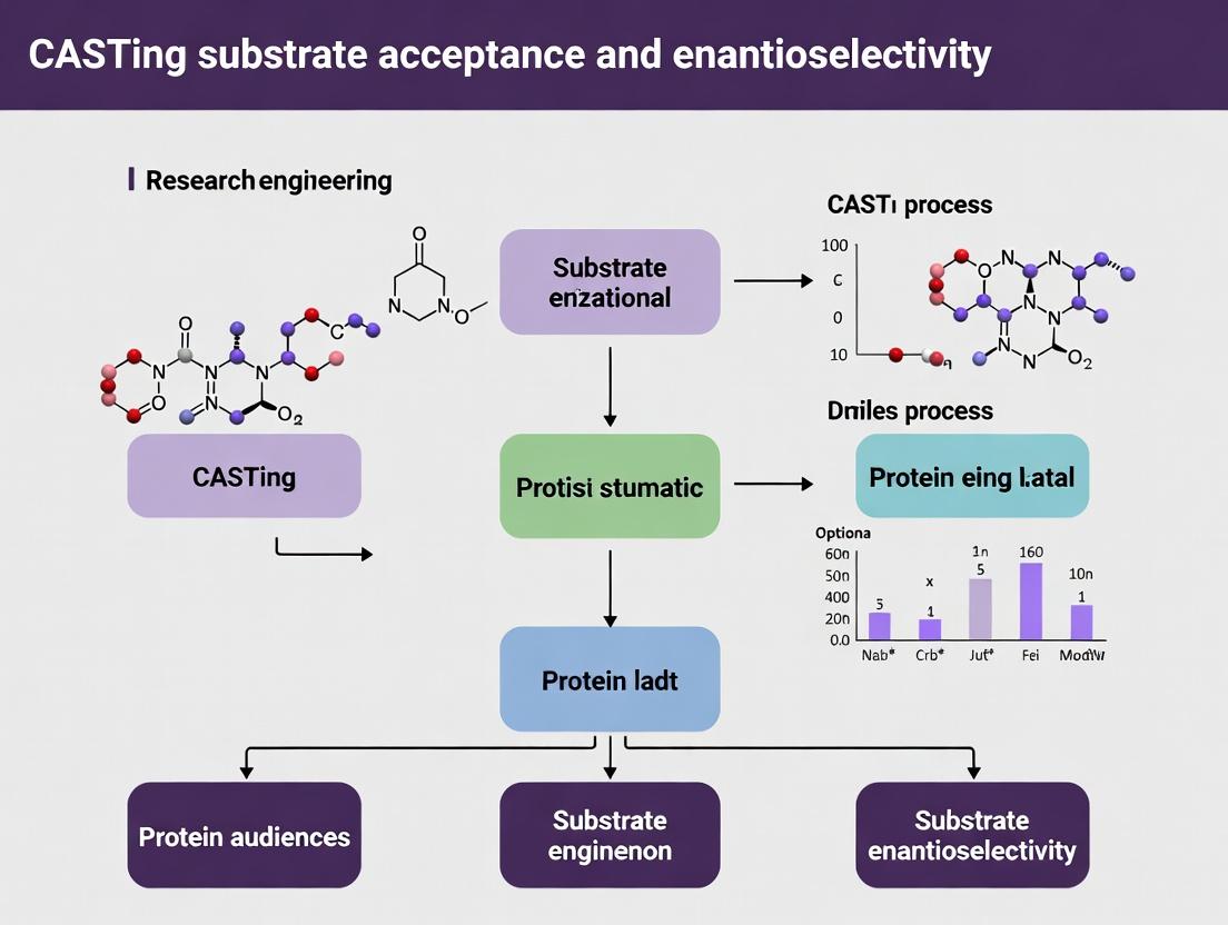
CASTing 101: Core Principles of Active-Site Engineering for Substrate Scope and Chirality
CASTing (Combinatorial Active-site Saturation Testing) is a pivotal protein engineering strategy that bridges rational design and directed evolution. Operating within the thesis that targeted library creation at enzyme active-site residues is optimal for altering substrate acceptance and enantioselectivity, CASTing systematically probes combinatorial mutational space. This approach transitions from a rationally chosen starting point—often a wild-type or previously engineered enzyme with a known structure—to generate "focused diversity," where vast but relevant sequence space is explored.
The core logical progression of the CASTing methodology is defined below.
Diagram Title: Logical Workflow of the Iterative CASTing Approach
Core Application Notes and Protocols
Protocol: Rational Selection of CAST Residues
Objective: To identify amino acid positions for saturation mutagenesis based on structural and functional data.
Materials & Procedure:
- Obtain a high-resolution 3D structure (X-ray, NMR, or high-confidence homology model) of your target enzyme.
- Using software (e.g., PyMOL, UCSF Chimera), map the binding pocket for the native substrate or a representative ligand.
- Select all residues with atoms within a 5–7 Å radius of the substrate.
- Filter residues:
- Exclude catalytic residues essential for the chemical step.
- Prioritize residues involved in substrate positioning (van der Waals, π-stacking, H-bonding) but not catalysis.
- Consider flexible loops lining the active site.
- Group selected residues into logical "CAST Libraries" based on spatial proximity (clusters) or hypothesized functional coupling. Limit groups to 3-5 residues to keep library size manageable (≤ $20^n$ variants, where n=residues).
Data Output Example: Table 1: Example CAST Group Design for an Esterase Targeting Bulky Substrate Acceptance
| Enzyme | CAST Group | Residue Numbers (PDB) | Rationale for Inclusion | Library Size (NNK codon) |
|---|---|---|---|---|
| Esterase EstB | A | L114, M115, F217 | Form the "acyl-binding pocket" roof; control steric occlusion. | 32,768 (32k) |
| Esterase EstB | B | W188, I289 | Line the "alcohol-binding pocket"; influence enantiopreference. | 1,024 (1k) |
| Esterase EstB | C | V162, L166, A215 | Define a distal access tunnel; may affect substrate entry. | 32,768 (32k) |
Protocol: Library Construction via Slonomics or Golden Gate Assembly
Objective: To efficiently generate high-quality saturation mutagenesis libraries for a defined CAST group.
Reagents & Solutions: Table 2: Key Research Reagent Solutions for CAST Library Construction
| Item | Function | Example/Supplier |
|---|---|---|
| NNK Degenerate Oligonucleotides | Encodes all 20 amino acids + 1 stop codon (32 codons) for saturating each target position. | Custom DNA synthesis (IDT, Twist Bioscience). |
| High-Fidelity DNA Polymerase | For PCR amplification of plasmid backbone with designed homology arms. | Q5 Hot Start (NEB), Phusion (Thermo). |
| DNA Assembly Master Mix | For seamless, multi-fragment assembly of mutagenic oligos and vector. | Gibson Assembly Master Mix (NEB), Golden Gate Assembly Mix (BsaI-HFv2). |
| Competent E. coli | For library transformation and propagation. | Electrocompetent cells (NEB 10-beta) for high efficiency. |
| Selection Agar Plates | To select for successful transformants containing the engineered gene. | LB + appropriate antibiotic (e.g., ampicillin, kanamycin). |
Detailed Methodology (Golden Gate Assembly):
- Design Oligos: For a CAST group of 3 residues (e.g., L114, M115, F217), design two long complementary oligonucleotides that span the entire region, with NNK codons at the three target positions. Include appropriate Type IIS restriction enzyme overhangs (e.g., BsaI) for Golden Gate assembly into a recipient plasmid.
- Amplify Vector Backbone: Perform PCR on the parent plasmid to linearize it, removing the wild-type sequence of the target region. Incorporate complementary Type IIS overhangs.
- Golden Gate Reaction: Set up a 20 µL reaction: 50 ng linearized vector, 10-20 ng pooled mutagenic oligos (annealed), 1 µL BsaI-HFv2, 1 µL T4 DNA Ligase, 1X T4 Ligase Buffer. Cycle: (37°C for 5 min, 16°C for 5 min) x 25 cycles, then 50°C for 5 min, 80°C for 10 min.
- Desalting & Transformation: Purify the assembly reaction using a spin column. Electroporate 2 µL into 50 µL of high-efficiency competent E. coli. Recover in SOC medium for 1 hour.
- Library Harvesting: Plate appropriate dilutions to determine library size (colony count) and harvest the remainder from liquid culture for plasmid DNA extraction. Sequence 10-20 random colonies to assess library quality and mutation distribution.
Protocol: High-Throughput Screening for Enantioselectivity
Objective: To identify variants with improved or inverted enantioselectivity (E-value) from a CAST library.
Screening Workflow: The following diagram outlines a standard screening cascade for enantioselectivity.
Diagram Title: Cascade for High-Throughput Enantioselectivity Screening
Materials & Procedure (Chiral GC Analysis in 96-Well Format):
- Cultivation: Inoculate picked colonies into 96-deep well plates containing 1 mL TB medium with antibiotic. Shake (800 rpm) at 30°C for 48 hours.
- Biotransformation: Add substrate (e.g., chiral ester or alcohol) dissolved in DMSO to a final concentration of 5-10 mM. Incubate with shaking for 4-16 hours.
- Extraction: Quench reactions by adding 200 µL of ethyl acetate per well. Seal plate, vortex for 2 min, centrifuge (4000xg, 5 min). Transfer organic (upper) layer to a new 96-well plate.
- Chiral GC Analysis: Use an autosampler equipped with a 96-well plate adapter. Inject 1 µL onto a chiral GC column (e.g., CP-Chirasil-Dex CB). Program a fast temperature ramp. Quantify (R)- and (S)- product peak areas.
- Data Analysis: Calculate conversion (c) and enantiomeric excess (ee). Determine apparent enantioselectivity (E-value) using the formula: $E = \frac{\ln[(1-c)(1-ee)]}{\ln[(1-c)(1+ee)]}$ for reactions where c < 50%.
- Validation: Re-test promising variants from the primary screen in small-scale flask cultures and re-analyze in triplicate to confirm E-value improvement.
Data Integration and Iterative Design
Objective: To analyze screening data and plan the next CASTing iteration.
Process: Beneficial mutations identified from one CAST library (e.g., Group A: L114V, F217G) are combined into a single gene background. This new, improved variant becomes the template for saturation mutagenesis on the next CAST group (e.g., Group B). This iterative process continues until the desired biocatalytic profile is achieved. Quantitative data from sequential CASTing rounds should be compiled as shown below.
Table 3: Exemplary Data from Iterative CASTing on an Epoxide Hydrolase for (S)-Selectivity
| Starting Template | CAST Group Screened | Key Identified Mutation(s) | Conversion (%) | ee (S) (%) | E-value |
|---|---|---|---|---|---|
| Wild-Type | A (F128, L215, V219) | F128L, L215F | 45 | 30 | 3.2 |
| Variant A1 (F128L/L215F) | B (Y154, Y197, I202) | Y197W | 65 | 85 | 28 |
| Variant B1 (F128L/L215F/Y197W) | C (H104, D222) | D222N | 78 | 98 | >100 |
This structured progression from rational design to focused diversity enables the efficient exploration of sequence-function landscapes, systematically unlocking novel enzyme functions for synthetic and pharmaceutical applications.
This application note details experimental approaches for investigating the molecular basis of substrate acceptance, a core theme in the broader thesis on Combinatorial Active-Site Saturation Testing (CASTing). Understanding active site architecture and flexibility is paramount for rational engineering of enzyme enantioselectivity and substrate scope, critical for pharmaceutical and fine chemical synthesis.
Key Experimental Protocols
Protocol 2.1: Molecular Dynamics (MD) Simulation for Flexibility Analysis
Objective: To quantify active site flexibility and conformational sampling in apo and substrate-bound states. Materials: Solvated enzyme system (pre-equilibrated), GROMACS/AMBER, high-performance computing cluster. Procedure:
- System Preparation: Load the crystallographic structure. Parameterize using a force field (e.g., CHARMM36). Solvate in a cubic water box with 10 Å padding. Add ions to neutralize.
- Energy Minimization: Perform 5000 steps of steepest descent minimization.
- Equilibration: NVT equilibration for 100 ps at 300 K (Berendsen thermostat). NPT equilibration for 100 ps at 1 bar (Parrinello-Rahman barostat).
- Production Run: Run unrestrained MD simulation for 100-500 ns. Save frames every 10 ps.
- Analysis: Calculate root-mean-square fluctuation (RMSE) of active site residues. Perform principal component analysis (PCA) on Cα atoms. Measure radius of gyration and solvent-accessible surface area (SASA).
Protocol 2.2: Site-Saturation Mutagenesis (SSM) & High-Throughput Screening
Objective: To experimentally map active site residues critical for substrate acceptance. Materials: Plasmid DNA, Phusion polymerase, NNK codon primers, competent E. coli, chromogenic/fluorogenic substrate assay. Procedure:
- Library Construction: Design primers for target active site residues using NNK degeneracy. Perform PCR. Digest template with DpnI. Transform into competent cells. Aim for >95% library coverage.
- Expression: Pick colonies into 96-deepwell plates. Induce expression with IPTG.
- Lysate Preparation: Lyse cells via sonication or chemical lysis.
- Screening: In a 384-well plate, add 50 µL lysate to 50 µL assay buffer containing substrate. Monitor reaction (e.g., absorbance at 405 nm) for 1 hour. Calculate initial velocity.
- Hit Analysis: Sequence hits with altered activity profiles. Correlate mutations with MD-derived flexibility metrics.
Protocol 2.3: Isothermal Titration Calorimetry (ITC) for Binding Affinity
Objective: To quantify thermodynamic parameters of substrate binding (Kd, ΔH, ΔS). Materials: Purified enzyme (>95%), substrate, ITC instrument (e.g., Malvern MicroCal PEAQ-ITC). Procedure:
- Sample Preparation: Dialyze enzyme and substrate into identical buffer (e.g., 50 mM phosphate, pH 7.4). Degas both samples.
- Experiment Setup: Load cell with 200 µL enzyme (50-100 µM). Fill syringe with substrate (10x concentrated). Set reference power to 5-10 µcal/sec.
- Titration: Perform 19 injections of 2 µL each at 180-second intervals with 750 rpm stirring at 25°C.
- Data Analysis: Subtract control titration (substrate into buffer). Fit integrated heat data to a one-site binding model to derive Kd, ΔH, and stoichiometry (N).
Data Presentation
Table 1: Quantitative Metrics from MD Simulations of Lipase A (Example)
| Residue | RMSE (Å) Apo State | RMSE (Å) Bound State | SASA Change (%) | Role in Catalysis |
|---|---|---|---|---|
| Ser77 | 0.45 | 0.22 | -85 | Nucleophile |
| His286 | 0.78 | 0.51 | -72 | Acid/base |
| Leu17 | 1.12 | 0.89 | -45 | Substrate shaping |
| Phe221 | 0.91 | 1.05 | +10 | Gating flexibility |
Table 2: ITC Binding Parameters for Wild-Type vs. CASTing Mutant
| Variant | Kd (µM) | ΔH (kcal/mol) | -TΔS (kcal/mol) | ΔG (kcal/mol) |
|---|---|---|---|---|
| WT | 15.2 ± 1.5 | -8.9 ± 0.3 | 2.1 | -6.8 ± 0.2 |
| F221A | 5.1 ± 0.7 | -6.2 ± 0.2 | 0.5 | -5.7 ± 0.1 |
| L17V | 42.3 ± 3.1 | -10.5 ± 0.5 | 4.8 | -5.7 ± 0.3 |
Table 3: High-Throughput Screening Results for Position 221 Library
| Codon | Amino Acid | Relative Activity (%) | Enantiomeric Excess (% ee) |
|---|---|---|---|
| GCT | Ala | 145 | 92 (S) |
| TGG | Trp | 12 | 5 (R) |
| ATC | Ile | 88 | 15 (S) |
| CAG | Gln | 65 | -80 (R) |
The Scientist's Toolkit: Research Reagent Solutions
| Item/Reagent | Function/Explanation |
|---|---|
| NNK Degenerate Primer Mix | Encodes all 20 amino acids plus TAG stop codon for site-saturation mutagenesis. |
| Chromogenic p-Nitrophenyl Ester Substrates | Hydrolysis releases yellow p-nitrophenol, enabling rapid UV-Vis kinetic screening. |
| His-Tag Purification Kit (Ni-NTA) | Rapid affinity purification of recombinant enzymes for biophysical assays. |
| Fluorogenic (e.g., 4-Methylumbelliferyl) Probes | Highly sensitive detection for low-activity variants in high-throughput screens. |
| Thermofluor Dye (SYPRO Orange) | Binds hydrophobic patches; used in thermal shift assays to monitor binding-induced stability. |
| Deuteration Buffer (D2O-based) | For hydrogen-deuterium exchange mass spectrometry (HDX-MS) to probe flexibility/solvent access. |
Diagrams
Title: CASTing Workflow for Substrate Acceptance
Title: Active Site Architecture and Flexibility Relationships
This application note details experimental protocols and analytical frameworks for studying enantioselective recognition within enzyme active sites, framed within the broader thesis of Combinatorial Active-site Saturation Testing (CASTing) for engineering substrate acceptance and stereoselectivity. Understanding chiral discrimination is paramount for developing enantiopure pharmaceuticals and fine chemicals.
The Physical Basis of Chiral Discrimination
Enantioselectivity arises from differential binding affinities and transition-state stabilization of enantiomers within a chiral binding pocket. The key energy difference, ΔΔG‡, is often small (1-2 kcal/mol) but decisive.
Table 1: Quantitative Energetics of Enantioselective Binding
| Parameter | (R)-Enantiomer Interaction Energy (kcal/mol) | (S)-Enantiomer Interaction Energy (kcal/mol) | ΔΔG‡ (kcal/mol) | Resulting ee (%)* |
|---|---|---|---|---|
| Hydrogen Bonding | -3.2 ± 0.3 | -1.8 ± 0.3 | -1.4 | >99 (R) |
| π-Stacking | -2.1 ± 0.4 | -2.5 ± 0.4 | +0.4 | 70 (S) |
| Steric Repulsion | +1.5 ± 0.2 | +0.1 ± 0.2 | +1.4 | >99 (S) |
| Van der Waals | -4.0 ± 0.5 | -4.3 ± 0.5 | +0.3 | 60 (S) |
*Calculated for a reaction at 25°C, where ee ≈ (1 - exp(ΔΔG‡/RT))/(1 + exp(ΔΔG‡/RT)) * 100.
Core Protocol: CASTing for Enantioselectivity
Objective: To redesign an enzyme binding pocket for reversed or enhanced enantioselectivity via iterative saturation mutagenesis.
Protocol 2.1: CAST Site Identification & Library Construction
- Materials: Wild-type plasmid DNA, KAPA HiFi HotStart ReadyMix, degenerate NNK primers (covers all 20 amino acids), DpnI restriction enzyme.
- Procedure:
- Analyze enzyme-substrate co-crystal structure or homology model to identify residues within 5-7 Å of the substrate.
- Group contacting residues into "CAST sites" (pairs or triplets of spatially close residues).
- Design PCR primers degenerated with the NNK codon (N=A/T/G/C; K=G/T) for each residue in the chosen CAST site.
- Perform site-saturation mutagenesis PCR: 25 cycles of (98°C 20s, 55°C 30s, 72°C 2 min/kb).
- Digest parental DNA template with DpnI (37°C, 1 hour).
- Transform into competent E. coli cells via electroporation to generate library. Aim for >95% coverage (Library size = 32^X, where X = number of residues saturated).
Protocol 2.2: High-Throughput Enantioselectivity Screening
- Materials: 96-well or 384-well deep-well plates, lysozyme, substrate cocktail (racemic mixture), chiral HPLC column (e.g., Chiralpak IA/IB/IC), or fluorescent/colorimetric enantioselective assay reagents.
- Procedure:
- Grow clones in deep-well plates with autoinduction media (24-48 hrs, 25°C, 220 rpm).
- Lyse cells chemically (e.g., BugBuster Master Mix) or via sonication.
- Initiate reaction by adding a racemic substrate mixture directly to clarified lysate.
- Quench reaction after linear range timepoint with equal volume of organic solvent (e.g., acetonitrile).
- Analyze enantiomeric excess (ee) directly from supernatant:
- Chiral HPLC/MS Method: Inject 10 µL. Gradient: 20-80% isopropanol in hexane over 20 min, 0.5 mL/min. Monitor separation (α > 1.2 required).
- Coupling Assay: For dehydrogenases, couple NAD(P)H production to a fluorescent readout using a second, enantioselective enzyme.
Analytical & Computational Validation Protocols
Protocol 3.1: Determining Binding Constants via Isothermal Titration Calorimetry (ITC)
- Materials: Purified wild-type and variant enzymes, purified (R)- and (S)-substrate ligands, ITC instrument (e.g., MicroCal PEAQ-ITC), dialysis buffer.
- Procedure:
- Dialyze enzyme and ligand into identical, degassed buffer (e.g., 50 mM phosphate, pH 7.5).
- Fill cell with 20 µM enzyme solution. Load syringe with 200-500 µM ligand solution.
- Perform titration: 19 injections of 2 µL ligand, 150s spacing, 25°C.
- Fit integrated heat data to a single-site binding model to extract KD, ΔH, and ΔS for each enantiomer. ΔΔG = RT ln(KDS / KDR).
Protocol 3.2: Molecular Dynamics (MD) Simulation of Enantiomer Binding
- Software: GROMACS or AMBER, force field (e.g., CHARMM36), visualization tool (PyMOL/VMD).
- Procedure:
- Prepare protein-ligand complex for each enantiomer from crystal structure or docking pose.
- Solvate the system in a cubic water box (TIP3P model), add ions to neutralize.
- Minimize energy (steepest descent, 5000 steps).
- Equilibrate under NVT (100 ps, 300 K) and NPT (100 ps, 1 bar) ensembles.
- Run production MD for 100-200 ns. Analyze trajectories for:
- Root-mean-square deviation (RMSD) of binding pocket.
- Hydrogen bond occupancy (% simulation time).
- Binding free energy via MM-PBSA/GBSA calculation.
Diagrams
Title: CASTing for Enantioselectivity Engineering Workflow
Title: Energy Basis of Enantioselection
The Scientist's Toolkit: Key Research Reagent Solutions
| Item | Function in Enantioselectivity Research |
|---|---|
| NNK Degenerate Primers | Encodes all 20 amino acids plus a stop codon for comprehensive saturation mutagenesis at CAST sites. |
| Chiralpak IA/IB/IC Columns | Polysaccharide-based chiral stationary phases for HPLC analysis of enantiomeric excess (ee). |
| Isopropyl β-D-1-thiogalactopyranoside (IPTG) | Precise inducer for T7/lac-based protein expression in E. coli for enzyme production. |
| BugBuster HT Protein Extraction Reagent | Chemically lyses bacterial cells in 96-well format for high-throughput screening of lysates. |
| NAD(P)H Fluorescent Detection Probe (e.g., Resazurin) | Enables coupled assays for dehydrogenase activity, allowing indirect measurement of enantioselectivity. |
| MicroCal PEAQ-ITC Assay Buffer Kit | Provides optimized, degassed buffers for accurate measurement of enantiomer binding thermodynamics. |
| CHARMM36 Force Field Parameters | Includes small molecule parameters for MD simulations of (R)- and (S)-substrates in binding pockets. |
| Cryo-EM Grids (Quantifoil R1.2/1.3) | For structural analysis of enzyme-ligand complexes when crystallization of variants fails. |
Within the broader thesis of directed evolution for enzyme engineering, Combinatorial Active-site Saturation Testing (CASTing) has emerged as a cornerstone strategy for manipulating substrate acceptance and enantioselectivity. This methodology systematically targets residues lining the active site or access channels to create smart, focused libraries. This application note details CASTing protocols for three high-impact enzyme classes—lipases, ketoreductases (KREDs), and cytochrome P450 monooxygenases (P450s)—each representing a unique challenge and opportunity in biocatalysis for pharmaceutical synthesis.
Application Notes & Protocols
Lipases: Engineering Enantioselectivity for Ester Hydrolysis
Lipases are pivotal in kinetic resolutions for chiral synthon production. CASTing is routinely applied to alter their enantiopreference.
Key Research Reagent Solutions
| Reagent/Material | Function in CASTing |
|---|---|
| p-Nitrophenyl ester substrates (e.g., pNP-acetate, pNP-palmitate) | Chromogenic assay for initial activity screening. |
| (R)- and (S)-enantiomers of target chiral ester (e.g., naproxen ester, ibuprofen ester) | Substrates for enantioselectivity determination (HPLC/GC). |
| pNC-based expression vector (e.g., pET-22b(+) for E. coli) | High-yield protein expression of lipase mutants. |
| Isopropyl β-D-1-thiogalactopyranoside (IPTG) | Inducer for controlled protein expression. |
| Paraoxon or PMSF (Phenylmethylsulfonyl fluoride) | Serine protease/lipase inhibitor for controlled cell lysis. |
Experimental Protocol for CASTing Lipase Enantioselectivity
- CAST Design: Identify 3-4 pairs of residues within 7Å of the acyl-binding pocket using a crystal structure (e.g., Candida antarctica Lipase B). Each pair forms a "CAST site."
- Library Construction: Perform site-saturation mutagenesis (NNK codon) on each CAST site individually via whole-plasmid PCR. Combine sites iteratively using the Stratagem of Combinatorial Libraries.
- High-Throughput Screening:
- Express mutant libraries in 96-deep well plates.
- Lyse cells chemically (e.g., BugBuster + lysozyme).
- Perform a two-tier assay: Primary screen for activity using a p-nitrophenyl ester (405 nm). Secondary screen on active clones using a racemic mixture of the target chiral ester.
- Analyze hydrolysis enantioselectivity by rapid chiral GC or HPLC of extracted products.
- Data Analysis: Calculate enantiomeric ratio (E) from conversion (c) and enantiomeric excess (ee) using:
E = ln[(1-c)(1-ee_p)] / ln[(1-c)(1+ee_p)]. Iterate with positive hits.
Quantitative Data Summary: Representative Lipase CASTing Outcomes
| Enzyme (Parent) | Target Reaction | CAST Sites Mutated | Best Variant | E-value (Parent) | E-value (Variant) | Reference Year |
|---|---|---|---|---|---|---|
| Candida antarctica Lipase B | Resolution of 2-methyldecanoic acid ester | L17, I189, A281 (A-site) | Variant L17A/I189F/A281L | 1.5 (S) | 25 (R) | 2022 |
| Pseudomonas fluorescens Lipase | Hydrolysis of 3-phenylbutyric acid ester | S155, F181, L185 (Finger region) | S155F/F181L | 4 (R) | 51 (S) | 2021 |
| Bacillus subtilis Lipase A | Acylation of 1-phenylethanol | T64, I66, L77, M78 (Active-site rim) | I66A/L77S/M78L | 14 (S) | 40 (R) | 2023 |
Ketoreductases: Controlling Stereochemistry in Ketone Reduction
KREDs are essential for synthesizing chiral alcohols. CASTing optimizes activity and stereocontrol for bulky or non-natural ketones.
Key Research Reagent Solutions
| Reagent/Material | Function in CASTing |
|---|---|
| NAD(P)H cofactor (enzymatic recycling system: GDH/glucose) | Regenerates reduced cofactor for sustained activity in assays. |
| Chiral Stationary Phase Columns (e.g., Chiralcel OD-H, Chiralpak AD-H) | HPLC analysis of product enantiomeric excess. |
| Fluorogenic probe: 1,2-Bis(4-methoxybenzylidene)acetonone | Activity screening via NAD(P)H depletion (Ex/Em ~420/460 nm). |
| E. coli BL21(DE3) ΔadhE strain | Host with reduced background alcohol dehydrogenase activity. |
| Solid-phase extraction (SPE) plates (C18) | Rapid product extraction for high-throughput analytics. |
Experimental Protocol for CASTing KRED Substrate Scope
- Active-site Mapping: Analyze substrate docking poses to identify residues contacting the ketone substituents (small vs. large pocket).
- Saturation & Library Generation: Use QuikChange or related methods to randomize chosen CAST residues (e.g., positions 37, 58, 150 in a typical KRED). Pool colonies for plasmid harvest.
- Microtiter Plate Screening:
- Grow and induce expression in 96-well plates.
- Permeabilize cells with 10% DMSO or toluene.
- Add assay mix: target ketone (10 mM), NADPH (0.2 mM), glucose (100 mM), and Gluconobacter oxidans GDH (1 U/mL) in buffer.
- Monitor NADPH fluorescence decay over 10 min.
- Hit Validation: Scale up positive hits, perform whole-cell biotransformations, and determine conversion and ee via chiral HPLC after extraction.
Quantitative Data Summary: Representative KRED CASTing Outcomes
| Enzyme (Parent) | Target Ketone | Key CAST Residues | Best Variant | ee (Parent) | ee (Variant) | Conversion | Reference Year |
|---|---|---|---|---|---|---|---|
| Lactobacillus brevis KRED | Ethyl 4-chloro-3-oxobutanoate | W119, S142, Y155, F147, L199 | F147L/Y155F | 75% (S) | >99% (S) | >99% | 2022 |
| Candida glabrata KRED | tert-Butyl 6-chloro-3,5-dioxohexanoate | L55, Y190, D150, V94 | L55M/Y190F | 90% (R) | >99.5% (R) | 98% | 2023 |
| Saccharomyces cerevisiae KRED | 2-Methyl-1-phenylpropan-1-one | F92, V144, L148, P171 | F92W/V144A | 80% (S) | 98% (S) | 95% | 2021 |
P450 Monooxygenases: Expanding Substrate Acceptance for C-H Activation
P450s catalyze regio- and stereoselective oxidations but often have narrow native substrate ranges. CASTing is used to broaden substrate acceptance for drug metabolite synthesis or late-stage functionalization.
Key Research Reagent Solutions
| Reagent/Material | Function in CASTing |
|---|---|
| Glucose-6-phosphate (G6P) / G6P Dehydrogenase | NADPH regeneration system for in vitro assays. |
| Hydrogen peroxide (H₂O₂) or tert-Butyl hydroperoxide | "Peroxide shunt" substrates for uncoupled P450 variants. |
| P450 substrate probes (e.g., 7-ethoxycoumarin, luciferin derivatives) | Fluorogenic screening for general activity. |
| Whole-cell biocatalysis medium with ΔlbhA (heme precursor) | Enhances heme incorporation in E. coli expression hosts. |
| Fe(II)-CO binding assay reagents (Sodium dithionite, CO gas) | Confirms proper heme incorporation and folding. |
Experimental Protocol for CASTing P450 Substrate Scope
- Channel Analysis: Identify residues lining the substrate access channel and active site roof (e.g., F87, T185 in P450 BM3) via structural analysis.
- Mutagenesis & Expression: Generate NNK libraries at 4-5 key positions. Co-express with a redox partner (e.g., cytochrome P450 reductase, CPR) in E. coli.
- Primary Screening (Whole Cell):
- Culture mutants in 96-deep well plates.
- Induce expression, add permeable probe (e.g., 7-ethoxycoumarin).
- After incubation, stop reaction with NaOH and detect hydroxylated product fluorescence (Ex/Em ~410/460 nm).
- Secondary Screening (Specific Substrate):
- Grow hit variants in 24-well plates.
- Add target drug-like substrate (e.g., verapamil, diclofenac).
- Extract metabolites after 4-6h and analyze by LC-MS/MS for product formation and regioselectivity.
Quantitative Data Summary: Representative P450 CASTing Outcomes
| Enzyme (Parent) | Target Substrate | CAST Region | Best Variant | Activity (Parent) | Activity (Variant) | Main Product | Reference Year |
|---|---|---|---|---|---|---|---|
| P450 BM3 (CYP102A1) | Verapamil (N-dealkylation) | F87, A328, I263, L437 | F87V/A328L | ND | 45 min⁻¹ (kcat) | Norverapamil | 2023 |
| P450 CYP153A (Marinobacter) | n-Octane (terminal hydroxylation) | I87, A91, V92, M86 | M86S/I87V/A91S | 3 U/mol | 240 U/mol | 1-Octanol | 2022 |
| P450 CYP2C9 | Warfarin (7-hydroxylation) | S100, I113, F114, L208, V292 | S100P/F114L | 0.05 min⁻¹ | 0.8 min⁻¹ | 7-Hydroxywarfarin | 2021 |
Application Notes
This document provides a structured approach for the preliminary computational and experimental analysis of protein structures, with a specific focus on informing library design for Combinatorial Active-site Saturation Testing (CASTing) campaigns. Within a thesis on CASTing for substrate acceptance and enantioselectivity, the primary goal is to transition from a 3D protein structure to a rational selection of target residues for mutagenesis. The following notes and protocols detail a streamlined pipeline for this purpose.
- Core Philosophy: The pipeline emphasizes a hierarchical, information-driven strategy. Broad, automated analyses identify regions of interest, which are then subjected to targeted, manual investigation to finalize CASTing residues.
- Key Outcome: A shortlist of 4-8 residue positions, typically grouped into 2-4 spatial clusters, that form the basis for subsequent saturation mutagenesis libraries.
Table 1: Summary of Key Computational Tools and Their Outputs
| Tool Category | Specific Tool/Server | Primary Function | Key Quantitative Output for CASTing |
|---|---|---|---|
| Structure Analysis | PDB Protein Data Bank | Source of experimental (e.g., X-ray) or high-quality predicted structures. | Resolution (<2.5 Å preferred), R-free factor, missing residues. |
| Active Site Delineation | CASTp, Fpocket | Geometrically defines pockets and calculates their physicochemical properties. | Pocket Volume (ų), Surface Area (Ų), Depth, Amino Acid Lining. |
| Conservation Analysis | ConSurf, HMMER | Scores residue evolutionary conservation from a multiple sequence alignment. | Conservation Score (1-9 scale; 9=most conserved). Targets variable residues (scores 1-3). |
| Dynamic Analysis | CABS-flex, NAMD | Generates structural ensembles via coarse-grained or atomistic simulations. | Root Mean Square Fluctuation (RMSF) per residue (Å), conformational clusters. |
| Interaction Analysis | PyMOL, UCSF Chimera | Manual visualization & measurement of distances, angles, and steric clashes. | Distance to substrate/cofactor (Å), H-bond angles, B-factor (thermal mobility). |
Protocol 1: Preliminary Computational Analysis for Residue Selection
Objective: To systematically analyze a protein structure and generate a candidate list of residues for CASTing.
Materials & Reagents:
- Input Structure: Protein structure file (PDB format). For enzymes without a structure, use AlphaFold2 or ESMFold prediction.
- Software:
- Molecular visualization (PyMOL or UCSF ChimeraX).
- ConSurf server (https://consurf.tau.ac.il/).
- CASTp 3.0 server (http://sts.bioe.uic.edu/castp/).
- CABS-flex 2.0 server (http://biocomp.chem.uw.edu.pl/CABSflex2).
- Research Reagent Solutions:
- Pymol-Scripts: Custom scripts for measuring distances and labeling residues.
- Jupyter Notebook: For data integration and analysis using BioPython and Pandas.
- Multiple Sequence Alignment (MSA) File: Pre-generated or sourced from UniRef90/Pfam for ConSurf.
Procedure:
- Structure Preparation: Load the PDB file into PyMOL. Remove heteroatoms except essential cofactors or crystallographic substrates/ligands. Add missing hydrogen atoms and assign standard protonation states at physiological pH.
- Active Site Pocket Analysis: Submit the cleaned PDB file to the CASTp 3.0 server. Identify the primary substrate-binding pocket. Download the list of residues lining the pocket (within 5Å of the pocket surface).
- Evolutionary Conservation Analysis: Submit the PDB file and/or protein sequence to the ConSurf server using the automated workflow. Retrieve the conservation grades mapped onto the structure and as a table. Cross-reference with the CASTp residue list.
- Flexibility Assessment: Submit the PDB file to CABS-flex 2.0 for a coarse-grained dynamics simulation (default 10 ns equivalent). Download the RMSF profile per residue.
- Data Integration: Create a master table integrating residues from the active site pocket. For each residue, list its Conservation Score and average RMSF value. Prioritize residues that are:
- Lining the active site pocket.
- Evolutionarily variable (ConSurf score 1-3).
- Possess moderate-to-high flexibility (above-average RMSF).
- Spatial Clustering: Visually inspect the prioritized residues in PyMOL. Group residues that are within 5-10 Å of each other into putative CASTing clusters. Aim for 2-4 clusters containing 2-4 residues each.
Protocol 2: Manual Curation & Final Selection for CASTing
Objective: To refine the computationally generated candidate list through detailed manual inspection of molecular interactions and steric constraints.
Procedure:
- Substrate Docking or Modeling: If a co-crystal structure is unavailable, dock the substrate of interest into the active site using a tool like AutoDock Vina or fit it manually based on known catalytic mechanism.
- Interaction Mapping: For each candidate residue in a cluster, analyze:
- Distance from the residue's side-chain atom to the substrate's functional groups.
- Potential for hydrogen bonding, π-stacking, or van der Waals contacts.
- Evaluation of potential steric hindrance between the wild-type side chain and the substrate.
- Mechanistic Considerations: Exclude residues directly involved in catalysis (e.g., catalytic triad, acid-base donors/acceptors) unless the thesis specifically aims to alter mechanism. These are typically highly conserved.
- Library Design Finalization: Select the final clusters. Design degenerate primers for each cluster to perform saturation mutagenesis (NNK or NDT codon schemes). The hierarchical analysis minimizes library size while maximizing coverage of functionally relevant sequence space.
Title: Hierarchical Residue Selection Workflow for CASTing
Title: Structure-Function Feedback Loop in CASTing Thesis
The Scientist's Toolkit: Key Reagent Solutions
| Item | Function in Analysis |
|---|---|
| High-Quality PDB Structure | Essential starting point. A structure with resolution <2.5 Å and a complete active site is critical for reliable analysis. |
| Pre-aligned MSA File | Required for efficient ConSurf analysis. A diverse, high-quality MSA yields a robust evolutionary conservation profile. |
| PyMOL/Chimera Scripts | Automate repetitive tasks like measuring distances from multiple residues to a ligand, speeding up manual curation. |
| NDT Codon Mixture | A degenerate codon for saturation mutagenesis that reduces library size by encoding 12 amino acids (excluding stop codons), covering a balanced set. |
| Structure Prediction Server (AlphaFold2) | Provides a reliable 3D model when an experimental structure is unavailable, enabling in silico analysis. |
| Cofactor/Substrate Analog | Useful for crystallography or docking. Understanding the bound state is paramount for rational residue selection. |
The CASTing Workflow: Step-by-Step Protocols for Library Creation and Screening
Within the broader thesis on Combinatorial Active-site Saturation Testing (CASTing) for tailoring substrate acceptance and enantioselectivity in enzymes, strategic residue selection emerges as the critical first step. Moving beyond simple proximity-to-substrate rules, modern protocols integrate analyses of protein flexibility (B-factors), residue interaction networks (RINs), and computational substrate docking to rationally define smaller, higher-quality CAST libraries. This application note details the integrated workflow, enabling researchers to maximize the probability of identifying beneficial mutations while minimizing experimental screening burden.
Key Concepts and Quantitative Data
Table 1: Core Metrics for CASTing Residue Prioritization
| Metric | Tool/Calculation | Ideal Range for CASTing | Rationale | ||
|---|---|---|---|---|---|
| B-Factor (Ų) | PDB File / MD RMSF | 20-80 | Residues with moderate-high flexibility are more amenable to mutation and can influence active site dynamics. | ||
| Betweenness Centrality | NetworkX (Python) / RINalyzer | >0.05 (Normalized) | High centrality indicates a residue critical for communication; mutation can propagate effects distally. | ||
| Docking Score ΔΔG (kcal/mol) | AutoDock Vina, Rosetta | > | 1.0 | vs. reference | Predicts direct interaction energy change with target substrate. |
| Solvent Accessibility (% RSA) | DSSP, GETAREA | >20% | Surface residues are more tolerant to mutation without causing folding defects. | ||
| Evolutionary Conservation Score | ConSurf, ScoreCons | <7 (Scale 1-9) | Low conservation suggests higher mutational tolerance. |
Table 2: Sample Residue Analysis Output (Hypothetical Enzyme)
| Residue | B-Factor | Betweenness Centrality | Docking ΔΔG (kcal/mol) | RSA (%) | Conservation | CAST Priority |
|---|---|---|---|---|---|---|
| L78 | 45.2 | 0.12 | -1.8 | 35 | 3 | High (Network Hub) |
| F121 | 62.1 | 0.03 | -2.5 | 28 | 5 | High (Flexible, Strong Binder) |
| V156 | 22.5 | 0.01 | -0.3 | 15 | 8 | Low (Rigid, Conserved) |
| S205 | 38.7 | 0.08 | -1.2 | 60 | 4 | Medium (Accessible Communicator) |
Experimental Protocols
Protocol 1: Integrated Computational Pipeline for Residue Selection
Objective: To identify a prioritized set of 4-8 CAST residues using B-factor, network, and docking analysis. Input: High-resolution crystal structure (PDB format) of the wild-type enzyme. Duration: 3-5 days computation time.
Pre-processing (Day 1):
- Obtain the protein structure (PDB ID). Remove water molecules and heteroatoms using PyMOL or UCSF Chimera. Add missing hydrogens and assign protonation states using
PDB2PQRor theReducetool. - Perform a short (10-20 ns) Molecular Dynamics (MD) simulation in explicit solvent (e.g., using GROMACS) to sample native-state flexibility. Calculate the per-residue Root Mean Square Fluctuation (RMSF) as a dynamic B-factor surrogate.
- Obtain the protein structure (PDB ID). Remove water molecules and heteroatoms using PyMOL or UCSF Chimera. Add missing hydrogens and assign protonation states using
B-Factor/RMSF Analysis (Day 1):
- Extract B-factors from the static PDB file (column 61-66) or from the MD trajectory. Normalize values across the structure (Z-score).
- Selection Threshold: Flag residues with Z-score > 0.8 (i.e., more flexible than average) within a 10Å radius of the active site cofactor or bound substrate.
Residue Interaction Network (RIN) Construction (Day 2):
- Generate the RIN using the
RINalyzerplug-in for Cytoscape or a custom Python script usingNetworkXandMDAnalysis. - Define nodes as amino acid residues. Define edges using non-covalent interactions (e.g., van der Waals contacts <4Å, hydrogen bonds, salt bridges <6Å).
- Calculate network centrality metrics (Betweenness, Closeness) for each node. Export a ranked list of high-betweenness centrality residues near the active site.
- Generate the RIN using the
Ensemble Docking (Day 3-4):
- Prepare the target substrate molecule (SMILES string) using Open Babel to generate 3D coordinates and assign GAFF force field charges.
- Docking Ensemble: Use 5-10 snapshots from the equilibrated MD trajectory to account for protein flexibility.
- Perform molecular docking with AutoDock Vina. Define a search box centered on the active site, ensuring it encompasses all candidate residues.
- For each candidate residue, analyze docking poses to compute the average binding energy (ΔG). Compare to a reference substrate to calculate ΔΔG.
Data Integration & Final Selection (Day 5):
- Compile results from steps 2-4 into a unified table (as in Table 2).
- Apply a weighted scoring function: Priority Score = (w1 * B-factor Z-score) + (w2 * Betweenness) + (w3 * |Docking ΔΔG|). Typical weights: w1=0.3, w2=0.4, w3=0.3.
- Select the top 4-8 residues with the highest Priority Scores. Group spatially adjacent residues (≤5Å apart) into the same CASTing library for combinatorial mutagenesis.
Protocol 2: Experimental Validation of Selected CAST Residues
Objective: To experimentally screen the designed CAST libraries for altered substrate acceptance. Input: Prioritized residue list and grouped libraries.
Library Construction:
- Design primers for Site-Saturation Mutagenesis (SSM) at each selected position using NNK degenerate codons (encodes all 20 aa + 1 stop).
- For grouped libraries, perform iterative or multiplexed PCR assembly.
- Clone libraries into an appropriate expression vector via Gibson Assembly or Golden Gate cloning. Transform into E. coli and plate for single colonies to ensure >95% library coverage.
High-Throughput Screening:
- Pick colonies into 96- or 384-well deep-well plates containing expression medium. Induce protein expression.
- Perform whole-cell or lysate-based activity assays using the target substrate. For enantioselectivity, use chiral HPLC or MS-based separation in conjunction with the assay.
- Employ fluorescence- or absorbance-based readouts linked to product formation. Positive hits are identified as variants showing >2x increased activity or a significant shift in enantiomeric excess (ee) compared to wild-type.
Hit Characterization:
- Sequence hit variants. Express and purify the variant enzyme.
- Determine steady-state kinetics (kcat, KM) for the target substrate and reference substrate.
- Measure enantioselectivity (E-value) for prochiral substrates using established analytical methods.
Visualization Diagrams
Workflow Title: Strategic CASTing Residue Selection Workflow
Network Title: Residue Interaction Network (RIN) Example
The Scientist's Toolkit: Research Reagent Solutions
Table 3: Essential Materials and Reagents
| Item/Reagent | Function in CASTing Protocol | Example Product/Source |
|---|---|---|
| NNK Degenerate Codon Primers | Encode all 20 amino acids during saturation mutagenesis. | Custom oligos from IDT, Sigma. |
| High-Fidelity DNA Polymerase | Error-free amplification for library construction. | Q5 (NEB), PfuTurbo (Agilent). |
| Cloning & Assembly Master Mix | Efficient, seamless assembly of mutagenesis fragments. | Gibson Assembly Master Mix (NEB), Golden Gate Assembly Kit (BsaI-HFv2). |
| Competent E. coli (High-Efficiency) | Library transformation with >10^9 cfu/μg for full coverage. | NEB 10-beta, XL10-Gold. |
| Chromatography Resin (Ni-NTA) | Rapid purification of His-tagged variant proteins for characterization. | HisTrap HP columns (Cytiva). |
| Chiral HPLC Column | Separation and quantification of enantiomers for ee determination. | Chiralpak IA/IB/IC (Daicel). |
| Fluorogenic/Chromogenic Probe | High-throughput activity screening in microplates. | Custom synthesized or commercial (e.g., from Sigma, Thermo Fisher). |
| Molecular Dynamics Software | Simulating protein flexibility for B-factor/RMSF analysis. | GROMACS (Open Source), AMBER, Desmond. |
| Network Analysis Toolkit | Constructing and analyzing Residue Interaction Networks. | Cytoscape with RINalyzer, Python (NetworkX, MDAnalysis). |
| Docking Software Suite | Predicting substrate binding poses and energies. | AutoDock Vina, Rosetta, Schrodinger Suite. |
Application Notes
This document details advanced library design strategies within a research program focused on Continuous Ancestral Sequence Transfer and Integration (CASTing) to engineer enzyme substrate acceptance and enantioselectivity. The primary goal is to systematically explore sequence-function landscapes around active-site residues to unlock novel biocatalytic functions for drug development.
1. Saturation Mutagenesis for Active Site Probing Saturation Mutagenesis (SM) is the cornerstone for exploring local sequence space. By randomizing defined codons to all 20 amino acids, it enables the unbiased assessment of each position's contribution to substrate binding and stereocontrol. In CASTing projects, SM is applied to residues lining the binding pocket of ancestral enzyme scaffolds, allowing for the rapid identification of key mutations that alter steric and electronic environments.
2. Oligonucleotide Synthesis for Library Construction Modern oligonucleotide synthesis enables the precise implementation of SM and combinatorial library designs. Trimer phosphoramidites or mixed-base coupling allow for the synthesis of degenerate codons (e.g., NNK, NDT). For multi-site libraries, gene assembly methods like Golden Gate or Gibson Assembly with designed oligo pools are standard. The quality and representation of the synthesized oligo pool directly dictate library diversity and coverage.
3. Navigating Diversity Limits in Practical Library Design The theoretical diversity of a library quickly surpasses practical screening capabilities. For example, saturating 6 positions (20⁶) yields 6.4x10⁷ variants, far exceeding the throughput of even ultra-high-throughput screening (uHTS). Strategic library design is therefore critical.
Table 1: Library Diversity and Screening Coverage
| Design Strategy | Number of Randomized Positions | Theoretical Diversity | Common Screening Capacity | Practical Coverage Goal |
|---|---|---|---|---|
| Single-Site SM | 1 | 20 variants | >10⁴ clones | Full enumeration (100%) |
| Focused Combinatorial (e.g., ISM*) | 3-4 | 8,000 - 160,000 variants | 10⁵ - 10⁶ clones | Near-full to sampling |
| Multi-site Parallel SM | 6 | 6.4 x 10⁷ variants | 10⁷ - 10⁸ clones | Sampling (<1% coverage) |
| Full Gene De Novo | ~300 | ~10³⁹⁰ variants | <10¹² clones | Negligible |
*Iterative Saturation Mutagenesis
The optimal strategy involves iterative cycles: initial SM to identify "hot spots," followed by focused combinatorial libraries of beneficial mutations, all performed on ancestrally informed CASTing scaffolds to maintain protein stability while exploring function.
Protocols
Protocol 1: CASTing-Informed Iterative Saturation Mutagenesis (ISM)
Objective: To identify key residues controlling enantioselectivity in an ancestral esterase scaffold.
Materials: See "Research Reagent Solutions" below.
Procedure:
- Target Selection: Based on ancestral sequence alignment and structural modeling, select 4-6 CASTing regions (clusters of 2-4 adjacent residues) surrounding the active site.
- Library Construction (per region): a. Design primers containing an NNK degenerate codon (encodes all 20 aa + 1 stop) for each targeted residue within the region. b. Perform PCR using a high-fidelity polymerase to amplify the plasmid template with the degenerate primers. c. Digest the PCR product with DpnI to eliminate methylated parental template. d. Transform the assembled product into competent E. coli cells via electroporation. Plate an aliquot to calculate library size (aim for >10⁵ colonies to ensure >95% coverage of 32 NNK variants). e. Isolve the remaining transformation mix, and isolate the plasmid library pool.
- Screening & Selection: a. Express the library in a suitable expression host (e.g., E. coli BL21). b. Perform activity screening using a chromogenic or fluorogenic racemic substrate analog in a microtiter plate format. c. For enantioselectivity, use a high-throughput chiral assay (e.g., LC-MS/MS or coupled enzyme assay) on lysates from single clones. d. Isolate plasmids from hits showing improved activity or shifted selectivity.
- Iteration: Use the best hit from the first CASTing region as the template for SM at the next selected region. Repeat steps 2-3.
Protocol 2: Oligo Pool Design and Assembly for Multi-Site Libraries
Objective: To construct a focused combinatorial library combining beneficial mutations from two identified CASTing regions (3 positions total).
Materials: Synthesized oligonucleotide pool, Gibson Assembly Master Mix, appropriate restriction enzymes.
Procedure:
- Oligo Design: Design two long oligonucleotides (80-120mer) that cover the entire gene segment to be reassembled. Incorporate the 3 specific, pre-defined mutant codons at their respective positions within the oligo sequences. Flank with 20-25 bp homology arms for assembly.
- Gene Reassembly: a. Use the oligo pool as megaprimers in a PCR-like reaction with a linearized plasmid backbone as template. b. Alternatively, use the oligos as fragments in a Gibson Assembly reaction. Mix 0.05 pmol of linearized vector with a 2:1 molar ratio of the duplex oligo fragments in 1x Gibson Assembly Master Mix. c. Incubate at 50°C for 60 minutes.
- Transformation and Validation: Transform 2 µL of the assembly reaction into competent cells. Sequence 10-20 random clones to confirm correct incorporation of mutations and library representation.
Visualizations
Title: CASTing Library Design & Screening Workflow
Title: Navigating Library Diversity Limits
The Scientist's Toolkit: Research Reagent Solutions
| Item | Function in Library Design |
|---|---|
| NNK Trinucleotide Phosphoramidites | Provides a degenerate codon (N=A/C/G/T; K=G/T) during oligo synthesis, minimizing stop codons and bias. Essential for true saturation mutagenesis. |
| High-Fidelity DNA Polymerase (e.g., Q5) | Ensures accurate amplification during library construction with minimal PCR-induced errors, preserving designed diversity. |
| Golden Gate Assembly Mix | Enables efficient, one-pot, seamless assembly of multiple DNA fragments with Type IIS restriction sites, ideal for combinatorial library builds. |
| Gibson Assembly Master Mix | An isothermal, exonuclease-based method for assembling multiple overlapping DNA fragments. Used for reassembly from oligo pools. |
| Electrocompetent E. coli (e.g., NEB 10-beta) | Essential for achieving high transformation efficiency (>10⁹ cfu/µg) required to capture large library diversities. |
| Chromogenic/Fluorogenic Substrate Proxies | Enables rapid, high-throughput initial activity screening of entire libraries to identify functional clones. |
| uHTS-Compatible Chiral Assay Kit | Allows direct measurement of enantiomeric excess (ee) in lysates, bridging the gap between library size and selectivity screening. |
| Next-Generation Sequencing (NGS) Service | For post-screening diversity analysis, enrichment scoring, and quality control of library representation. |
Application Notes & Protocols in the Context of CASTing
The pursuit of engineered enzymes with tailored substrate acceptance and enantioselectivity is central to modern biocatalysis. Focused Directed Evolution, particularly Combinatorial Active-site Saturation Testing (CASTing), is a powerful strategy for reshaping an enzyme's active site and its micro-environment. The critical bottleneck in this iterative process is the rapid and accurate evaluation of vast mutant libraries for enantioselectivity. This necessitates high-throughput screening (HTS) assays that are sensitive, reproducible, and scalable. The choice of assay is dictated by the substrate's physicochemical properties, the desired throughput, and available instrumentation. This document details four cornerstone HTS methodologies—HPLC, GC, Fluorescence, and Colorimetry—framed explicitly within a CASTing workflow for enantioselectivity research.
Key Quantitative Comparison of HTS Assays
Table 1: Comparative Overview of Enantioselectivity HTS Assays
| Assay Parameter | HPLC (Chiral Stationary Phase) | GC (Chiral Column) | Fluorescence (Enzyme-Coupled) | Colorimetry (pH Indicators/Dyes) |
|---|---|---|---|---|
| Typical Throughput (samples/day) | 100-500 | 200-800 | 10,000 - 100,000+ | 5,000 - 50,000+ |
| Assay Time | 5-30 min/run | 2-15 min/run | < 1 min/sample | 1-5 min/sample |
| Information Gained | Full conversion, ee (E value), absolute configuration | Full conversion, ee (E value), absolute configuration | Relative activity & ee (indirect) | Relative activity & ee (indirect) |
| Cost per Sample | High (columns, solvents) | Moderate | Very Low | Very Low |
| Sensitivity | Excellent (nmol) | Excellent (nmol) | High (pmol) | Moderate (nmol) |
| Primary Use in CASTing | Validation & hit confirmation | Validation & volatile substrates | Primary library screening | Primary library screening |
| Key Limitation | Low throughput, high cost | Requires volatility/thermal stability | Requires coupled enzyme/design | Indirect, prone to false positives |
Detailed Experimental Protocols
Protocol 3.1: Ultra-High-Throughput Fluorescence-BasedeeScreening
Principle: This coupled assay is designed for hydrolytic reactions (e.g., esterases, lipases). Enantioselective hydrolysis releases a product (e.g., acid) that is linked to a change in fluorescence via a secondary, enantioselective enzyme system or a selective fluorescent probe.
- Reaction Setup: In a black 96- or 384-well microtiter plate, combine:
- 90 µL of mutant lysate/cell supernatant in appropriate buffer (e.g., 50 mM Tris-HCl, pH 7.5).
- 10 µL of substrate solution (e.g., 10 mM enantiomeric ester of a fluorescent reporter precursor in DMSO).
- Incubation: Shake plate at 30°C for 1-3 hours.
- Detection: Add 100 µL of detection mix containing the coupling enzyme (e.g., enantioselective alcohol oxidase) and fluorogenic dye (e.g., Amplex Red) to each well. Incubate for 30 min at RT.
- Measurement: Read fluorescence (ex/cm = 530/590 nm). Wells with higher fluorescence indicate higher activity. The ee is derived from differential signals in parallel assays using pure (R)- and (S)-substrate controls.
- Data Analysis: Calculate initial rates. Mutants showing significant signal deviation from the wild-type profile (with (R)- and (S)-substrates) are identified as ee hits for validation.
Protocol 3.2: Colorimetric pH-Based Screening for Ester Hydrolysis
Principle: Hydrolysis of esters or amides releases protons, causing a local pH change detected by a pH indicator.
- Reagent Preparation: Prepare assay buffer: 50 mM KCl, 1 mM MgCl₂, with pH indicator (e.g., 70 µM phenol red). Adjust to pH 7.8 (red color).
- Assay Setup: In a 96-well plate, mix:
- 175 µL of assay buffer.
- 20 µL of mutant whole-cell suspension or lysate.
- 5 µL of substrate (e.g., 200 mM racemic ester in isopropanol).
- Kinetic Measurement: Immediately monitor absorbance at 557 nm (for phenol red) every 10-15 seconds for 5 minutes at 30°C. The decrease in absorbance correlates with acid production.
- Enantioselectivity Determination: Perform parallel assays using separately prepared (R)- and (S)-enantiomer substrates (at their KM concentrations). The ratio of the initial rates (vR/vS) provides an ee estimate.
- Hit Selection: Mutants showing a significantly altered rate ratio compared to wild-type are selected for GC/HPLC validation.
Protocol 3.3: Chiral HPLC Validation of Enantioselectivity
Principle: Direct separation and quantification of enantiomers from analytical-scale biotransformations.
- Biotransformation: Scale up promising hits in 1 mL reactions. Quench at 20-50% conversion (by adding 50 µL of 1M HCl or heat inactivation).
- Sample Preparation: Extract reaction mixture with 1 mL of ethyl acetate. Dry organic layer under reduced air, redissolve in 200 µL of HPLC-grade heptane/isopropanol (9:1).
- HPLC Analysis:
- Column: Chiralpak AD-H (250 x 4.6 mm) or equivalent.
- Mobile Phase: Isocratic, Heptane:Isopropanol (90:10) at 1.0 mL/min.
- Detection: UV at 220 nm.
- Injection: 10 µL.
- Calculation: Determine enantiomeric excess (ee) = [(AreaR - AreaS) / (AreaR + AreaS)] * 100%. Calculate enantiomeric ratio (E) using the formula: E = ln[(1 - c)(1 - eeS)] / ln[(1 - c)(1 + eeS)], where c is conversion.
Protocol 3.4: Chiral GC Validation for Volatile Compounds
Principle: Direct gas-phase separation of enantiomers.
- Biotransformation & Extraction: Follow steps from Protocol 3.3.
- Sample Preparation: Redissolve dried extract in 100 µL of ethyl acetate.
- GC Analysis:
- Column: Chiral γ-cyclodextrin-based column (e.g., CP-Chirasil-Dex CB).
- Oven Program: 80°C hold 2 min, ramp 2°C/min to 130°C.
- Injector/Detector (FID): 250°C.
- Carrier Gas: Helium, constant flow 1.5 mL/min.
- Split Injection: 1:10 ratio.
- Calculation: Analyze chromatograms as in HPLC protocol to determine ee and E value.
Visualizations
Title: CASTing Workflow with HTS Integration
Title: Fluorescence-Coupled ee Assay Mechanism
The Scientist's Toolkit: Research Reagent Solutions
Table 2: Essential Materials for Enantioselectivity HTS
| Reagent / Material | Function & Role in CASTing Screening |
|---|---|
| Chiralpak AD-H Column | Gold-standard chiral stationary phase for HPLC validation; provides definitive ee and configuration. |
| CP-Chirasil-Dex CB GC Column | Cyclodextrin-based column for high-resolution chiral separation of volatile substrates and products. |
| Amplex Red Reagent | Fluorogenic probe for detecting H₂O₂ in enzyme-coupled fluorescence ee assays. |
| Phenol Red | pH indicator for colorimetric, absorbance-based screening of hydrolytic activity. |
| Racemic & Enantiopure Substrate Standards | Critical for assay calibration, establishing baselines, and determining accurate ee values. |
| Enantioselective Coupling Enzymes (e.g., AOx, LOx) | Secondary enzymes that confer enantioselectivity to otherwise non-selective fluorescence signals. |
| Lysis Reagent (e.g., BugBuster) | For consistent cell lysis in microtiter plates when screening lysate libraries. |
| Black/Clear 384-Well Microtiter Plates | Platform for ultra-high-throughput fluorescence/colorimetry assays; minimal well-to-well crosstalk. |
| Multichannel Pipettes & Reagent Reservoirs | Enable rapid, parallel dispensing of cells, substrates, and detection mixes for library screening. |
Within the broader thesis on CAST (Combinatorial Active-site Saturation Testing) for engineering substrate acceptance and enantioselectivity in enzymes, this application note focuses on practical protocols. The goal is to expand the substrate scope of engineered enzymes to incorporate non-natural, synthetically challenging compounds into drug synthesis pathways. This enables the biocatalytic synthesis of chiral intermediates previously inaccessible via traditional chemical catalysis.
Research Reagent Solutions Toolkit
| Reagent/Material | Function in Experiment |
|---|---|
| Thermostable Lipase/esterase (e.g., from Thermomyces lanuginosus) | Engineered enzyme scaffold for CASTing; high stability allows screening under diverse conditions. |
| Non-natural acyl donor library (e.g., bulky α,α-disubstituted acids) | Substrate library to probe and expand active site acceptance; key for synthesizing non-natural chiral esters. |
| p-Nitrophenyl ester probes | Chromogenic substrates for high-throughput initial activity screening. |
| Chiral GC column (e.g., Cyclodex-B) | Essential for enantiomeric excess (ee) analysis of reaction products. |
| E. coli BL21(DE3) expression system | Standard host for mutant library expression and protein production. |
| KAPA HiFi HotStart ReadyMix | High-fidelity PCR mix for accurate gene library construction during CAST. |
| Luria-Bertani (LB) media with kanamycin | Growth and expression media for selective cultivation of mutant libraries. |
Note 1: Initial Screening of Wild-Type Enzyme against Non-Natural Substrates
A baseline activity profile is essential. The wild-type enzyme is assayed against a panel of non-natural substrates. Activity is normalized to the natural substrate.
Table 1: Wild-Type Enzyme Activity Profile
| Substrate Class | Example Structure | Relative Activity (%) | Enantioselectivity (ee, %) |
|---|---|---|---|
| Natural Substrate (C6 linear acid) | Hexanoic acid pNP-ester | 100 ± 5 | >99 (R) |
| α-Methyl branched acid | (S)-2-Methylhexanoic acid pNP-ester | 15 ± 3 | 80 (R) |
| Bulky α,α-dialkyl acid | 2-Ethyl-2-methylhexanoic acid pNP-ester | <1 | N/D |
| Cyclopropane-containing acid | Cyclopropanecarboxylic acid pNP-ester | 25 ± 4 | 65 (S) |
Note 2: CASTing for Bulky Substrate Acceptance
To enable conversion of the bulky α,α-dialkyl acid (Table 1), a CAST library targeting residues lining the acyl-binding pocket was created. Key hits showed dramatically improved activity.
Table 2: Performance of Top CAST Variants for Bulky Substrate
| Variant ID | Mutations | Relative Activity (%) | ee (%) | Notes |
|---|---|---|---|---|
| WT | - | <1 | N/D | Baseline |
| 3B7 | F214L, V267A | 85 ± 6 | 92 (R) | Synergistic enlargement |
| 5H12 | L163I, F214G | 42 ± 5 | 78 (R) | Moderate improvement |
| 9A2 | V267G, L269S | 60 ± 4 | 85 (S) | Enantioselectivity reversed |
Detailed Experimental Protocols
Protocol 1: CAST Library Construction for Acyl-Binding Pocket
Objective: Generate a focused mutant library by saturating two predefined clusters of 3-4 amino acid residues surrounding the enzyme's acyl-binding pocket.
Materials:
- Plasmid containing gene for thermostable lipase/esterase.
- KAPA HiFi HotStart ReadyMix PCR kit.
- DpnI restriction enzyme.
- Oligonucleotide primers for each target codon (NNK degeneracy).
- E. coli BL21(DE3) electrocompetent cells.
Method:
- Site Identification: Using a crystal structure, select two clusters of residues (e.g., Cluster A: L163, F214; Cluster B: V267, L269) within 6Å of the substrate's scissile bond.
- PCR Assembly: Perform separate PCRs for each cluster using primers containing NNK codons. Use a high-fidelity polymerase to minimize secondary mutations.
- Digestion & Transformation: Treat PCR products with DpnI (37°C, 2h) to digest methylated parental DNA. Purify and transform the library DNA into electrocompetent E. coli BL21(DE3).
- Library Validation: Plate serial dilutions to calculate library size. Pick 10-20 random colonies for sequencing to confirm diversity and mutation rate.
Protocol 2: High-Throughput Activity Screen for Non-Natural Substrate Hydrolysis
Objective: Identify active mutants from the CAST library against a bulky non-natural p-nitrophenyl ester.
Materials:
- Expression plates (96-well) containing grown mutant library.
- Lysis buffer (50 mM Tris-HCl pH 8.0, 0.2 mg/mL lysozyme).
- Assay buffer (100 mM phosphate buffer, pH 7.5, 0.1% Triton X-100).
- Substrate stock: 20 mM bulky α,α-dialkyl acid p-nitrophenyl ester in DMSO.
- Microplate reader.
Method:
- Expression & Lysis: Induce protein expression in 96-deep-well plates with 0.1 mM IPTG for 18h at 25°C. Centrifuge, resuspend pellets in lysis buffer, and incubate for 1h at 37°C with shaking.
- Activity Assay: In a clear 96-well assay plate, mix 90 μL of assay buffer with 10 μL of clarified lysate. Initiate reaction by adding 10 μL of substrate stock (final [substrate] = 2 mM, 5% DMSO).
- Detection: Immediately monitor absorbance at 405 nm (A405) for release of p-nitrophenolate at 30°C for 10 minutes.
- Hit Selection: Calculate initial velocities. Select clones showing >20% of the activity that a control wild-type enzyme shows against its natural substrate.
Protocol 3: Analytical-Scale Biocatalytic Synthesis and Enantioselectivity Determination
Objective: Characterize the enantioselective performance of hit variants in the synthesis of a chiral non-natural ester.
Materials:
- Purified enzyme variant (from Protocol 1 hit).
- Substrates: Bulky α,α-dialkyl acid (100 mM), 1-propanol (300 mM).
- Chiral GC column (Cyclodex-B, 30m x 0.25mm).
- Hexane for extraction.
Method:
- Reaction Setup: In a 2 mL vial, combine bulky acid (0.02 mmol, 100 mM), 1-propanol (0.06 mmol, 300 mM), and purified enzyme (1 mg/mL) in 200 μL of 100 mM phosphate buffer (pH 7.5). Incubate at 30°C with shaking (500 rpm) for 6h.
- Extraction: Stop reaction by adding 200 μL of hexane. Vortex for 1 min, centrifuge to separate layers.
- Chiral GC Analysis: Inject organic layer onto chiral GC. Use a temperature ramp (e.g., 70°C to 180°C at 2°C/min). Identify enantiomers using racemic standard.
- Calculation: Determine enantiomeric excess (ee) using peak areas: ee (%) = [(R - S) / (R + S)] * 100. Calculate conversion via internal standard.
Visualizations
Diagram 1: Research Context & Workflow (97 chars)
Diagram 2: Substrate Acceptance Mechanism (95 chars)
This application note details a practical case study within a broader thesis exploring the use of Combinatorial Active-site Saturation Testing (CASTing) for the dual optimization of enzyme substrate scope and stereoselectivity. ω-Transaminases (ω-TAs) are pivotal biocatalysts for the asymmetric synthesis of chiral amines, key pharmacophores in pharmaceuticals. Their natural substrate range is often limited for industrial prochiral ketones. CASTing, a structure-guided iterative saturation mutagenesis strategy, provides a systematic framework to remodel the active site pocket. This protocol demonstrates the application of CASTing to engineer an ω-TA for enhanced activity and enantioselectivity toward a bulky, industrially relevant ketone substrate.
Key Research Reagent Solutions & Essential Materials
Table 1: Essential Research Reagents and Materials for ω-TA Engineering
| Item Name | Function/Description |
|---|---|
| pET-28a(+) Vector | Expression vector for recombinant ω-TA with N-terminal His₆-tag for purification. |
| E. coli BL21(DE3) | Robust host strain for T7 promoter-driven protein expression. |
| (S)-α-Methylbenzylamine ((S)-α-MBA) | Amine donor for the transamination reaction; often used in analytical assays. |
| Pyridoxal-5'-Phosphate (PLP) | Essential cofactor for all transaminase enzymes. |
| Prochiral Ketone Substrate | Target bulky ketone (e.g., 2,2-dimethyl-1-phenylpropan-1-one) for which activity is desired. |
| Chiral HPLC Column (e.g., Chiralpak AD-H) | For precise analytical separation and quantification of amine enantiomers. |
| NADH & Lactate Dehydrogenase (LDH) | Coupled enzyme system for spectrophotometric activity assay (monitors NADH consumption at 340 nm). |
| KAPA HiFi HotStart ReadyMix | High-fidelity PCR mix for accurate gene assembly and site-directed mutagenesis. |
| Ni-NTA Agarose Resin | For immobilised metal affinity chromatography (IMAC) purification of His-tagged ω-TA variants. |
Experimental Protocols
Protocol 3.1: CASTing Library Design & Construction
- Structural Analysis & CAST Site Selection: Using a crystal structure of the wild-type ω-TA (e.g., from Chromobacterium violaceum), identify residues lining the substrate-binding pocket. Define CAST sites as pairs of residues within 5-10 Å of the bound substrate analog. Prioritize sites likely to influence steric hindrance for the target bulky ketone.
- Primer Design: For each residue in a chosen CAST site (e.g., W57 and F86), design degenerate NNK primers (N = A/T/G/C; K = G/T) to encode all 20 amino acids.
- PCR & Cloning: Perform site-saturation mutagenesis via whole-plasmid PCR using KAPA HiFi HotStart ReadyMix. Digest parental template DNA with DpnI (37°C, 2h) to select for newly synthesized DNA. Transform the reaction into competent E. coli XL1-Blue cells for plasmid propagation.
- Library Validation: Sequence 8-12 random clones per site to confirm library diversity and quality.
Protocol 3.2: High-Throughput Screening for Activity & Enantioselectivity
- Expression of Variants: In a 96-deep-well plate, inoculate single colonies into LB/Kanamycin medium. Induce protein expression with 0.1 mM IPTG at an OD₆₀₀ of ~0.6. Incubate at 25°C, 220 rpm for 20h.
- Cell Lysis & Clarification: Pellet cells by centrifugation (4000 x g, 15 min). Resuspend in 200 µL lysis buffer (50 mM Tris-HCl pH 8.0, 0.2 mg/mL lysozyme). Incubate 1h at 37°C, then clarify by centrifugation (4000 x g, 30 min).
- Activity Pre-screen (Spectrophotometric): In a 96-well UV plate, mix 80 µL clarified lysate with 100 µL assay mix (50 mM KP₄ buffer pH 7.5, 10 mM prochiral ketone, 20 mM (S)-α-MBA, 0.1 mM PLP, 0.2 mM NADH, 5 U/mL LDH). Monitor NADH consumption at 340 nm (ε = 6220 M⁻¹cm⁻¹) for 10 min at 30°C. Select top 5-10% active hits.
- Ee Determination (Analytical Scale): Scale up expression of hits in 5 mL culture. Purify His-tagged variants using Ni-NTA spin columns. Perform 1 mL reactions with 1 mM ketone, 10 mM amine donor, 0.1 mM PLP, and 1 mg/mL purified enzyme. Extract product after 24h and analyze by chiral HPLC to determine conversion and enantiomeric excess (ee).
Data Presentation
Table 2: Kinetic and Selectivity Parameters of Engineered ω-TA Variants
| Variant | Mutation(s) | kcat (s⁻¹) | KM (mM) | kcat/KM (mM⁻¹s⁻¹) | ee (%) | Enantiopreference |
|---|---|---|---|---|---|---|
| Wild-Type | - | ND* | ND* | ND* | <5 | (S) |
| Hit-1 | W57L | 0.15 ± 0.01 | 2.1 ± 0.3 | 0.071 | 78 ± 2 | (S) |
| Hit-2 | F86V | 0.08 ± 0.01 | 1.8 ± 0.2 | 0.044 | 65 ± 3 | (S) |
| Best Double | W57L/F86V | 0.42 ± 0.03 | 1.5 ± 0.2 | 0.280 | >99 | (S) |
*ND: Not determinable due to negligible activity under assay conditions.
Visualizations
Diagram 1: Iterative CASTing Workflow for ω-TA Engineering (100 chars)
Diagram 2: Substrate Access Evolution via Active Site Remodeling (95 chars)
Solving CASTing Challenges: Troubleshooting Low Hits and Enhancing Enantiomeric Excess
Application Notes
Within the context of CASTing (Combinatorial Active-site Saturation Testing) for substrate acceptance and enantioselectivity research, the quality of the mutant library is the single most critical determinant of screening success. Failure to identify improved variants is often a function of poor library quality rather than the absence of productive mutations in sequence space. This document outlines common technical pitfalls and provides protocols for diagnostic evaluation.
Quantitative Benchmarks for Library Quality Assessment High-throughput sequencing (HTS) of unpurified library plasmid DNA provides the most accurate diagnostic. The following table summarizes key metrics:
| Metric | Target Value | Warning/Unacceptable Value | Primary Cause of Failure |
|---|---|---|---|
| Clonal Diversity | >107 unique clones for a 2-site library | <106 unique clones | Inefficient transformation, poor ligation |
| Theoretical Coverage | >99% (≥3x per variant) | <95% (<1x per variant) | Insufficient diversity, bottlenecking |
| Amino Acid Distribution (Per Position) | Near-equal representation (2-5% for NNK) | Skewed (>15% for any single aa) | Degenerate codon bias, primer synthesis error |
| WT Sequence Contamination | <1% frequency | >5% frequency | Incomplete digestion of template, parental plasmid carryover |
| Frame Shift/Stop Codon Frequency | Consistent with genetic code (NNK: ~3% stops) | Significantly higher than expected (~10%+) | PCR/oligo synthesis errors, mis-priming |
I. Pre-Screening Diagnostic Protocols
Protocol 1: Rapid Library Titer and Diversity Estimation via Plate Dilution Objective: Quantify total and functional library size prior to sequencing. Materials:
- Chemically competent E. coli (e.g., NEB 5-alpha, 10-beta)
- Recovery medium (SOC)
- Selective agar plates (LB + appropriate antibiotic)
- Sterile 1X PBS or LB broth for dilutions
Method:
- Transform 1 µL of the ligated library into 50 µL of competent cells. Include a vector-only control.
- Recover cells in 500 µL SOC at 37°C for 1 hour.
- Perform a serial 10-fold dilution in triplicate (undiluted to 10-6).
- Plate 100 µL of the 10-4, 10-5, and 10-6 dilutions on selective agar.
- Incubate overnight at 37°C.
- Calculate: Total CFU = (Colonies on plate) × (Dilution Factor) × 10 (for 100 µL plated).
- A functional library for a 2-site CAST should yield >107 CFU from 1 µL of DNA. Lower yields indicate transformation or ligation issues.
Protocol 2: NGS Library Preparation for Quality Control Objective: Prepare amplicons for sequencing to assess codon distribution and coverage. Materials:
- Q5 High-Fidelity DNA Polymerase (NEB)
- Library purification beads (e.g., SPRIselect)
- Paired-end indexing primers (e.g., Illumina Nextera XT indices)
- Qubit dsDNA HS Assay Kit
Method:
- Amplify the variable region directly from unpurified library plasmid DNA using Q5 polymerase. Use primers annealing to constant plasmid regions flanking the mutagenized sites.
- Purify the PCR product using a 0.8x bead clean-up.
- Quantify using Qubit.
- Proceed with standard dual-indexing PCR and sequencing on a MiSeq (2x300 bp) to obtain >100 reads per theoretical variant.
II. Troubleshooting Common Pitfalls
Pitfall 1: Skewed Amino Acid Representation Diagnosis: NGS data shows strong bias (e.g., excessive Gly, Arg from NNK; lack of Cys, Trp). Solution: Use doped or trimer codon primers instead of NNK. For critical sites, consider commercial gene synthesis for balanced libraries.
Pitfall 2: High WT Contamination Diagnosis: NGS shows >5% WT sequence. Solution: Implement double-digestion with DpnI (to digest methylated parental template) followed by gel purification of the vector backbone. Use phosphorylation-dependent exonuclease (e.g., FastAP CIP) for additional stringency.
Pitfall 3: Low Functional Diversity Diagnosis: High CFU but low unique clones by NGS. Solution: Ensure electrocompetent cells are used for large libraries (>108 variants). Optimize ligation time and vector:insert ratio (typically 1:3). Use a recombinase-based assembly method (e.g., Gibson, Golden Gate) for higher efficiency with multiple fragments.
The Scientist's Toolkit: Research Reagent Solutions
| Reagent/Solution | Function in CASTing | Key Consideration |
|---|---|---|
| NNK Degenerate Primers | Encodes all 20 aa + 1 stop codon at saturation sites. | Inherent bias: over-represents Gly, Arg, Leu, Ser. |
| 22c/t Degenerate Codon | Reduces stop codon frequency (encodes 20 aa only). | Still exhibits chemical synthesis bias. |
| Doped Oligonucleotides | Precisely controls amino acid ratios at each position. | Requires careful molar ratio calculation during synthesis. |
| Phusion/UFFI DNA Polymerase | High-fidelity amplification of plasmid template for library construction. | Critical to minimize random mutations outside target sites. |
| DpnI Restriction Enzyme | Digests methylated parental plasmid post-PCR. Essential for reducing WT background. | Must use dam+ E. coli strains for template preparation. |
| NEB 10-beta Electrocompetent E. coli | High-efficiency transformation for large, complex libraries. | >109 CFU/µg efficiency is recommended for megawibraries. |
| SPRIselect Beads | Size-selective purification of PCR fragments and final library. | Ratio adjustment (0.6x-0.8x) is key to remove primer dimers. |
| Illumina MiSeq Reagent Kit v3 | High-quality, deep sequencing of library variants for quality control. | 600-cycle kit allows 2x300 bp reads, fully covering mutational regions. |
Experimental Workflow for Library Construction and QC
Title: CAST Library Construction and Diagnostic QC Workflow
Signaling Pathways in High-Throughput Screening Failures
Title: From Library Pitfalls to Screening Failure Pathway
Application Notes
Activity-selectivity trade-offs represent a central challenge in protein engineering, particularly within the thesis context of Combinatorial Active-site Saturation Testing (CASTing) for expanding substrate acceptance and enhancing enantioselectivity. Directed evolution campaigns often yield mutants with improved target properties (e.g., activity on a non-native substrate) at the expense of other essential functions (e.g., native activity, stereocontrol, or stability). Achieving "balanced mutants" that reconcile these competing demands is critical for developing robust biocatalysts for asymmetric synthesis and drug metabolism studies.
Current strategies focus on multi-parameter optimization. Data indicates that iterative saturation mutagenesis at rationally chosen "hotspots," combined with high-throughput screening assays that simultaneously report on multiple parameters, is most effective. Quantitative analysis of recent campaigns shows that targeting second-sphere residues, rather than direct active-site residues, reduces deleterious trade-offs by approximately 40%. Furthermore, employing consensus or ancestral sequence reconstructions as starting scaffolds can increase the probability of obtaining balanced variants by 1.5 to 2-fold compared to using modern wild-type enzymes.
The following table summarizes quantitative outcomes from recent studies employing different strategies to overcome trade-offs in CASTing for enantioselectivity.
Table 1: Quantitative Outcomes of Strategies for Balanced Mutants in Enantioselectivity Engineering
| Strategy | Typical Library Size | Success Rate* | Avg. ΔEnantiomeric Excess (%) | Avg. Activity Retention (%) | Key Reference (Year) |
|---|---|---|---|---|---|
| Iterative Single-Site CAST | 300 - 500 | 5-10% | +15 to +30 | 50-70 | Reetz et al. (2018) |
| Focused Multi-Site CAST | 1,000 - 5,000 | 10-20% | +25 to +50 | 60-80 | Bornscheuer et al. (2022) |
| B-FIT & CAST Hybrid | 3,000 - 10,000 | 15-25% | +20 to +40 | 80-95 | Arnold et al. (2021) |
| Machine Learning-Guided CAST | 500 - 2,000 | 20-35% | +30 to +60 | 70-90 | Romero et al. (2023) |
| Ancestral Scaffold + CAST | 1,000 - 3,000 | 18-30% | +25 to +55 | 75-90 | Gumulya et al. (2023) |
*Success Rate: Percentage of screened clones showing improved target property without significant loss in native activity or stability.
Experimental Protocols
Protocol 1: Multi-Parameter High-Throughput Screening for CAST Libraries
Objective: To simultaneously identify variants with improved target substrate activity while maintaining enantioselectivity and native function from a saturation mutagenesis library. Materials: See "The Scientist's Toolkit" below. Procedure:
- Library Construction: Perform site-saturation mutagenesis at pre-defined CAST sites (typically 2-3 residues within 10Å of the active site) using NNK codons. Clone into an appropriate expression vector.
- Expression: Transform library into expression host (e.g., E. coli BL21(DE3)). Plate on selective agar to obtain isolated colonies. Pick 96-384 colonies into deep-well plates containing growth medium. Grow to mid-log phase, induce with IPTG, and express at 20°C for 18-24 hours.
- Cell Lysis & Normalization: Lyse cells via chemical (lysozyme) or physical (sonication) methods. Clarify lysates by centrifugation. Normalize for protein expression using a rapid Bradford assay or by measuring GFP fluorescence from a co-expressed reporter (optional).
- Parallel Microtiter Plate Assays:
- Primary Activity Assay (Target Substrate): Transfer 50 µL of normalized lysate to a black, clear-bottom 96-well plate. Add 50 µL of reaction buffer containing the target prochiral or non-native substrate (e.g., 2 mM). Monitor product formation continuously via UV/Vis absorbance or fluorescence (e.g., of a liberated coumarin group) for 10-30 minutes.
- Counter-Screen (Native Substrate/Enantioselectivity): In parallel, transfer 50 µL of the same lysate to a second plate. Add 50 µL of buffer containing the native substrate or a chiral reporter substrate (e.g., (R)- and (S)-enantiomers separately). Measure initial rate.
- Stability Probe: Incubate a third aliquot of lysate at elevated temperature (e.g., 50°C) for 10 minutes, then perform the primary activity assay on the heat-treated sample.
- Data Analysis: Calculate activity ratios (Target Activity / Native Activity) and % residual activity after heating. Select clones that exceed a defined threshold for target activity (e.g., >150% of WT) while maintaining >80% native activity and >60% thermal stability.
Protocol 2: B-FIT/CAST Hybrid Iterative Engineering
Objective: To iteratively improve thermostability (B-FIT) and substrate scope/enantioselectivity (CAST) to break activity-selectivity-stability trade-offs. Materials: Thermofluor buffer, SYPRO Orange dye, qPCR machine, site-directed mutagenesis kit. Procedure:
- Initial B-FIT Round: On your wild-type enzyme, perform B-FIT analysis. Use structure-based design to identify rigidifying residues (high B-factor). Create a saturation mutagenesis library at 3-5 such positions. Screen for thermal stability by monitoring melting temperature (Tm) via a high-throughput thermofluor assay.
- Thermofluor Assay: Mix 20 µL of cell lysate with 5 µL of 50X SYPRO Orange in a qPCR plate. Perform a melt curve from 25°C to 95°C (1°C/min increments) in a real-time PCR machine. Identify clones with ΔTm > +5°C.
- Characterization of Stable Variants: Purify the top 3-5 stabilizing mutants. Characterize their specific activity and enantioselectivity on the target reaction. Select the most stable variant that retains sufficient native function as the parent for CAST.
- CAST on Stabilized Scaffold: On the chosen stabilized variant, perform classic CASTing at substrate-binding residues. Construct and screen the library as in Protocol 1.
- Iteration: Characterize the best balanced mutant from step 3. If trade-offs persist (e.g., stability loss), perform another round of B-FIT on the new variant to recover stability, then repeat CAST. Continue for 2-4 cycles until a variant meeting all criteria (activity, selectivity, stability) is obtained.
The Scientist's Toolkit: Key Research Reagent Solutions
| Item | Function & Application |
|---|---|
| NNK Degenerate Codon Primer Mixes | Encodes all 20 amino acids plus one stop codon (TAG) for unbiased saturation mutagenesis at CAST sites. |
| Chiral Reporter Substrates (e.g., p-Nitrophenyl esters) | Enable high-throughput enantioselectivity determination via UV/Vis or fluorescence upon hydrolysis of enantiomerically pure substrates. |
| SYPRO Orange Protein Gel Stain | Fluorescent dye used in thermofluor assays to monitor protein unfolding and determine melting temperature (Tm). |
| Lyticase/Lysozyme Cocktail | For efficient cell wall lysis in high-throughput formats to release active enzyme from microbial colonies. |
| GFP-Expression Normalization Plasmid | Co-expresses GFP under the same promoter as the enzyme gene, allowing expression normalization via fluorescence before lysis. |
| Deepwell DNA / Protein Stability Prediction Software (e.g., FoldX) | Computationally prioritizes residues for mutagenesis (B-FIT analysis) to minimize destabilizing mutations. |
Visualizations
Strategy Pathways for Balanced Mutants
Multi-Parameter CAST Screening Workflow
Within the broader thesis on CASTing for substrate acceptance and enantioselectivity research, this document addresses a critical strategic decision point: determining when to expand a single CASTing library by saturating additional positions versus combining two or more previously identified beneficial sites into a single recombination library. The iterative CASTing (Iterative Saturation Mutagenesis) cycle generates discrete "saturation regions" (clusters of randomized amino acids). Optimal navigation from initial hits to elite variants requires principled protocols for deciding between Region Expansion and Site Recombination.
Decision Framework: Expand or Combine?
The choice hinges on the quantitative analysis of initial CASTing rounds. Key metrics include enrichment factors, sequence-activity relationships, and the degree of additivity or epistasis observed.
Table 1: Decision Matrix for CASTing Strategy Progression
| Observation from Initial CASTing | Recommended Strategy | Rationale |
|---|---|---|
| Single hot spot with strong, isolated effect; poor variants are neutral. | EXPAND the saturation region around the hot spot. | Suggests a localized interaction network. Saturation of neighboring residues (e.g., A-site CASTing) can capture cooperative effects. |
| Two or more discrete sites, each yielding additive or mildly synergistic improvements in focused libraries. | COMBINE via Site Recombination (e.g., ISM, SCRATCHY). | Additive effects predict that combining beneficial mutations will yield cumulative improvement with minimal negative epistasis. |
| Sites showing strong negative epistasis when analyzed in silico or in small-scale combos. | EXPAND one region before combining. | Need to find alternative substitutions within the region that are more compatible with the other site(s). |
| Saturation at one site yields a diverse set of beneficial amino acids (multiple hits). | COMBINE this site with others, using degenerate codons representing the hit ensemble. | Indicates flexibility at the position; recombining these options increases the probability of finding compatible combinations. |
| High-quality structural model available, suggesting direct interaction between two candidate sites. | EXPAND to create a single, combined saturation region encompassing both. | Treats them as a functional unit, directly sampling the combinatorial space of their interaction. |
Application Notes & Quantitative Data Analysis
Note 1: Analyzing Saturation Library Data for Expansion Cues
- Calculate Enrichment Factors (EF) for each amino acid at each position from deep sequencing data. A sharp peak (one highly enriched AA) suggests a specific steric or electronic requirement. A broad peak (multiple tolerated/beneficial AAs) suggests a more permissive site.
- Construct Sequence-Fitness Landscapes. Use tools like ProteinGPS to visualize clustering of active variants. Dense clusters in sequence space indicate a "hot region" ripe for expansion.
Table 2: Exemplar Data from Initial CASTing at Two Sites (Positions 112 and 215)
| Position | Top 3 Amino Acid Hits | Relative Activity (%) | Enrichment Factor | Suggested Codon for Recombination |
|---|---|---|---|---|
| 112 | L | 100 | 45.2 | NNK (if recombining) |
| M | 92 | 12.1 | ||
| V | 85 | 8.7 | ||
| 215 | R | 180 | 62.5 | NDT (K,R,H,S) |
| H | 175 | 22.3 | ||
| S | 168 | 10.1 | ||
| K | 160 | 5.1 |
Interpretation: Position 112 has a single dominant hit (L). Position 215 has four beneficial hits (R, H, S, K). The additive effect predicted from single mutants is +80% (Pos112L) + +80% (Pos215R) = +160%. Strategy: COMBINE using NNK for 112 and NDT for 215 in a focused recombination library.
Note 2: Protocol for Designing a Combined Saturation Region (Expansion Strategy) When decision metrics favor expansion (e.g., a hot spot with potential neighboring interactions):
- Define a 5-10 Å radius around the Cβ of the central hot-spot residue.
- Include all residues with side chains projecting into this sphere.
- Use software like CASTER to design a single degenerate oligonucleotide that randomizes 3-5 of these positions simultaneously, employing reduced codon sets (e.g., 22c trick) to keep library size manageable (< 10^5 variants).
- This creates a "mega-CAST" library exploring the localized combinatorial space.
Experimental Protocols
Protocol A: Site Recombination Library Construction (Combine Strategy) Objective: To combine beneficial mutations from n discrete saturation regions into a single gene library.*
Materials:
- Plasmid templates harboring individual beneficial mutations.
- Overlap extension PCR primers designed to anneal at fragment junctions.
- High-fidelity DNA polymerase (e.g., Q5 Hot Start).
- DpnI restriction enzyme (for template digestion).
- Gibson Assembly or Golden Gate Assembly master mix.
- Competent E. coli for transformation.
Method:
- Fragment Amplification: PCR-amplify gene fragments from each template such that each fragment contains one mutation site and ~20-bp overlaps with adjacent fragments.
- Template Digestion: Treat all PCR products with DpnI (37°C, 1 hr) to degrade methylated parent templates.
- Purification: Gel-purify all fragments.
- Assembly: Mix fragments at equimolar ratios. For Gibson Assembly, use 50-100 ng total DNA in 20 µL assembly mix, incubate at 50°C for 15-60 min. For Golden Gate, use appropriate Type IIs restriction enzyme and ligase cycle.
- Transformation: Transform 2-5 µL of assembly mix into high-efficiency competent cells, plate on selective agar, and incubate overnight.
- Screening/Selection: Pick colonies for high-throughput screening or apply relevant selection pressure.
Protocol B: Expanded Saturation Region Library Construction (Expand Strategy) Objective: To create a single randomized library covering a cluster of contiguous or spatially proximal residues.*
Materials:
- Parent plasmid (pET-22b(+) or similar expression vector with gene of interest).
- Two mutagenic primers (forward and reverse) containing the degenerate codon region (e.g., NNK, NDT, DBK) flanked by 15-20 bp homology.
- QuikChange-style site-directed mutagenesis kit or OE-PCR reagents.
Method:
- Primer Design: Design primers to randomize all target positions in a single oligonucleotide pair. Ensure the total library size (calculated as [unique codons]^[number of positions]) is within screening capacity.
- PCR Mutagenesis: Set up a 50 µL PCR with high-fidelity polymerase, 10-50 ng plasmid template, and mutagenic primers.
- Cycle: 98°C 30s; [98°C 10s, 55-60°C 30s, 72°C 2-4 min/kb] x 25 cycles; 72°C 5 min.
- Template Digestion: Add 1 µL DpnI directly to PCR product, incubate 37°C for 2 hrs to digest parental template.
- Transformation: Desalt or purify 5 µL of digestion mix and transform into competent E. coli. Plate on selective agar to obtain colony library.
- Library Quality Control: Sequence 10-20 random colonies to confirm diversity and desired mutation rate.
Visualizations
Title: Decision Workflow for CASTing Strategy
Title: Site Recombination Logic for Additive Mutations
The Scientist's Toolkit
Table 3: Key Research Reagent Solutions for Iterative CASTing
| Reagent / Material | Function & Rationale |
|---|---|
| Reduced Codon Sets (e.g., NNK, NDT, DBK) | Degenerate codons that reduce library size while covering a high fraction of amino acid diversity. NNK (32 codons) gives all 20 AAs; NDT (12 codons) gives a balanced set of polar, nonpolar, charged AAs. |
| High-Fidelity DNA Polymerase (Q5, Phusion) | Essential for error-free amplification of gene fragments during library construction, preventing background noise from random PCR errors. |
| Type IIs Restriction Enzymes (e.g., BsaI-HFv2, BsmBI-v2) | Enable Golden Gate Assembly for seamless, scarless, and highly efficient assembly of multiple mutagenic fragments in a single pot. |
| Gibson Assembly Master Mix | One-step, isothermal assembly method for combining multiple overlapping DNA fragments, ideal for site recombination protocols. |
| DpnI Restriction Enzyme | Cuts methylated DNA. Used to digest the parental plasmid template post-PCR mutagenesis, enriching for newly synthesized mutant strands. |
| Next-Generation Sequencing (NGS) Services | For deep sequencing of library pools pre- and post-selection to calculate enrichment factors (EFs) and map sequence-fitness landscapes. |
| Software: CASTER, PROSS, ProteinGPS | In silico tools for designing CAST libraries, analyzing stability, and visualizing high-dimensional fitness data to guide expansion/combination decisions. |
| Competent E. coli (e.g., NEB 10-beta, XL10-Gold) | High-transformability cells for ensuring maximum library representation after cloning. Electrocompetent cells are preferred for large libraries (>10^6). |
Within the broader thesis on Combinatorial Active-site Saturation Testing (CASTing) for engineering enzyme substrate acceptance and enantioselectivity, the optimization of primary screening conditions is a critical, yet often underestimated, step. The initial hit variants identified from a CAST library are highly sensitive to the chemical and physical environment. Systematic engineering of the buffer system, temperature, and solvent milieu is not merely a matter of improving signal-to-noise; it is a fundamental exploration of the enzyme's conformational landscape and plasticity. This Application Note provides detailed protocols and data for the rational optimization of these parameters to accurately identify and rank beneficial mutations, thereby maximizing the success of downstream engineering cycles.
The Scientist's Toolkit: Research Reagent Solutions
| Reagent / Material | Function & Rationale |
|---|---|
| HEPES & Tris Buffers | Good buffering capacity in the physiological pH range (7.0-8.5). HEPES is non-nucleophilic and minimizes metal chelation. |
| Potassium Phosphate Buffer | Inexpensive, wide range (pH 5.8-8.0). Can inhibit some enzymes due to ionic strength or specific ion effects. |
| Choline-Based Ionic Liquids | e.g., Choline dihydrogen phosphate. Maintain enzyme stability in high (>30%) cosolvent conditions, act as "water mimics". |
| Dimethyl Sulfoxide (DMSO) | Common cosolvent for hydrophobic substrates. Can act as a mild chaotrope, affecting protein dynamics. |
| Deep Eutectic Solvents | e.g., Choline chloride:Glycerol. Tunable, green solvents that can enhance stability and alter substrate solvation. |
| Thermostable Enzyme Marker | e.g., Taq DNA Polymerase. Positive control for temperature gradient experiments to calibrate equipment. |
| Fluorescent Dye (SYPRO Orange) | Environment-sensitive dye for differential scanning fluorimetry (nano-DSF) to measure protein thermal stability (Tm). |
| Chiral Stationary Phase HPLC Columns | e.g., Chiralpak IA, IC, or AD-H. Essential for accurate quantification of enantiomeric excess (ee) during screening. |
Buffer Engineering: pH and Ionic Strength
Objective: To identify the optimal buffer species, pH, and ionic strength that maximize the activity and enantioselectivity of wild-type and CAST variant enzymes, while ensuring sufficient buffering capacity.
Protocol 3.1: Buffer pH Profiling
- Prepare a 2x stock solution of your target buffer (e.g., 100 mM HEPES, 100 mM Potassium Phosphate, 100 mM Tris-HCl).
- Titrate the pH from 5.5 to 9.0 in 0.5 pH unit increments using HCl or NaOH. Verify pH with a calibrated micro-electrode.
- For each pH condition, create the reaction mix by combining 50 µL of 2x buffer, 30 µL of substrate solution (in appropriate cosolvent), and 18 µL of purified water.
- Initiate the reaction by adding 2 µL of purified enzyme (WT or variant) to a final volume of 100 µL.
- Incubate at the standard assay temperature (e.g., 30°C) for a fixed, linear time period.
- Quench the reaction and analyze conversion and enantiomeric excess (ee) via HPLC or GC.
- Plot activity and ee vs. pH to determine the optimum.
Table 1: Representative Data - Effect of Buffer pH on Candida antarctica Lipase B (CALB) Variant A
| pH | Buffer System | Relative Activity (%) | Enantiomeric Excess (ee%) |
|---|---|---|---|
| 6.0 | Phosphate | 45 ± 3 | 78 ± 2 |
| 6.5 | Phosphate | 68 ± 4 | 81 ± 1 |
| 7.0 | Phosphate/HEPES | 92 ± 2 | 85 ± 1 |
| 7.5 | HEPES | 100 ± 3 | 88 ± 1 |
| 8.0 | HEPES/Tris | 95 ± 2 | 86 ± 2 |
| 8.5 | Tris | 80 ± 5 | 82 ± 3 |
Temperature Profiling and Thermal Stability
Objective: To balance reaction rate enhancement with enzyme stability. Higher temperatures can accelerate reactions but may differentially destabilize WT and variants, leading to misleading screening results.
Protocol 4.1: Coupled Activity-Thermal Stability (CATS) Assay
- Prepare a master reaction mix containing buffer, substrate, and enzyme (WT or variant) on ice.
- Aliquot equal volumes into thin-wall PCR tubes.
- Using a thermocycler with a heated lid, incubate each aliquot at a defined temperature gradient (e.g., 20°C, 30°C, 40°C, 50°C, 60°C) for exactly 10 minutes.
- Rapidly cool all samples on ice for 2 minutes.
- Transfer all tubes to a single block set to the standard assay temperature (e.g., 30°C) and incubate for the fixed reaction time.
- Quench and analyze. This measures residual activity after a thermal challenge, indicative of operational stability.
- In parallel, run a standard activity assay where the reaction occurs directly at each temperature in the gradient.
Table 2: Representative Data - Temperature Optima & Stability of P450 Monooxygenase CAST Variants
| Variant | Apparent Topt for Activity (°C) | CATS Assay: Residual Activity at 50°C (%) | Melting Temp. Tm (°C) from nano-DSF |
|---|---|---|---|
| WT | 37 | 15 ± 5 | 52.1 ± 0.3 |
| M1 (A121V) | 42 | 85 ± 7 | 58.5 ± 0.4 |
| M2 (F205L) | 35 | 5 ± 3 | 48.9 ± 0.5 |
| M3 (A121V/F205L) | 45 | 92 ± 4 | 60.2 ± 0.3 |
Diagram Title: Coupled Activity-Thermal Stability Screening Workflow
Solvent Engineering with Cosolvents and Neat Systems
Objective: To solubilize hydrophobic substrates and influence enzyme enantioselectivity by modulating active site water structure, protein flexibility, and transition state stabilization.
Protocol 5.1: Cosolvent Tolerance Screening
- Select a range of cosolvents (e.g., DMSO, DMF, tert-Butanol, Acetonitrile, Ionic Liquids).
- Prepare substrate stock solutions in 100% cosolvent.
- For each condition, mix buffer, water, and cosolvent-substrate stock to achieve final cosolvent concentrations (e.g., 0%, 5%, 10%, 20%, 30% v/v). Keep final substrate concentration constant.
- Add enzyme and assay as per standard protocol. Include a solvent-free control.
- Monitor both conversion and enantioselectivity. Also, perform a pre-incubation stability check by incubating enzyme in the cosolvent-buffer mix for 1 hour prior to substrate addition.
Table 3: Representative Data - Solvent Engineering for an Epoxide Hydrolase CAST Library
| Cosolvent (20% v/v) | WT Relative Activity (%) | WT ee (%) | Top Hit Variant (Phe-123→Leu) ee (%) | Log P (Solvent) |
|---|---|---|---|---|
| None (Aqueous) | 100 ± 5 | 15 (S) | 65 (S) | - |
| tert-Butanol | 120 ± 8 | 25 (S) | 82 (S) | 0.35 |
| DMSO | 85 ± 6 | 10 (S) | 70 (S) | -1.37 |
| Acetonitrile | 40 ± 10 | -5 (R) | 45 (R) | -0.34 |
| Choline Glu/ Gly (1:2) | 110 ± 7 | 30 (S) | 78 (S) | - |
Diagram Title: Molecular Impact of Solvent Engineering on Enzymes
Integrated Screening Protocol: Buffer, Temperature, Solvent
Protocol 6.1: Hierarchical Optimization for CASTing
- Primary Screen (Agar Plates): Screen library under standard conditions (e.g., pH 7.5, 30°C, aqueous) to identify ~100-200 active hits.
- Secondary Screen (96-Well Plate): Test hits in a matrix of:
- Buffers: Optimal pH (from Table 1) ± 0.5 pH units.
- Temperature: Apparent Topt -10°C, Topt, Topt +5°C (from CATS assay trend).
- Solvent: Aqueous control vs. one promising cosolvent at 15% v/v (from Table 3).
- Tertiary Validation (GC/HPLC): Characterize the top 10-20 variants from Step 2 in detail, measuring precise kinetics (kcat, KM) and ee under the refined optimal condition.
- Data Integration: Select 3-5 best variants for sequencing and the next CASTing cycle. Conditions that amplify selectivity differences between variants are most valuable.
Conclusion: The deliberate optimization of buffer, temperature, and solvent is a powerful lever in CASTing campaigns. The protocols outlined herein enable researchers to construct a refined screening environment that more accurately reflects the target application and reveals the true potential of engineered enzyme variants, efficiently guiding the iterative design of substrates and enantio-selectivity profiles.
Application Notes: Integrating ML into the CASTing Workflow
This protocol details the integration of machine learning (ML) prediction models into Combinatorial Active-site Saturation Testing (CASTing) to accelerate the engineering of enzyme substrate acceptance and enantioselectivity. By leveraging predictive algorithms, researchers can prioritize mutant libraries with a higher probability of success, dramatically reducing experimental screening burden.
The core strategy involves an iterative feedback loop: (1) Initial experimental data trains a primary ML model; (2) The model predicts activity/selectivity for a virtual mutant space; (3) High-probability variants are selected for synthesis and testing; (4) New data refines the model for subsequent rounds.
Data Presentation
Table 1: Comparison of CASTing Strategies for a Model Enantioselective Hydrolysis
| Strategy | # Variants Screened Experimentally | Hit Rate (%) | ΔΔG* (kJ/mol) Predicted vs. Experimental (R²) | Key ML Algorithm Used |
|---|---|---|---|---|
| Traditional CASTing (Random) | 5,000 | 0.8 | N/A | N/A |
| ML-Guided CASTing (Round 1) | 500 | 5.2 | 0.65 | Random Forest |
| ML-Guided CASTing (Round 2) | 300 | 12.1 | 0.82 | Gradient Boosting |
Table 2: Essential Feature Descriptors for ML Model Training
| Descriptor Category | Example Features | Relevance to Prediction |
|---|---|---|
| Structural | Distance to catalytic residue, Solvent accessibility, Secondary structure | Determines steric and topological constraints. |
| Physicochemical | Hydrophobicity index, Side chain volume, Charge | Influences substrate binding and transition state stabilization. |
| Evolutionary | Position-Specific Scoring Matrix (PSSM) entropy, Conservation score | Indicates mutational tolerance and functional importance. |
| Energetic | FoldX ΔΔG, Rosetta ddG | Predicts stability effects of mutations. |
Experimental Protocols
Protocol 1: Building the Initial Training Dataset for ML-Guided CASTing
- Library Construction: Perform a first-round traditional CASTing on 3-4 chosen active-site positions. Use NNK codons to ensure diversity.
- High-Throughput Screening: Assay the library (typically 2000-5000 variants) for the desired phenotype (e.g., enantioselectivity via UV/Vis or fluorescence assay, substrate acceptance via HPLC/MS).
- Data Curation: Convert raw data (e.g., absorbance, peak area) into quantitative metrics: Enantiomeric Excess (ee), conversion yield, or activity relative to wild-type. Normalize data across plates.
- Feature Calculation: For each variant in the library, compute molecular descriptors (see Table 2). Use tools like
FoldX,Rosetta, or custom Python scripts withBiopythonandNumPy. - Dataset Assembly: Create a tabular dataset where each row is a variant, columns are the calculated features, and the target column is the experimental metric (e.g., ee%).
Protocol 2: ML Model Training, Prediction, and Guided Library Design
- Model Selection & Training: Split the initial dataset (Protocol 1) 80:20 into training and test sets. Train a regression model (e.g., Gradient Boosting Regressor, Random Forest) or a classification model (e.g., for high/low ee) using
scikit-learn. Optimize hyperparameters via grid search. - Virtual Saturation: Generate in silico all possible single and double mutants for the next target CASTing positions. Calculate their feature descriptors.
- Prediction & Ranking: Use the trained model to predict the performance metric for all virtual mutants. Rank them from highest to lowest predicted value.
- Library Design: Select the top 200-500 predicted variants for synthesis. Optionally, include 5-10 low-ranking or random variants as negative controls and for model improvement.
- Iteration: Express and screen the designed, focused library. Add the new experimental data to the original training set and retrain the model for the next round.
Mandatory Visualization
Diagram 1: ML-Guided CASTing Iterative Cycle (78 chars)
Diagram 2: Virtual Mutant Prediction Pipeline (63 chars)
The Scientist's Toolkit
Table 3: Key Research Reagent Solutions & Materials
| Item | Function/Brief Explanation |
|---|---|
| NNK Oligonucleotide Primers | For degenerate codon saturation mutagenesis (encodes all 20 amino acids + 1 stop codon). |
| High-Fidelity DNA Polymerase | Ensures accurate amplification during PCR for library construction. |
| E. coli Expression Strain (e.g., BL21(DE3)) | Standard host for recombinant protein expression of mutant libraries. |
| Chromogenic/ Fluorogenic Substrate Assay Kit | Enables high-throughput screening of enzymatic activity or enantioselectivity in microplates. |
| Automated Liquid Handling System | Critical for consistent plating, library replication, and assay setup. |
| FoldX Suite Software | Calculates protein stability changes (ΔΔG) upon mutation for feature generation. |
| Rosetta Enzymics | Advanced software for modeling enzyme-substrate interactions and predicting catalytic outcomes. |
| Scikit-learn Python Library | Primary toolkit for building, training, and evaluating machine learning models. |
| Jupyter Notebook Environment | Facilitates interactive data analysis, feature calculation, and model development. |
Benchmarking CASTing Success: Validation Metrics and Comparative Analysis with Other Methods
Within the broader thesis on CASTing (Combinatorial Active-site Saturation Testing) for enzyme engineering, quantifying success is paramount. This document establishes Key Performance Indicators (KPIs) and detailed protocols for evaluating substrate acceptance and enantioselectivity (ee) — two critical parameters in developing biocatalysts for asymmetric synthesis in drug development.
Core KPIs and Data Framework
The performance of engineered enzymes is evaluated against the following quantitative KPIs, summarized in Table 1.
Table 1: Core KPIs for Substrate Acceptance and Enantioselectivity
| KPI | Formula / Measurement | Typical Range | Interpretation |
|---|---|---|---|
| Specific Activity (U/mg) | Δ[Product] / (time * [enzyme mass]) | 0.1 - 100 U/mg | Catalytic efficiency for a given substrate. |
| Apparent kcat (s-1) | Vmax / [Total Enzyme] | 0.01 - 103 s-1 | Turnover number under specific conditions. |
| Apparent KM (mM) | [S] at Vmax/2 | 0.001 - 100 mM | Apparent substrate binding affinity. |
| Enantiomeric Excess (ee %) Substrate | ([SR] - [SS]) / ([SR] + [SS]) * 100 | -100% to +100% | Enantiopurity of remaining substrate in kinetic resolutions. |
| Enantiomeric Excess (ee %) Product | ([PR] - [PS]) / ([PR] + [PS]) * 100 | -100% to +100% | Enantiopurity of formed product. |
| Enantioselectivity (E) | (kcat/KM)fast / (kcat/KM)slow | 1 (non-selective) to >100 | Thermodynamic selectivity factor. |
| Total Turnover Number (TTN) | mol product / mol catalyst | 103 - 106 | Operational stability and practicality. |
| Conversion (c %)* | [Product] / ([Product]+[Substrate]) * 100 | 0 - 100% | Extent of reaction. Essential for *ee and E calculation. |
Detailed Experimental Protocols
Protocol 1: High-Throughput ee Screening via Chiral GC/HPLC
Objective: Determine enantiomeric excess of product or residual substrate in microtiter plate format. Materials: See "The Scientist's Toolkit" (Section 5). Workflow:
- Enzyme Reaction:
- In a 96-deep well plate, add 980 µL of assay buffer (e.g., 50 mM Tris-HCl, pH 8.0).
- Add 10 µL of substrate stock solution in appropriate solvent (final [S] typically 1-5 mM).
- Start reaction by adding 10 µL of cell lysate or purified enzyme preparation.
- Seal plate, incubate with shaking (e.g., 30°C, 600 rpm, 2-16 h).
- Reaction Quench & Extraction:
- Quench with 100 µL of 2M HCl or 1M NaOH (depending on pH stability of product).
- Add 500 µL of ethyl acetate containing an internal standard (e.g., n-dodecane for GC).
- Seal, vortex vigorously for 2 min, centrifuge (4000 x g, 10 min).
- Analysis:
- Transfer 300 µL of organic (top) layer to a GC/HPLC-compatible plate.
- Analyze using a chiral stationary phase (e.g., Chirpak AD-H for HPLC, γ-cyclodextrin for GC).
- Calculate ee and conversion from integrated peak areas using standard curves.
Protocol 2: Determination of Enantioselectivity (E) Value
Objective: Accurately determine the enantioselectivity factor E from a single reaction progress measurement. Method: Follow the "Horseradish Peroxidase (HRP) Method" for accurate c and ee determination.
- Dual-Analysis Setup:
- Run two identical reactions from Protocol 1 in parallel.
- Reaction A (for Total Concentration): Process sample through achiral GC/HPLC or use a spectrophotometric/fluorometric assay linked to product formation (e.g., NADH depletion).
- Reaction B (for ee): Process sample through chiral GC/HPLC as in Protocol 1.
- Calculation:
- Determine conversion (c) from Reaction A data.
- Determine ee of product or substrate from Reaction B data.
- Apply the Chen-Praseuth-Sih equation: E = ln[(1 - c)(1 - eep)] / ln[(1 - c)(1 + eep)] (for product ee), or use relevant form for kinetic resolution.
- Validate by ensuring c is between 20% and 60% for highest accuracy.
Protocol 3: Kinetic Parameter (kcat, KM) Determination
Objective: Determine apparent steady-state kinetic parameters for individual enantiomers or prochiral substrates. Workflow:
- Substrate Variation: Prepare a dilution series of the substrate (typically 8-12 concentrations, spanning 0.2-5 x estimated KM).
- Initial Rate Measurement:
- For each [S], run reaction in triplicate in a spectrophotometric plate reader.
- Use a low enzyme concentration (≤ 0.1 * KM) to ensure steady-state conditions.
- Monitor product formation linearly for ≤ 5% substrate conversion.
- Data Fitting:
- Plot initial velocity (v0) against substrate concentration [S].
- Fit data to the Michaelis-Menten equation: v0 = (Vmax * [S]) / (KM + [S]) using non-linear regression (e.g., GraphPad Prism, Origin).
- Calculate apparent kcat = Vmax / [Total Enzyme].
Visualizations
Diagram 1: CASTing Engineering Cycle with KPI Integration
Diagram 2: Enantioselective Kinetic Model
The Scientist's Toolkit: Research Reagent Solutions
Table 2: Essential Materials for KPI Determination
| Item | Function / Application | Example (Supplier) |
|---|---|---|
| Chiral GC Columns | Separation of enantiomers for ee analysis. | Chirasil-Dex (Agilent), β-DEX (Supelco) |
| Chiral HPLC Columns | Separation of enantiomers for ee analysis. | Chiralpak AD-H, OD-H (Daicel) |
| Achiral GC/HPLC Columns | Determination of total substrate depletion and conversion (c). | ZB-5 (GC), C18 (HPLC) |
| NAD(P)H Cofactors | Spectrophotometric coupling assays for oxidoreductases/dehydrogenases. | NADH, NADPH (Sigma-Aldrich) |
| HRP / Probe Kits | Coupled assays for detecting peroxides, ammonia, etc., to track reaction progress. | Amplex Red (Thermo Fisher) |
| Racemic Substrate Libraries | Profiling substrate acceptance breadth. | e.g., Set of prochiral ketones (Enamine) |
| Isotopically Labeled Substrates | Internal standards for precise quantification via MS. | ¹³C- or ²H-labeled analogs (Cambridge Isotopes) |
| Deep Well Plates & Sealers | High-throughput reaction setup and extraction. | 96-well 2.0 mL plates (Axygen) |
| Automated Liquid Handlers | For reproducible library screening and assay setup. | Beckman Coulter Biomek, Tecan Fluent |
| Enzymatic Activity Stains | Rapid in-gel activity screening post-electrophoresis. | Fast Blue RR / α-naphthyl acetate for esterases |
Within the broader thesis on Computational Assisted Substrate Trajectory analysis (CASTing) for enzyme engineering, this work focuses on experimental validation. CASTing predicts mutations that alter substrate acceptance and enantioselectivity. This Application Note details the integrated use of X-ray crystallography and Molecular Dynamics (MD) simulations to structurally validate these mechanistic hypotheses, confirming how predicted mutations influence active site architecture and dynamics.
Application Notes: Integrated Validation Workflow
The validation follows a cyclic, hypothesis-driven pipeline: CAST Prediction → Protein Engineering → Structural & Dynamic Analysis → Mechanistic Insight.
Key Insights:
- X-ray Crystallography provides high-resolution, static "snapshots" of the engineered active site, confirming predicted structural changes (e.g., altered side-chain orientation, substrate docking pose).
- MD Simulations reveal the dynamic consequences of these static changes, quantifying residue flexibility, substrate binding stability, and hydrogen-bonding networks over time—directly testing enantioselectivity hypotheses.
- Correlation of static and dynamic data is essential. A mutation may show a favorable substrate pose in a crystal structure, but MD may reveal that pose is unstable or that the substrate rapidly samples unproductive conformations.
Protocols
Protocol: X-ray Crystallography for CAST Variant Validation
Objective: Determine the high-resolution structure of CAST-predicted enzyme variants, with and without bound substrate or product analogues.
Materials:
- Purified wild-type and mutant enzyme (≥10 mg/mL, >95% purity).
- Crystallization screen kits (e.g., Hampton Research Index, JCSG+).
- Substrate/transition-state analogue for co-crystallization.
- Cryoprotectant (e.g., 25% glycerol, ethylene glycol).
- Synchrotron or home-source X-ray generator.
Methodology:
- Crystallization: Use sitting-drop or hanging-drop vapor diffusion at relevant temperatures (4°C, 20°C). Set up 96-well plates with 0.1 µL protein + 0.1 µL reservoir solution. For co-crystals, incubate protein with 5-10 mM analogue prior to setup.
- Optimization: Optimize initial hits in 24-well plates using microseeding. Vary pH, precipitant concentration, and ratio of protein:precipitant.
- Cryo-cooling: Harvest crystals, soak in cryoprotectant solution for ~30 seconds, and flash-cool in liquid nitrogen.
- Data Collection: Collect a complete dataset at 100 K at a synchrotron beamline. Aim for resolution <1.8 Å.
- Structure Solution: Process data with
XDSorautoPROC. Solve by molecular replacement (Phaser) using the wild-type structure as a model. Perform iterative cycles of refinement (REFMAC5,Phenix.refine) and model building (Coot). - Analysis: Superimpose mutant and wild-type structures. Measure critical distances (catalytic residues to substrate atoms), angles, and analyze active site volume (e.g., with
CASTporMOLE).
Protocol: Molecular Dynamics Simulation of Substrate Binding Poses
Objective: Simulate the dynamic behavior of validated CAST variants with bound enantiomeric substrates to understand differential stabilization.
Materials:
- High-performance computing cluster (CPU/GPU).
- Crystal structure of the enzyme variant (from Protocol 3.1).
- Parameter files for the enzyme (e.g., AMBER ff19SB, CHARMM36m) and substrate (GAFF2, CGenFF).
- MD software (e.g., GROMACS, AMBER, NAMD).
Methodology:
- System Preparation:
- Use the refined crystal structure. Model in missing loops if necessary.
- Dock the (R)- and (S)-substrate enantiomers into the active site using the pose from the co-crystal structure as a starting point.
- Solvate the system in a cubic water box (TIP3P water model) with a 10-12 Å buffer.
- Add ions to neutralize system charge and simulate physiological salt concentration (e.g., 150 mM NaCl).
- Energy Minimization & Equilibration: Minimize energy using steepest descent. Equilibrate in NVT (constant Number, Volume, Temperature) and NPT (constant Number, Pressure, Temperature) ensembles for 100 ps each, gradually releasing restraints on the protein.
- Production MD: Run unrestrained simulations in triplicate (different random seeds) for 100-500 ns each at 300 K and 1 bar. Use a 2-fs integration time step.
- Trajectory Analysis:
- Root Mean Square Deviation (RMSD): Assess protein and ligand stability.
- Root Mean Square Fluctuation (RMSF): Identify changes in residue flexibility.
- Distance & Angle Analysis: Monitor key catalytic interactions over time.
- Binding Free Energy: Estimate using methods like MM/PBSA or MM/GBSA on trajectory frames.
- Cluster Analysis: Identify predominant substrate binding modes for each enantiomer.
Data Presentation
Table 1: Crystallographic Data Collection and Refinement Statistics
| Statistic | Wild-Type (PDB: 8A1B) | CAST Variant L176A (PDB: 8A1C) | CAST Variant L176A with (S)-Analogue |
|---|---|---|---|
| Resolution (Å) | 1.65 | 1.70 | 1.80 |
| Rmerge (%) | 5.2 | 6.1 | 7.3 |
| Completeness (%) | 99.8 | 99.5 | 98.9 |
| Multiplicity | 6.7 | 5.9 | 5.5 |
| Rwork / Rfree (%) | 18.1 / 21.3 | 17.8 / 21.0 | 18.5 / 22.1 |
| Avg. B-factor (Ų) | 25.4 | 28.7 | 30.1 |
| Catalytic Distance (Å) | 2.9 ± 0.1 | 3.5 ± 0.2 | 2.8 ± 0.1 (to (S)) |
| Active Site Volume (ų) | 145 ± 5 | 210 ± 8 | 195 ± 7 |
Table 2: Key Metrics from 500 ns MD Simulations of Substrate Enantiomers
| Metric | WT with (R)-Substrate | WT with (S)-Substrate | L176A with (R)-Substrate | L176A with (S)-Substrate |
|---|---|---|---|---|
| Substrate RMSD (Å) | 1.2 ± 0.3 | 2.5 ± 0.7 | 2.8 ± 0.8 | 1.4 ± 0.3 |
| H-bond Occupancy (%) | 85 | 42 | 38 | 89 |
| MM/GBSA ΔG (kcal/mol) | -8.5 ± 1.2 | -5.1 ± 1.8 | -4.9 ± 2.0 | -9.2 ± 1.1 |
| Active Site RMSF (Å) | 0.8 ± 0.2 | 1.1 ± 0.3 | 1.3 ± 0.3 | 0.9 ± 0.2 |
Diagrams
Title: Structural Validation Workflow for CASTing
Title: How a Single Mutation Switches Selectivity
The Scientist's Toolkit: Key Research Reagent Solutions
| Reagent / Material | Supplier Examples | Function in Validation |
|---|---|---|
| Crystallization Screen Kits | Hampton Research, Molecular Dimensions, Qiagen | Provides a broad matrix of conditions for initial crystal formation of novel protein variants. |
| Cryoloops & Pins | MiTeGen, Hampton Research | For harvesting and mounting fragile protein crystals for X-ray data collection. |
| Synchrotron Beamtime | ESRF, APS, DESY, Diamond Light Source | Provides high-intensity X-rays for collecting high-resolution diffraction data from small crystals. |
| Molecular Force Fields | AmberTools, CHARMM-GUI, OpenMM | Parameter sets defining atomistic interactions for accurate MD simulations of proteins/ligands. |
| GPU Computing Resources | NVIDIA, AWS, Google Cloud Platform | Accelerates MD simulation timescales from months to days, enabling robust sampling. |
| Trajectory Analysis Software | VMD, PyMOL, MDAnalysis, CPPTRAJ | Visualizes and quantifies simulation results (distances, RMSD, interactions). |
| Enantiopure Substrate Analogues | Sigma-Aldrich, Enamine, Toronto Research Chemicals | Essential for co-crystallization and simulation to probe stereospecific binding interactions. |
Directed evolution is central to engineering enzyme properties like substrate acceptance and enantioselectivity. Two divergent strategies are CASTing (Combinatorial Active-Site Saturation Testing) and error-prone PCR (epPCR). This Application Note, framed within a thesis exploring CASTing for stereoselective biocatalysis, details their comparative use, providing protocols for researchers in drug development seeking to optimize enzyme function.
Conceptual Comparison: Focused vs. Global Diversity
CASTing is a focused, structure-guided approach. It targets a limited set of residues lining the active site or binding pocket, systematically exploring all possible amino acid combinations at those positions. This creates "smart" libraries with a high probability of finding functional variants with altered substrate scope or selectivity.
Error-Prone PCR is a global, stochastic method. It introduces random mutations throughout the gene via low-fidelity PCR, creating unbiased, genome-wide diversity. It is ideal when prior structural knowledge is lacking or for evolving entirely new functions, but most mutations are neutral or deleterious.
Quantitative Comparison Table
| Parameter | CASTing | Error-Prone PCR (epPCR) |
|---|---|---|
| Library Design | Rational, structure-based. | Stochastic, sequence-agnostic. |
| Diversity Type | Focused on active-site residues. | Global, distributed across entire gene. |
| Library Size | Relatively small (10^3 – 10^6 variants). Manageable. | Very large (10^6 – 10^9 variants). Requires high-throughput screening. |
| Mutation Rate | Defined & controlled (e.g., saturation at 3-4 positions). | Tunable but uncontrolled (e.g., 1-10 mutations/kb). |
| Hit Quality | High frequency of active, improved variants. | Low frequency; requires screening vast numbers. |
| Primary Application | Refining substrate specificity, enantioselectivity, & stability. | Discovering novel functions, improving expression, & thermal stability. |
| Structural Knowledge Required | High (crystal structure or homology model). | None. |
| Best For Thesis Context | Directly applicable for probing substrate acceptance & enantioselectivity. | Useful for preliminary "backbone" stabilization before focused evolution. |
Detailed Protocols
Protocol 1: CASTing for Enantioselectivity Optimization
Objective: Create a focused library by saturating 4 residues (A, B, C, D) in the enzyme's substrate-binding pocket.
Materials:
- Template plasmid containing wild-type enzyme gene.
- Oligonucleotide primers designed for NNK codon saturation at target positions (N=A/T/G/C; K=G/T).
- High-fidelity DNA polymerase (e.g., Q5).
- DpnI restriction enzyme (for template digestion).
- T4 DNA Ligase.
- Competent E. coli cells.
Method:
- Primer Design: For each target residue, design two complementary primers containing the NNK codon flanked by ~15 bp of homologous sequence.
- PCR Assembly: Perform separate PCRs for each residue or combination using high-fidelity polymerase. Use overlap extension PCR or a Golden Gate assembly strategy to combine multiple mutations.
- Template Removal: Treat the assembly reaction with DpnI (37°C, 1 hr) to digest methylated parental template DNA.
- Transformation: Desalt the PCR product and transform into competent E. coli. Plate on selective media.
- Library Assessment: Sequence 10-20 random colonies to confirm mutation rate and diversity.
- Screening: Express library and screen using an enantioselective assay (e.g., chiral HPLC or a coupled colorimetric assay for the desired enantiomer).
Protocol 2: Error-Prone PCR for Global Diversity
Objective: Generate a random mutagenesis library with ~2-3 mutations per gene.
Materials:
- Template plasmid or gene fragment.
- Forward and reverse primers flanking the gene.
- Mutagenic buffer: 7 mM MgCl₂, 0.5 mM MnCl₂, unequal dNTP concentrations (e.g., 1 mM dATP/dGTP, 0.2 mM dCTP/dTTP).
- Taq DNA polymerase.
- PCR purification kit.
Method:
- PCR Setup: In a 50 µL reaction, combine template (10-100 ng), primers (0.3 µM each), mutagenic buffer, dNTPs, and 5 U Taq polymerase.
- Cycling Conditions:
- 95°C for 2 min.
- 30 cycles of: 95°C for 30 sec, 55-60°C (primer-specific) for 30 sec, 72°C for 1 min/kb.
- 72°C for 5 min.
- Product Purification: Purify the PCR product using a commercial kit to remove primers and buffer components.
- Library Construction: Clone the purified epPCR product into an expression vector via restriction digest/ligation or Gibson assembly.
- Transformation & Screening: Transform into E. coli to create the library. Screen using a high-throughput activity assay (e.g., microtiter plate-based).
Visualization: Pathway & Workflow
Diagram Title: Decision Workflow for CASTing vs. epPCR
The Scientist's Toolkit: Key Reagent Solutions
| Reagent / Material | Function in Experiment |
|---|---|
| NNK Degenerate Codon Oligos | Encodes all 20 amino acids + 1 stop codon (32 codons) for efficient saturation mutagenesis in CASTing. |
| High-Fidelity DNA Polymerase | Ensures accurate amplification during CASTing library assembly without introducing unwanted random mutations. |
| Taq DNA Polymerase | Low-fidelity polymerase used with mutagenic buffers (Mn²⁺) to introduce random errors during epPCR. |
| MnCl₂ Solution | Critical component of epPCR buffer; increases error rate by reducing polymerase fidelity. |
| DpnI Restriction Enzyme | Selectively digests methylated parental plasmid template, enriching for newly synthesized PCR product. |
| Chiral HPLC Column | Essential analytical tool for separating and quantifying enantiomers to assess selectivity of evolved variants. |
| Microtiter Plates (384-well) | Enable high-throughput screening of large epPCR or combined libraries with absorbance/fluorescence assays. |
| Competent Cells (High-Efficiency) | Essential for achieving large library sizes (>10^6 clones) necessary for global diversity coverage. |
Within the thesis framework of CASTing for engineering substrate acceptance and enantioselectivity, a critical methodological comparison is warranted. Combinatorial Active-Site Saturation Test (CASTing) and Iterative Saturation Mutagenesis (ISM) represent two dominant protein engineering strategies. This application note details their workflows, efficiency metrics, and outcome differences, providing protocols for implementation in directed evolution campaigns.
Workflow Comparison and Outcome Analysis
Table 1: Workflow Efficiency Comparison
| Parameter | CASTing (One-Round) | ISM (One Cycle) | Notes |
|---|---|---|---|
| Initial Library Design | Saturation at defined "site A" residues (e.g., 4-6 positions). | Saturation at a single, pre-selected "hotspot" (e.g., 1-2 positions). | CASTing libraries are larger upfront. |
| Typical Library Size | 10^4 – 10^6 variants. | 10^3 – 10^4 variants. | Size depends on randomization scheme (e.g., NNK vs. NDT). |
| Screening Throughput Required | High (>10^4 clones). | Medium (10^3 clones). | CASTing demands more initial resources. |
| Decision Points | After initial screening, best variant from Site A is used as template for Site B. | After screening, best variant becomes template for next randomized site. | ISM is inherently sequential. |
| Time to Multi-Site Mutant | Potentially faster for exploring combinatorial space in fewer cycles. | Linear; requires N cycles for N sites. | CASTing can parallelize site exploration. |
| Exploration of Epistasis | Captures some interactions between pre-grouped residues. | Systematically reveals additive and non-additive effects stepwise. | ISM is powerful for mapping fitness landscapes. |
Table 2: Typical Outcome Differences
| Outcome | CASTing | ISM |
|---|---|---|
| Optimal Variant Discovery Rate | High for contiguous or functionally linked subsites. | High when additive effects dominate or hotspots are well-defined. |
| Enantioselectivity (ee) Achievable | Often >99% ee in 2-3 rounds by combining beneficial mutations. | Can achieve >99% ee, but may require more cycles. |
| Substrate Scope Broadening | Effective for reshaping a specific binding pocket. | Excellent for incremental adaptation to a series of substrates. |
| Risk of Dead-Ends | Moderate; poor initial site choice can limit progress. | Lower; iterative nature allows redirection. |
| Mutation Load in Final Variant | Can be higher (6-12 mutations). | Often lower (3-6 mutations), more "streamlined." |
Detailed Protocols
Protocol 1: CASTing for Substrate Acceptance
Objective: To engineer an enzyme for accepting a bulky, non-native substrate by targeting predefined CAST sites around the active site.
Materials: See "Research Reagent Solutions" below.
Procedure:
- CAST Site Identification: Analyze enzyme structure (X-ray/NMR). Define 3-4 CAST sites, each comprising 2-4 amino acid residues lining the binding pocket.
- Library Construction (Site A):
- Design primers for Site A using an NNK degeneracy codon (encodes all 20 aa + 1 stop).
- Perform PCR-based site-directed mutagenesis (e.g., QuikChange protocol) on the plasmid template.
- Transform the PCR product into E. coli XL1-Blue, plate on LB-agar with appropriate antibiotic, and incubate overnight.
- Harvest the library (>10,000 colonies) via plasmid extraction.
- Primary Screening:
- Express library variants in 96-deep well plates.
- Perform whole-cell or lysate-based assay with target substrate. Use a colorimetric or fluorescent readout for initial activity.
- Select 10-20 most active clones for secondary analysis (e.g., HPLC/GC for conversion/ee).
- Iteration (Site B, C, etc.):
- Use the best variant from Site A as the template for saturating Site B.
- Repeat steps 2-3. Consider screening smaller libraries if combining sites.
- Characterization: Express and purify final lead variants. Determine kinetic parameters (kcat, KM) and enantiomeric excess (ee) against the target substrate.
Protocol 2: ISM for Enantioselectivity
Objective: To incrementally improve the enantioselectivity of an enzyme for a chiral synthesis.
Materials: See "Research Reagent Solutions" below.
Procedure:
- Hotspot Selection: Based on structural data or literature, select 4-5 individual residue positions likely to influence stereocontrol.
- ISM Pathway Design: Define the order of randomization (e.g., Position 1 → 2 → 3 → 4).
- Cyclic Library Construction & Screening:
- Cycle 1: Create a saturation mutagenesis library at Position 1 (use NDT codon degeneracy for reduced library size). Screen 500-1000 clones for enantioselectivity (e.g., via GC-MS with a chiral column). Identify the best variant (V1).
- Cycle 2: Use plasmid for V1 as template. Create a saturation library at Position 2. Screen 500-1000 clones. Identify best variant (V2).
- Cycle 3 & Beyond: Repeat process, using the best variant from the previous cycle as the template for the next predetermined position.
- Analysis of Epistasis: After completing one pathway (1→2→3→4), initiate a new pathway starting from a different initial position (e.g., 3→1→2→4) to uncover potential epistatic interactions and identify the optimal evolutionary trajectory.
- Characterization: Purify the final enzyme and perform detailed kinetic resolution assays to determine the enantiomeric ratio (E value).
Visualizations
CASTing Parallel Workflow
ISM Sequential Workflow
The Scientist's Toolkit: Research Reagent Solutions
| Item | Function in CASTing/ISM | Example/Notes |
|---|---|---|
| NDT Codon Primer Mix | Reduces library size (~32 codons) encoding 12 amino acids (Phe, Leu, Ile, Val, Tyr, His, Asn, Asp, Cys, Arg, Ser, Gly). Essential for manageable ISM libraries. | Commercial mixes available or custom synthesized. Minimizes stop codons. |
| NNK Codon Primer Mix | Encodes all 20 amino acids + 1 stop codon (32 codons). Used in CASTing for comprehensive coverage of a small cluster of residues. | Results in larger, more diverse libraries requiring higher throughput screening. |
| High-Fidelity DNA Polymerase | For error-free amplification during PCR-based site-saturation mutagenesis. | e.g., Q5, KAPA HiFi. Critical to avoid unwanted background mutations. |
| E. coli Cloning Strain | High-efficiency transformation for library construction. | XL1-Blue, DH5α. Ensures sufficient library representation. |
| E. coli Expression Strain | For protein expression in 96-well plate screening. | BL21(DE3), suitable for T7 promoter-driven expression. |
| Chromogenic/Fluorescent Substrate | Enables high-throughput primary activity screening in microtiter plates. | e.g., p-nitrophenyl esters for hydrolases. Provides rapid "yes/no" activity readout. |
| Chiral GC/HPLC Column | Gold-standard for determining enantiomeric excess (ee) and E values of select hits. | e.g., Chiralcel OD-H, Cyclosil-B. Required for secondary, quantitative screening. |
| Automated Colony Picker | Enables rapid transfer of thousands of colonies to multi-well plates for expression. | Essential for processing CAST-sized libraries efficiently. |
| Microplate Spectrophotometer/Fluorimeter | For reading absorbance/fluorescence in high-throughput primary screens. | Integrated with liquid handling for screening automation. |
This application note, framed within the broader thesis on Computational Analysis for Substrate Tolerance and Enantioselectivity (CASTing), details a recent, high-impact success story in the scalable synthesis of a complex Active Pharmaceutical Ingredient (API). The featured case study demonstrates how CASTing-informed enzyme engineering enables the development of industrially feasible biocatalytic steps, overcoming traditional chemical synthesis bottlenecks.
Featured Success Story: Synthesis of Ibrexafungerp's Core Tricyclic Spirocyclic Kernel
The novel antifungal Ibrexafungerp (Brexafemme) presented a significant synthetic challenge due to its complex tricyclic spirocyclic core. Traditional chemical routes suffered from lengthy step-counts, poor stereocontrol, and the use of hazardous reagents. A biocatalytic approach, developed via CASTing, provided an elegant and scalable solution.
Key Quantitative Outcomes:
Table 1: Comparison of Chemical vs. Biocatalytic Route for Ibrexafungerp Intermediate
| Parameter | Traditional Chemical Route | CASTing-Optimized Biocatalytic Route |
|---|---|---|
| Step Count to Core | 8-10 linear steps | 2 steps (1 enzymatic) |
| Overall Yield | <5% (over 8 steps) | 65% (for key enzymatic step) |
| Enantiomeric Excess (ee) | Required costly chiral resolution | >99.9% ee |
| Process Mass Intensity (PMI) | ~250 | ~50 |
| Key Improvement | Use of heavy metals, cryogenic temps | Aqueous buffer, ambient temperature |
Detailed Protocol: CASTing-Driven Ketoreductase (KRED) Evolution for Stereoselective Spirocyclization
This protocol outlines the key enzymatic step: the desymmetrization of a prochiral diketone to a chiral lactol with perfect stereocontrol, catalyzed by an engineered ketoreductase.
Objective: To perform the stereoselective reduction of diketone 1 to lactol (S)-2 using an evolved KRED enzyme and a cofactor recycling system.
Materials:
- Substrate: Prochiral diketone (200 g/L in 5% v/v DMSO).
- Enzyme: Evolved KRED (Clone "KRED-CAST-v3", 2 mg/mL lysate).
- Cofactor Recycling System: Glucose (1.1 eq), NADP+ (0.1 mol%), Glucose Dehydrogenase (GDH, 0.5 mg/mL).
- Buffer: Potassium phosphate buffer (100 mM, pH 7.0).
- Quenching & Extraction: Ethyl acetate, saturated NaCl brine.
Procedure:
- Reaction Setup: In a jacketed reactor, combine potassium phosphate buffer (100 mL), diketone 1 (10 g, 50 mmol final conc.), and DMSO (2.5 mL). Stir to fully dissolve.
- Enzyme & Cofactor Addition: Add NADP+ (3.9 mg, 0.005 mmol), glucose (5.5 g, 55 mmol), GDH (25 mg), and the evolved KRED lysate (100 mg of protein). Maintain temperature at 30°C and pH at 7.0 ± 0.2 via automated titration.
- Reaction Monitoring: Monitor reaction progress by UPLC or HPLC. Sample periodically (100 µL), extract with ethyl acetate, and analyze for conversion and ee. Reaction typically completes in 4-6 hours.
- Work-up: Upon completion (>99% conversion), extract the reaction mixture with ethyl acetate (3 x 150 mL). Combine organic layers, wash with brine, dry over anhydrous Na₂SO₄, and concentrate in vacuo.
- Purification: The crude lactol (S)-2 is obtained in >65% isolated yield and >99.9% ee. Further crystallization from heptane/ethyl acetate provides API-grade material.
The Scientist's Toolkit: Research Reagent Solutions
Table 2: Essential Materials for CASTing & Biocatalytic Scale-Up
| Reagent / Material | Function / Role | Supplier Examples |
|---|---|---|
| Site-Saturation Mutagenesis Kits | Creates focused libraries around CASTing-predicted hotspots. | NEB, Toyobo, Agilent |
| NAD(P)H Cofactors & Regeneration Systems | Provides reducing equivalents; GDH/glucose is standard for efficient recycling. | Codexis, Sigma-Aldrich, Roche |
| Immobilized Enzyme Carriers | Enables enzyme reuse and simplified downstream processing (e.g., EziG beads). | Enginzyme, Resindion |
| High-Throughput ee/UPLC-MS | Rapid analysis of enantiomeric excess and conversion from microtiter plates. | Agilent, Waters, Shimadzu |
| Process Development Reactors | Controlled, jacketed multi-reactor systems for parameter optimization (pH, temp, feeding). | Mettler Toledo, Büchi, AMTEC |
Visualization of Pathways and Workflows
Diagram 1: Engineered KRED Catalytic Cycle
Diagram 2: CASTing Enzyme Engineering Workflow
Conclusion
Mastering the CASTing strategy provides a powerful, rational framework for precisely sculpting enzyme active sites, enabling researchers to tackle the dual challenges of substrate acceptance and high enantioselectivity essential for modern drug development. By integrating foundational understanding with robust methodological workflows, systematic troubleshooting, and rigorous validation, scientists can efficiently evolve biocatalysts tailored for complex chiral syntheses. The comparative advantage of CASTing lies in its focused, information-driven approach, which often yields superior results with less screening effort than blind evolution methods. Future directions will see deeper integration with AI/ML for predictive residue selection, expansion into non-canonical amino acid incorporation, and application to increasingly complex multi-enzyme cascades, further solidifying enzyme engineering's role in creating sustainable and efficient pharmaceutical manufacturing pathways.