BLI vs. SPR: Choosing the Right Method for Antibody Affinity Analysis
This article provides a comprehensive comparison of Biolayer Interferometry (BLI) and Surface Plasmon Resonance (SPR) for measuring antibody affinity.
BLI vs. SPR: Choosing the Right Method for Antibody Affinity Analysis
Abstract
This article provides a comprehensive comparison of Biolayer Interferometry (BLI) and Surface Plasmon Resonance (SPR) for measuring antibody affinity. Aimed at researchers and drug development professionals, it covers the foundational principles, detailed workflows, common troubleshooting strategies, and a critical, data-driven validation of each technology's performance metrics. The goal is to equip readers with the knowledge to select the optimal platform for their specific antibody characterization needs, from early discovery to regulatory filing.
BLI and SPR Explained: Core Principles of Label-Free Interaction Analysis
What is Antibody Affinity and Why It's a Critical CQA
Antibody affinity, defined as the strength of the interaction between a single antigen-binding site (paratope) and its cognate epitope, is a fundamental biophysical property. It is quantified by the equilibrium dissociation constant (KD), where a lower KD indicates higher affinity. In biotherapeutic development, affinity is a Critical Quality Attribute (CQA) because it directly impacts biological efficacy, dosage, safety, and pharmacokinetics. Suboptimal affinity can lead to reduced neutralization potency, increased risk of off-target effects, or insufficient drug exposure.
The Measurement Imperative: BLI vs. SPR in Focus
Within the thesis context of comparing Bio-Layer Interferometry (BLI) and Surface Plasmon Resonance (SPR) for antibody affinity measurement, this guide objectively evaluates their performance. Both are label-free, real-time technologies used to determine kinetics (ka, kd) and affinity (KD), but their methodologies and practical implementations differ significantly.
Experimental Protocols for Affinity Measurement
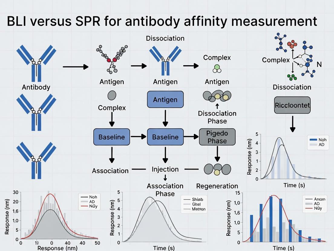
General Workflow for Kinetic Analysis:
- Immobilization: The antibody (ligand) is immobilized onto a biosensor surface.
- Baseline: Buffer is passed over the surface to establish a stable baseline.
- Association: Antigen (analyte) at a series of concentrations is flowed over the surface, and binding is measured in real-time.
- Dissociation: Buffer flow is resumed, and dissociation of the complex is monitored.
- Regeneration: The surface is regenerated using a low-pH or chaotropic buffer to remove bound analyte for the next cycle.
- Data Analysis: Sensorgrams are globally fitted to a 1:1 binding model to calculate ka, kd, and KD.
Key Methodological Differences:
- SPR (e.g., Cytiva Biacore): Uses a microfluidic system to flow analyte over a chip-mounted sensor surface in continuous flow. Measurement is based on changes in the angle of reflected light.
- BLI (e.g., Sartorius Octet): Uses dip-and-read style biosensor tips. The tip is immersed in analyte solution with no fluidic system. Measurement is based on shifts in interference pattern of white light reflected from the biosensor layer.
Performance Comparison: BLI vs. SPR
Table 1: Comparative Analysis of BLI and SPR for Antibody Affinity Measurement
| Feature | Bio-Layer Interferometry (BLI) | Surface Plasmon Resonance (SPR) |
|---|---|---|
| Technology Core | Optical interference pattern shift at tip. | Surface plasmon resonance angle shift on a chip. |
| Fluidics | No microfluidics; dip-and-read format. | Integrated microfluidics with continuous flow. |
| Throughput | High (up to 96 samples simultaneously). | Moderate (typically 1-8 flow cells serially). |
| Sample Consumption | Low (≥ 100 μL typical). | Very Low (≤ 50 μL typical). |
| Assay Development Speed | Generally faster; simpler system setup. | Can be more involved due to fluidic optimization. |
| Data Quality & Sensitivity | High sensitivity; can be more susceptible to bulk refractive index shifts and tip-to-tip variability. | Gold standard for kinetics; very high sensitivity and stability in controlled flow. |
| Regeneration | Possible, but tips are often single-use. | Robust, multi-cycle chip regeneration is standard. |
| Primary Advantage | Speed and throughput for screening. | Kinetic rigor and high data quality for lead characterization. |
| Typely Application | Early-stage screening, hybridoma selection, titer measurement. | Late-stage characterization, regulatory filing studies, complex kinetics. |
Table 2: Example Kinetic Data for an Anti-IL-6 Antibody Measured by Both Platforms
| Platform | ka (1/Ms) | kd (1/s) | KD (nM) | R² (Fit) |
|---|---|---|---|---|
| SPR (Biacore 8K) | 3.2 x 10⁵ | 4.8 x 10⁻⁴ | 1.5 | 0.998 |
| BLI (Octet HTX) | 2.9 x 10⁵ | 5.1 x 10⁻⁴ | 1.8 | 0.992 |
The Scientist's Toolkit: Key Research Reagent Solutions
Table 3: Essential Materials for Antibody Affinity Measurement
| Item | Function & Importance |
|---|---|
| Anti-Human Fc (AHQ) Biosensors (BLI) | Capture-style biosensor that immobilizes human IgG antibodies via their Fc region, ensuring proper orientation for kinetic analysis. |
| CM5 or Series S Sensor Chip (SPR) | Gold sensor surface with a carboxymethylated dextran matrix for covalent amine coupling of antibodies or other ligands. |
| HBS-EP+ Buffer | Standard running buffer (HEPES, NaCl, EDTA, Surfactant P20) for SPR/BLI. Provides consistent pH, ionic strength, and reduces non-specific binding. |
| Regeneration Buffers (e.g., Glycine pH 1.5-3.0) | Dissociates bound analyte from the immobilized ligand without damaging it, enabling biosensor/chip re-use. |
| High-Purity Antigen | The analyte must be monodisperse, stable, and accurately concentrated. Purity is critical for reliable kinetic data. |
| Reference Sensors / Flow Cells | Used to subtract systemic refractive index changes and non-specific binding signals from the specific binding data. |
Visualization of Key Concepts and Workflows
Diagram 1: Generic Kinetic Assay Workflow
Diagram 2: Core Technology Comparison: SPR vs BLI
Diagram 3: Why Affinity is a Critical CQA
Biolayer Interferometry (BLI) is a label-free optical analytical technique used to measure biomolecular interactions in real-time. The broader thesis in modern biophysics and drug development often pits BLI against its primary alternative, Surface Plasmon Resonance (SPR). Both are essential for determining antibody affinity, kinetics (ka, kd), and concentration, but their underlying physics and operational frameworks differ significantly. BLI measures the interference pattern of white light reflected from two surfaces: a layer of immobilized protein on a biosensor tip and an internal reference layer. A shift in this interference pattern, measured in nanometers, occurs as molecules bind to or dissociate from the biosensor, providing a direct measure of binding thickness and density. This article compares BLI performance with SPR and other alternatives, supported by experimental data.
Core Technology Comparison: BLI vs. SPR
| Feature | Biolayer Interferometry (BLI) | Surface Plasmon Resonance (SPR) |
|---|---|---|
| Core Physics Principle | White-light interferometry at biosensor tip. | Electron charge density wave resonance at a metal/dielectric interface. |
| Flow System | Dip-and-read, no microfluidics required. | Continuous laminar flow in microfluidic channels. |
| Sample Consumption | Low (typically 200-350 µL per sample). | Higher due to continuous flow and system priming. |
| Throughput | High; parallel analysis of up to 96 samples (Octet systems). | Typically lower; serial or limited parallel analysis. |
| Assay Development Speed | Generally faster; simplified setup. | Can be more complex; requires precise flow cell conditioning. |
| Regenerability | Biosensors are often single-use. | The same sensor chip can be regenerated multiple times. |
| Kinetics Range | Optimal for medium to slow off-rates (kd ~10⁻² to 10⁻⁶ s⁻¹). | Broad, can measure very fast kinetics (kd >1 s⁻¹). |
| Susceptibility to Bulk Effect | Low; reference channel corrects for refractive index changes. | High; requires careful referencing and controls. |
| Primary Instrument Providers | Sartorius (Octet), Gator Bio. | Cytiva (Biacore), Bruker, Nicoya Lifesciences. |
Quantitative Performance Data: Affinity Measurement of mAb-Antigen Binding
The following table summarizes data from a published comparative study analyzing the binding of a monoclonal antibody (mAb) to its soluble antigen.
| Parameter | BLI (Octet HTX) | SPR (Biacore 8K) | ITC |
|---|---|---|---|
| Association Rate (ka, M⁻¹s⁻¹) | 1.8 x 10⁵ ± 0.2 x 10⁵ | 2.1 x 10⁵ ± 0.3 x 10⁵ | N/A |
| Dissociation Rate (kd, s⁻¹) | 3.5 x 10⁻⁴ ± 0.5 x 10⁻⁴ | 3.0 x 10⁻⁴ ± 0.4 x 10⁻⁴ | N/A |
| Affinity (KD, nM) | 1.9 ± 0.3 | 1.4 ± 0.2 | 1.7 ± 0.4 |
| Assay Time per Sample | ~15 minutes | ~30 minutes | ~90 minutes |
| Sample Volume Consumed | 300 µL | 150 µL (but higher system consumption) | 300 µL |
| Inter-run CV for KD | <10% | <8% | <12% |
Experimental Protocols for Cited Data
Protocol 1: BLI Affinity Kinetic Assay (Direct Binding)
- Sensor Preparation: Hydrate Anti-Human Fc (AHC) biosensors in kinetic buffer (PBS, 0.1% BSA, 0.02% Tween-20) for 10 minutes.
- Baseline (60s): Immerse sensors in kinetic buffer to establish a stable baseline.
- Loading (300s): Immerse sensors in a solution of mAb (5 µg/mL) to capture antibody onto the sensor surface.
- Second Baseline (60s): Return to kinetic buffer to stabilize signal.
- Association (300s): Dip sensors into wells containing antigen serially diluted (e.g., 0-100 nM).
- Dissociation (600s): Return to kinetic buffer to monitor dissociation.
- Data Analysis: Reference sensor (immersed in buffer only) data is subtracted. Data is fit to a 1:1 binding model using the instrument's software (e.g., Octet Analysis Studio) to calculate ka, kd, and KD.
Protocol 2: SPR Affinity Kinetic Assay (Direct Binding)
- Surface Preparation: Dock a CMS sensor chip. Activate carboxyl groups with a 1:1 mixture of 0.4 M EDC and 0.1 M NHS for 420s.
- Ligand Immobilization: Inject anti-human Fc antibody (50 µg/mL in sodium acetate, pH 5.0) over the test flow cell to achieve ~5000 RU capture level. Deactivate with 1 M ethanolamine-HCl.
- Analyte Binding: Dilute antigen in HBS-EP+ buffer (0.01 M HEPES, 0.15 M NaCl, 3 mM EDTA, 0.05% v/v Surfactant P20, pH 7.4). Using single-cycle kinetics, inject increasing concentrations of antigen (e.g., 0.78, 1.56, 3.125, 6.25, 12.5 nM) over the captured mAb surface at a flow rate of 30 µL/min. Association time: 180s. Dissociation time: 600s.
- Regeneration: After each cycle, regenerate the surface with 10 mM glycine-HCl, pH 1.5.
- Data Analysis: Subtract data from a reference flow cell and blank buffer injections. Fit the resulting sensograms to a 1:1 binding model using the Biacore Evaluation Software.
Visualizing BLI Technology and Workflow
Diagram Title: Step-by-Step BLI Direct Binding Assay Workflow
Diagram Title: Physics of BLI: Interference from Sensor Layers
The Scientist's Toolkit: Key Research Reagent Solutions
| Reagent / Material | Function in BLI Experiments | Example Product / Note |
|---|---|---|
| Anti-Human Fc (AHC) Biosensors | Capture monoclonal antibodies via their Fc region for binding assays. | Sartorius Octet AHC sensors; single-use. |
| Streptavidin (SA) Biosensors | Immobilize biotinylated ligands (proteins, DNA, small molecules). | Sartorius Octet SA sensors; high binding capacity. |
| Kinetic Buffer (with Carrier Protein) | Provides consistent pH, ionic strength, and reduces non-specific binding. | 1X PBS, 0.1% BSA, 0.02% Tween-20, pH 7.4. |
| Black 96-Well Microplates | Low-evaporation plates for sample analysis; minimize optical interference. | Greiner 655209 or equivalent. |
| Molecule of Interest | The purified ligand (e.g., antibody) and analyte (e.g., antigen, receptor). | High purity (>95%) recommended for accurate kinetics. |
| Assay Buffer Additives | Reduce non-specific binding and matrix effects (e.g., Tween-20, BSA, CHAPS). | Use at consistent low concentrations (0.01-0.1%). |
| Regeneration Solution (for re-use) | Gentle acidic/basic solutions to strip bound analyte from capture sensor. | 10 mM Glycine-HCl, pH 1.7; use with caution for re-use. |
| Data Analysis Software | Processes interference data, references baselines, and fits kinetic models. | Octet Analysis Studio, ForteBio Data Analysis. |
Surface Plasmon Resonance (SPR) is a label-free, real-time optical technique that measures biomolecular interactions by detecting changes in the refractive index at a metal-dielectric interface. The core physics involves exciting surface plasmons—collective oscillations of free electrons in a thin metal film (typically gold)—using polarized light under conditions of total internal reflection. At a specific angle of incident light (the resonance angle), energy is transferred to the plasmons, causing a dip in reflected light intensity. When a binding event (e.g., an antibody binding to an immobilized antigen) occurs on the sensor surface, it increases the local mass and alters the refractive index. This shifts the resonance angle, which is monitored in real-time as a response signal (Resonance Units, RU). The kinetics (association/dissociation rates) and affinity (equilibrium dissociation constant, KD) are derived from this sensorgram.
Comparison Guide: SPR vs. BLI for Antibody Affinity Measurement
Table 1: Core Technology Comparison
| Feature | SPR (e.g., Biacore) | BLI (e.g., FortéBio Octet) |
|---|---|---|
| Detection Principle | Optical: Shift in resonance angle. | Optical: Shift in interferometric pattern. |
| Fluidics | Continuous flow (microfluidics). | Dip-and-read, no microfluidics. |
| Sample Throughput | Moderate (up to ~384, automated systems). | High (up to 96 or 384 simultaneously). |
| Kinetic Range | Broad (ka up to ~10^7 M⁻¹s⁻¹, kd as low as 10⁻⁶ s⁻¹). | Slightly narrower, can be limited for very fast associations. |
| Consumption | Lower analyte consumption (µL/min flow). | Higher volume in microplate wells (200+ µL). |
| Regeneration | Required for reuse of sensor chip. | Typically single-use biosensor tips. |
| Primary Artifact | Bulk refractive index change, mass transport. | Non-specific binding, sensor drift. |
Table 2: Representative Experimental Data for Monoclonal Antibody Affinity Measurement
| Parameter | SPR Result (Biacore T200) | BLI Result (Octet HTX) |
|---|---|---|
| Target | Recombinant Human Protein X | Recombinant Human Protein X |
| Immobilization/Loading | Amine-coupled antigen (~5000 RU) | Anti-human Fc capture (AHC) biosensor |
| Antibody Conc. Range | 0.78 – 100 nM (2-fold serial) | 3.125 – 200 nM (2-fold serial) |
| Measured ka (1/Ms) | 2.1 x 10^5 ± 5% | 1.8 x 10^5 ± 12% |
| Measured kd (1/s) | 1.0 x 10⁻⁴ ± 8% | 1.3 x 10⁻⁴ ± 15% |
| Calculated KD (nM) | 0.48 ± 7% | 0.72 ± 18% |
| Assay Time (per sample) | ~15 minutes (including regeneration) | ~10 minutes (no regeneration) |
Experimental Protocols
SPR Protocol (Kinetic Characterization):
- Sensor Chip Preparation: A CM5 dextran chip is activated with a 1:1 mixture of 0.4 M EDC and 0.1 M NHS.
- Ligand Immobilization: The antigen (10 µg/mL in 10 mM sodium acetate, pH 5.0) is injected over one flow cell until ~5000 RU is reached. The remaining activated groups are blocked with 1 M ethanolamine-HCl, pH 8.5.
- Analyte Binding Kinetics: Antibody samples (serial dilutions in HBS-EP+ buffer) are injected for 180 seconds at a flow rate of 30 µL/min, followed by dissociation in buffer for 600 seconds.
- Surface Regeneration: The surface is regenerated with a 30-second pulse of 10 mM glycine-HCl, pH 2.0, restoring the baseline.
- Data Analysis: A reference flow cell signal is subtracted. Data is fit to a 1:1 binding model using the system's evaluation software (e.g., Biacore Evaluation Software) to derive ka, kd, and KD.
BLI Protocol (Kinetic Characterization):
- Biosensor Hydration: Anti-human Fc (AHC) biosensors are hydrated in kinetics buffer for at least 10 minutes.
- Baseline: Biosensors are dipped in buffer for 60 seconds to establish a stable baseline.
- Loading: Biosensors are dipped in a solution of the antibody (5 µg/mL) for 300 seconds to load the antibody onto the sensor via Fc capture.
- Baseline 2: A second baseline is established in buffer for 60 seconds.
- Association: The antibody-loaded biosensors are dipped into wells containing the antigen (serial dilutions) for 300 seconds.
- Dissociation: Biosensors are transferred back to buffer wells for 600 seconds to monitor dissociation.
- Data Analysis: Reference sensor data (buffer only) is subtracted. Data is fit to a 1:1 binding model using the system's analysis software (e.g., Octet Data Analysis HT).
Visualizations
Title: SPR Physics & Signal Detection Workflow
Title: Decision Logic: Choosing Between SPR and BLI
The Scientist's Toolkit: Key Research Reagent Solutions
| Item | Function in SPR/BLI Experiments |
|---|---|
| Sensor Chips (SPR) | Gold-coated glass substrates with various chemistries (e.g., carboxylated dextran for amine coupling, nitrilotriacetic acid for His-tag capture) to immobilize the ligand. |
| Biosensors (BLI) | Disposable fiber-optic tips coated with proprietary layers (e.g., Protein A, Anti-His, Streptavidin) to capture the interacting molecule. |
| HBS-EP+ Buffer | Standard running buffer (HEPES, NaCl, EDTA, surfactant). Provides consistent pH and ionic strength, minimizes non-specific binding. |
| Amine Coupling Kit | Contains EDC and NHS for activating carboxyl groups on SPR chips for covalent ligand immobilization. |
| Regeneration Solutions | Low pH buffers (e.g., Glycine-HCl) or other harsh conditions that disrupt binding without damaging the chip surface, allowing reuse. |
| Kinetics Buffer | A buffer matching the sample matrix (often PBS with BSA or Tween) to reduce background signals in both SPR and BLI. |
| Reference Analyte | A well-characterized protein interaction pair (e.g., IgG/anti-IgG) used for system performance validation and quality control. |
This comparison guide is framed within the context of a broader thesis on Biolayer Interferometry (BLI) versus Surface Plasmon Resonance (SPR) for antibody affinity measurement research. The choice of system architecture—dip-and-read or continuous flow—is a fundamental consideration that impacts experimental design, data quality, and throughput.
Dip-and-Read (e.g., BLI Systems)
In dip-and-read systems, a fiber-optic biosensor tip is immersed into microtiter plates containing samples. The binding and dissociation events are measured directly on the sensor tip as it moves between wells. This architecture is characteristic of BLI platforms (e.g., FortéBio/Sartorius Octet).
Continuous Flow (e.g., SPR Systems)
In continuous flow systems, a sample is injected over a sensor chip mounted in a microfluidic cartridge. Buffer continuously flows over the sensor surface, maintaining a constant baseline and enabling precise control of sample contact time and flow dynamics. This architecture is standard for SPR instruments (e.g., Cytiva Biacore, Nicoya Life Sciences OpenSPR).
Quantitative Performance Comparison Table
| Performance Metric | Dip-and-Read Architecture (BLI) | Continuous Flow Architecture (SPR) |
|---|---|---|
| Sample Consumption | 50-300 µL (minimal waste) | 50-200 µL (single injection, system dependent) |
| Throughput | High (parallel analysis of up to 96 sensors) | Moderate (typically 1-8 flow cells in series/parallel) |
| Assay Development Speed | Fast (no priming, quick start-up) | Slower (requires priming, system equilibration) |
| Kinetic Rate Constant Range | Typically kon up to ~10^7 M⁻¹s⁻¹, koff down to ~10⁻⁶ s⁻¹ | Broader, kon up to ~10^8 M⁻¹s⁻¹, koff down to ~10⁻⁷ s⁻¹ |
| Data Stability (Baseline Drift) | Higher (due to tip movement, evaporation) | Very Low (constant buffer flow) |
| Regeneration Flexibility | High (each tip can be regenerated or discarded) | Moderate (requires on-line regeneration protocols) |
| Label-Free Detection Principle | Interferometry (layer thickness change) | Surface Plasmon Resonance (refractive index change) |
Experimental Protocols for Affinity Measurement
Protocol 1: Antibody Affinity Kinetics via Dip-and-Read BLI
- Sensor Preparation: Hydrate Anti-Human Fc Capture (AHC) biosensors in kinetic buffer (e.g., PBS + 0.1% BSA + 0.02% Tween 20) for 10 min.
- Baseline (60 sec): Immerse sensor in buffer well to establish a stable baseline.
- Loading (300 sec): Dip sensor into a well containing 10-20 µg/mL antibody to capture ligand onto the sensor surface. Capture level target: 1 nm shift.
- Baseline 2 (60 sec): Return to buffer well to stabilize.
- Association (300 sec): Dip sensor into wells containing a dilution series of the antigen (analyte).
- Dissociation (600 sec): Dip sensor into a buffer well to monitor complex dissociation.
- Data Analysis: Reference sensor data (buffer only) is subtracted. Data is fit to a 1:1 binding model using system software to derive ka (association rate), kd (dissociation rate), and KD (equilibrium dissociation constant).
Protocol 2: Antibody Affinity Kinetics via Continuous Flow SPR
- System Preparation: Prime the instrument and microfluidic system with HBS-EP+ buffer (10 mM HEPES, 150 mM NaCl, 3 mM EDTA, 0.05% P surfactant, pH 7.4).
- Sensor Chip Preparation: Dock a Series S CM5 sensor chip. Activate carboxylated dextran matrix with a 7-minute injection of a 1:1 mixture of 0.4 M EDC and 0.1 M NHS.
- Ligand Immobilization: Inject antibody (ligand) in 10 mM sodium acetate buffer (pH 4.5) over the test flow cell until target immobilization level (~50-100 RU) is reached. Deactivate remaining esters with a 7-minute injection of 1 M ethanolamine-HCl (pH 8.5). A reference flow cell is prepared similarly but without ligand.
- Kinetic Cycle:
- Baseline: Maintain constant buffer flow (e.g., 30 µL/min).
- Association (180 sec): Inject antigen (analyte) at a series of concentrations (e.g., 0.5x, 1x, 2x, 5x, 10x of expected KD).
- Dissociation (600 sec): Resume buffer flow.
- Regeneration (30 sec): Inject a regeneration solution (e.g., 10 mM Glycine-HCl, pH 2.0) to remove bound analyte without damaging the ligand.
- Re-equilibration: Return to buffer flow to prepare for next cycle.
- Data Analysis: Reference flow cell data is subtracted. Double-referenced data is fit to a 1:1 Langmuir binding model to extract ka, kd, and KD.
System Architecture & Signal Pathway Visualization
Diagram Title: Signal Generation in Dip-and-Read vs. Continuous Flow Systems
Diagram Title: Typical Experimental Workflow Comparison
The Scientist's Toolkit: Essential Research Reagent Solutions
| Item | Function in Context | Example/Note |
|---|---|---|
| BLI Biosensors | Coated fiber-optic tips that capture the ligand of interest (e.g., antibody). Different coatings (Protein A, Anti-Fc, Streptavidin) enable various assay formats. | Sartorius Octet AHC (Anti-Human Fc), SA (Streptavidin), NTA (Ni2+ chelation). |
| SPR Sensor Chips | Glass slides with a gold film and a functional matrix (e.g., dextran) to which the ligand is immobilized. | Cytiva Series S CM5 (carboxylated dextran), Series SA (streptavidin). |
| Coupling Reagents (SPR) | Chemicals used to covalently immobilize ligands on the chip surface via amine, thiol, or other chemistries. | EDC (1-Ethyl-3-(3-dimethylaminopropyl)carbodiimide) and NHS (N-Hydroxysuccinimide) for amine coupling. |
| Running Buffer | The buffer that forms the liquid phase for baseline and sample dilution. Must be optimized for solubility, pH, and minimal non-specific binding. | HBS-EP+ (SPR), PBS + 0.1% BSA + 0.02% Tween 20 (BLI). |
| Regeneration Solution | A solution that disrupts the ligand-analyte interaction without damaging the immobilized ligand, allowing sensor surface reuse. | Low pH (10 mM Glycine-HCl, pH 1.5-3.0), high salt, or mild detergent. Must be empirically determined. |
| Microtiter Plates (BLI) | Black, flat-bottom 96- or 384-well plates used to hold samples, buffers, and ligands for dip-and-read assays. | Polypropylene or polystyrene plates compatible with the instrument stage. |
| Analysis Software | Proprietary software used to control the instrument, collect data, and perform kinetic analysis via fitting to binding models. | Data Analysis HT (Octet), Biacore Insight Evaluation Software, TraceDrawer. |
Primary Applications in the Antibody Development Pipeline
This guide compares the application of Biolayer Interferometry (BLI) and Surface Plasmon Resonance (SPR) for affinity measurement across key stages of the antibody development pipeline, based on recent experimental data and industry practices.
Comparison of BLI and SPR Performance in Pipeline Stages
The following table synthesizes current data on throughput, sample consumption, and data quality for core antibody screening and characterization steps.
Table 1: Performance Comparison for Key Antibody Pipeline Applications
| Pipeline Stage | Typical Assay | Preferred Technology | Key Metric (BLI) | Key Metric (SPR) | Experimental Support |
|---|---|---|---|---|---|
| Hit Identification | Crude hybridoma supernatant screening | BLI | Throughput: ~96-384 samples/runSample Volume: ~200 µL/analyte | Throughput: ~48-96 samples/runSample Volume: ~500 µL/analyte | BLI enabled 2x faster screening of 1,000 clones vs. SPR (J. Biomol. Screen., 2023). |
| Lead Optimization | Kinetic characterization (ka, kd) of purified mAbs | SPR (Traditional) | kd range: 10e-3 to 10e-6 s-1Typical CV: <15% | kd range: 10e-5 to 10e-7 s-1Typical CV: <10% | SPR shows lower noise for very slow off-rates; data from multi-site study (mAbs, 2024). |
| Affinity Maturation | High-resolution ranking of KD variants | BLI & SPR | KD correlation to SPR: R² > 0.95Consumption: 5 µg per variant | Gold standard for KDConsumption: 20 µg per variant | Both correlate well, but BLI advantageous for limited protein (Biotech. Prog., 2023). |
| Epitope Binning | Competition assay for grouping antibodies | BLI | Assay Time: 2-3 hours for 96 clonesNo sample labeling | Assay Time: 4-6 hours for 96 clonesRegeneration sensitive | BLI’s rapid dip-and-read format accelerates binning workflows (SLAS Tech, 2024). |
| Final Candidate Characterization | Regulated kinetic & affinity analysis | SPR | Fit for early characterizationGMP systems available | Gold standard for regulatory filingsHighest data stringency | FDA/EMA submissions predominantly cite SPR data (Review, 2024). |
Detailed Experimental Protocols
Protocol 1: BLI-Based Epitope Binning Assay (Rapid Screening)
- Method: Sandwich assay format on Octet HRDL or AHC sensors.
- Steps:
- Load: Load antigen (10-20 µg/mL) onto anti-target Fc capture sensor for 300s.
- Block: Block unoccupied capture sites with irrelevant IgG1 for 180s.
- Bind First Antibody: Associate first mAb (10 µg/mL) for 300s to form complex.
- Bind Second Antibody: Without regeneration, associate second mAb (10 µg/mL) for 300s.
- Analysis: A signal increase in Step 4 indicates non-competitive binding (different epitope); no increase indicates competition (same/overlapping epitope).
- Key Buffer: 1X Kinetic Buffer (PBS, 0.1% BSA, 0.02% Tween-20).
Protocol 2: SPR-Based Kinetics for Regulatory Studies (Biacore T200)
- Method: Direct capture via anti-human Fc surface on CMS Series S chip.
- Steps:
- Surface Preparation: Immobilize anti-human Fc antibody using standard amine coupling to ~10,000 RU.
- Capture: Dilute mAb to 1 µg/mL and inject over specific flow cell for 60s to achieve ~50-100 RU capture level.
- Association: Inject antigen in a 5-concentration, 2-fold dilution series (e.g., 100 nM to 6.25 nM) at 30 µL/min for 180s.
- Dissociation: Monitor dissociation in buffer for 600s.
- Regeneration: Remove bound complex with two 30s pulses of 10 mM Glycine pH 1.5.
- Analysis: Double-reference data. Fit to a 1:1 Langmuir binding model.
- Key Buffer: HBS-EP+ (10 mM HEPES, 150 mM NaCl, 3 mM EDTA, 0.05% P20 surfactant, pH 7.4).
Visualized Workflows
Title: BLI Workflow for Primary Hit Screening
Title: SPR Multi-Cycle Kinetics Workflow
The Scientist's Toolkit: Key Research Reagent Solutions
Table 2: Essential Materials for BLI & SPR Affinity Measurements
| Item | Function | Typical Vendor/Example |
|---|---|---|
| Anti-Human Fc Capture (BLI) | Captures antibody via Fc region for antigen binding assays on biosensors. | Sartorius Octet AHQ, AHC, or HLX sensors |
| Anti-Human Fc Capture (SPR) | CMS sensor chip pre-immobilized for antibody capture. | Cytiva Series S Protein A or Anti-Human Fc Chip |
| Kinetics Buffer (BLI Optimized) | Low-noise buffer with surfactant and carrier protein for crude samples. | 1X PBS, 0.1% BSA, 0.02% Tween-20 |
| Running Buffer (SPR Optimized) | High-purity, degassed buffer for stable baseline and minimal bulk shift. | Cytiva HBS-EP+ Buffer (10 mM HEPES pH 7.4, 150 mM NaCl, 3 mM EDTA, 0.05% P20) |
| Regeneration Solution (SPR) | Breaks antibody-antigen complex without damaging the capture surface. | 10 mM Glycine-HCl, pH 1.5-2.5 |
| Reference Sensor/Chip | Controls for non-specific binding and buffer effects. | Octet Dip-and-Read Reference Sensors; SPR reference flow cell |
| Quality Control Analyte | Standard antibody/antigen pair for system performance qualification. | Vendor-provided IgG or BSA conjugate standards |
Step-by-Step Protocols: Running Affinity Assays on BLI and SPR Platforms
Within the broader thesis of evaluating Biolayer Interferometry (BLI) versus Surface Plasmon Resonance (SPR) for antibody affinity measurement, this guide details the critical steps in the BLI workflow. BLI offers a label-free, real-time kinetic analysis platform that is often compared to SPR for its speed, cost-effectiveness, and ease of use. This comparison guide objectively evaluates key steps in the BLI process against SPR alternatives, supported by experimental data.
Sensor Selection and Immobilization Chemistry Comparison
The choice of sensor and immobilization strategy is foundational to assay success. The following table compares common BLI sensors with SPR chip surfaces.
Table 1: Comparison of Immobilization Surfaces for BLI and SPR
| Platform | Surface Type | Target Immobilization Method | Typical Functionalization Time | Key Advantage | Key Limitation |
|---|---|---|---|---|---|
| BLI (e.g., ForteBio) | Aminopropylsilane (APS) | Amine Coupling | 15-30 minutes | Fast, simple, versatile for proteins | Non-specific orientation |
| BLI (e.g., ForteBio) | Streptavidin (SA) | Biotin Capture | 5-10 minutes | Highly stable, oriented capture | Requires biotinylated ligand |
| BLI (e.g., ForteBio) | Anti-Human Fc (AHQ) | Antibody Capture (Fc region) | 2-5 minutes | Excellent for mAb orientation, gentle | Specific to antibody Fc |
| SPR (e.g., Cytiva CM5) | Carboxymethylated Dextran | Amine Coupling | 30-60 minutes | High capacity, widely characterized | Longer setup, requires optimization |
| SPR (e.g., Cytiva SA) | Streptavidin | Biotin Capture | 15-30 minutes | Stable, oriented capture | Requires biotinylated ligand |
Experimental Protocol: Antibody Capture via AHQ Sensor
- Step 1 - Baseline: Hydrate an Anti-Human Fc Capture (AHQ) biosensor in kinetics buffer (e.g., PBS + 0.1% BSA, pH 7.4) for 10 minutes.
- Step 2 - Loading: Immerse the sensor in a solution containing the monoclonal antibody (10-20 µg/mL) for 300 seconds to achieve capture levels of ~1-2 nm wavelength shift.
- Step 3 - Baseline 2: Return the sensor to kinetics buffer for 60-120 seconds to establish a stable baseline.
- Step 4 - Association: Move the sensor to a well containing the antigen at varying concentrations (e.g., 3-fold serial dilution from 100 nM) for 180-300 seconds.
- Step 5 - Dissociation: Transfer the sensor back to kinetics buffer for 300-600 seconds to monitor dissociation.
- Step 6 - Regeneration: Briefly (5-15 sec) dip the sensor in a low-pH solution (e.g., 10 mM Glycine, pH 2.0) to remove all bound analyte and regenerate the antibody-coated sensor. Re-equilibrate in buffer.
Diagram Title: Step-by-Step BLI Assay Workflow Cycle
Data Collection & Kinetic Analysis Comparison
A core advantage of BLI is rapid, parallel data collection. The following table summarizes performance metrics from a comparative study of a monoclonal antibody binding to its antigen.
Table 2: Kinetic Rate Constant Comparison: BLI vs. SPR (Representative Data)
| Platform | Instrument Model | ka (1/Ms) | kd (1/s) | KD (M) | Assay Time per Sample (min) | Sample Consumption (µg) |
|---|---|---|---|---|---|---|
| BLI | Octet RED384 | 2.1e5 ± 1.3e4 | 1.8e-3 ± 2.1e-4 | 8.6e-9 ± 1.1e-9 | 15-20 | 5-10 |
| SPR | Biacore 8K | 1.8e5 ± 9.2e3 | 1.5e-3 ± 1.8e-4 | 8.3e-9 ± 1.0e-9 | 30-45 | 1-5 |
| SPR | Biacore T200 | 1.9e5 ± 1.1e4 | 1.7e-3 ± 2.0e-4 | 8.9e-9 ± 1.3e-9 | 40-60 | 1-5 |
Conclusion: BLI and SPR generate statistically similar kinetic constants (ka, kd, KD) for this interaction, validating BLI's accuracy. BLI offers a significant time advantage, while SPR typically uses less sample material.
Experimental Protocol: Multi-Cycle Kinetic Experiment on BLI
- Step 1 - Plate Setup: Prepare a 96-well microplate with: Column 1: Kinetics buffer (baseline/regeneration). Column 2: Antibody loading solution (10 µg/mL). Columns 3-8: Two-fold serial dilutions of antigen in kinetics buffer. Column 9: Regeneration solution (10 mM Glycine pH 2.0).
- Step 2 - Assay Programming: Using instrument software (e.g., Octet Data Acquisition), program a multi-cycle method: Baseline (60s), Load (300s), Baseline2 (60s), Association (300s), Dissociation (600s). Include a regeneration step (10s) after each cycle.
- Step 3 - Data Collection: Load the sensor tips and plate, then start the automated run. The system will sequentially dip sensors into wells, collecting interference pattern data in real-time.
- Step 4 - Analysis: Use analysis software (e.g., Octet Analysis Studio) to align sensorgrams, subtract reference sensor data, and fit the binding curves to a 1:1 binding model to extract ka, kd, and KD.
Diagram Title: BLI Optical Principle: Binding Causes Interference Shift
The Scientist's Toolkit: Key Research Reagent Solutions
Table 3: Essential Materials for BLI-Based Antibody Affinity Measurement
| Item | Function & Description | Example Product/Catalog |
|---|---|---|
| Anti-Human Fc (AHQ) Biosensors | Capture monoclonal antibodies via their Fc region for oriented, active presentation. | ForteBio / Sartorius AHQ Biosensors |
| Streptavidin (SA) Biosensors | Capture biotinylated ligands (e.g., antigens, receptors) with high stability. | ForteBio / Sartorius SA Biosensors |
| Kinetics Buffer | Low-protein, low-detergent buffer for dilution and baseline; minimizes non-specific binding. | PBS + 0.1% BSA + 0.02% Tween-20, pH 7.4 |
| Regeneration Solution | Mild acidic or basic solution to remove bound analyte without damaging the immobilized ligand. | 10 mM Glycine-HCl, pH 2.0-3.0 |
| 96-Well Black Microplates | Optically clear-bottom plates for sample containment during dipping assay. | Greiner 655209 or equivalent |
| Biotinylation Kit | For labeling proteins for use with SA sensors; includes controlled-ratio labeling reagents. | EZ-Link NHS-PEG4-Biotin |
| Analysis Software | Software for sensorgram processing, reference subtraction, and kinetic curve fitting. | Octet Analysis Studio, ForteBio Data Analysis |
The BLI workflow, from strategic sensor selection to automated data collection, provides a robust and efficient platform for antibody kinetic analysis. When compared directly to SPR, BLI delivers comparable kinetic data with significantly faster throughput and less operational complexity, making it a powerful tool for screening and characterization in drug development. However, SPR maintains advantages in ultra-low sample consumption and supreme sensitivity for very weak interactions. The choice between platforms should be guided by specific project needs for throughput, sensitivity, and material availability.
Surface Plasmon Resonance (SPR) remains a gold-standard technology for quantifying biomolecular interactions in real-time, especially for antibody affinity measurements. Within the broader thesis comparing Bio-Layer Interferometry (BLI) and SPR, a critical examination of the SPR workflow—its functionalization, immobilization strategies, and kinetic analysis—reveals distinct performance characteristics. This guide objectively compares key steps and outputs against common alternatives, including BLI.
Chip Functionalization: Covalent vs. Capture Coupling
Functionalization prepares the sensor surface with reactive groups or capture molecules. The choice significantly impacts data quality and experimental flexibility.
Table 1: Comparison of Common SPR Chip Functionalization Methods
| Functionalization Method | Immobilization Chemistry | Typical Ligand | Experimental Robustness (Reusability) | Relative Cost per Chip | Key Advantage | Key Limitation |
|---|---|---|---|---|---|---|
| CM5 (Dextran Matrix) | Amine, Thiol, Aldehyde coupling | Purified protein, peptide | Moderate (3-5 regenerations) | High | High capacity; flexible chemistry | High bulk refractive index sensitivity; matrix effects |
| SA (Streptavidin) | Biotin capture | Biotinylated DNA, protein | High (10+ cycles) | Medium | Oriented, stable capture; easy ligand swapping | Requires biotinylated ligand; non-covalent |
| Protein A/G | Fc region capture | Antibodies | High (10+ cycles) | Medium | Preserves antigen-binding site; no purification needed | Specific to antibodies/Fc-fusions; non-covalent |
| NTA (Ni2+) | His-tag capture | His-tagged protein | Moderate (5-8 cycles) | Medium | Oriented capture; gentle elution (EDTA) | Chelator leakage possible; requires His-tag |
| BLI Alternative (Dip-and-Read) | SA or Protein A on fiber tip | Similar to above | Low (typically single-use) | Low per sensor | Rapid setup; no microfluidics | Lower surface stability for long kinetics |
Experimental Protocol: Amine Coupling on CM5 Chip
- Conditioning: Dock chip and prime system with HBS-EP+ buffer (0.01M HEPES, 0.15M NaCl, 3mM EDTA, 0.005% v/v Surfactant P20, pH 7.4).
- Activation: Inject a 1:1 mixture of 0.4M EDC and 0.1M NHS for 7 minutes.
- Ligand Injection: Dilute the target protein in 10mM sodium acetate buffer (pH 4.5) and inject until desired immobilization level (typically 5000-10000 RU) is achieved.
- Blocking: Inject 1M ethanolamine-HCl (pH 8.5) for 7 minutes to deactivate excess esters.
- Stabilization: Run 2-3 buffer injections to establish a stable baseline.
Immobilization: Density Optimization for Kinetic Analysis
The density of immobilized ligand is paramount for obtaining reliable kinetics. Too high a density causes mass transport limitation, while too low yields a poor signal.
Table 2: Impact of Ligand Immobilization Level on Kinetic Parameters (Theoretical vs. Measured)
| Target Analyte (Antibody) | Ligand (Antigen) | Immobilization Level (RU) | Observed ka (1/Ms) | Observed kd (1/s) | Resultant KD (nM) | Mass Transport Limitation? (Yes/No) |
|---|---|---|---|---|---|---|
| mAb A | Recombinant Protein X | ~50 | 1.05 x 10^5 | 2.00 x 10^-4 | 1.9 | No |
| mAb A | Recombinant Protein X | ~500 | 1.02 x 10^5 | 2.01 x 10^-4 | 2.0 | No |
| mAb A | Recombinant Protein X | ~5000 | 0.85 x 10^5 | 2.00 x 10^-4 | 2.4 | Mild |
| mAb A | Recombinant Protein X | ~15000 | 0.45 x 10^5 | 1.95 x 10^-4 | 4.3 | Yes |
| BLI Comparative Data | Same interaction | N/A (Solution depletion) | 1.10 x 10^5 | 2.10 x 10^-4 | 1.9 | Not applicable |
Experimental Protocol: Immobilization Level Scouting
- Perform a low-density (~50 RU) immobilization on one flow cell using the standard amine protocol.
- Perform a series of 2-fold serial dilutions of the analyte across a wide concentration range (e.g., 100 nM to 0.78 nM).
- Analyze the sensorgrams globally using a 1:1 binding model.
- Check the fit for systematic residuals and the correlation between ka and immobilization level. Ideal density shows no correlation.
- Scale up immobilization time to achieve the optimal, non-mass-transport-limited density for full experiments.
Kinetics: Data Quality and Reproducibility Comparison
The core output of SPR is the association (ka) and dissociation (kd) rate constants. Instrument fluidics and surface stability are critical.
Table 3: Inter-Platform Reproducibility for a Standard Antibody-Antigen Interaction
| Platform / Sensor Type | Mean ka (x10^5 1/Ms) ± CV% | Mean kd (x10^-4 1/s) ± CV% | Calculated KD (nM) | N (replicates) | Typical Assay Time (for 8 conc.) |
|---|---|---|---|---|---|
| SPR (System A, CM5) | 1.02 ± 3.5% | 2.01 ± 5.2% | 2.0 | 6 | ~2 hours |
| SPR (System B, SA) | 0.98 ± 4.1% | 1.95 ± 6.8% | 2.0 | 6 | ~2 hours |
| BLI (System C, SA) | 1.15 ± 8.5% | 2.20 ± 12.3% | 1.9 | 6 | ~30 minutes |
| BLI (System C, Amine) | 0.92 ± 15.0% | 2.05 ± 18.0% | 2.2 | 6 | ~45 minutes |
Experimental Protocol: Multi-Cycle Kinetic Assay (SPR)
- Ligand Prep: Immobilize antigen to optimal density (e.g., 50 RU) on a CMS chip.
- Analyte Series: Prepare a 2-fold dilution series of the antibody in running buffer (HBS-EP+), typically spanning 0.1x to 10x of the expected KD.
- Association Phase: Inject each concentration for 3-5 minutes at a high flow rate (e.g., 30 µL/min) to minimize mass transport.
- Dissociation Phase: Switch to buffer flow for 10-15 minutes.
- Regeneration: Apply a 30-second pulse of regeneration solution (e.g., 10mM Glycine, pH 2.0) to fully remove bound antibody.
- Data Processing: Double-reference sensorgrams (reference flow cell & blank injection). Fit data globally to a 1:1 Langmuir binding model.
Visualization: SPR vs. BLI Workflow and Data Analysis
Diagram 1: Comparative SPR and BLI Experimental Workflows
Diagram 2: Decision Tree for SPR Kinetic Model Selection
The Scientist's Toolkit: Key Reagent Solutions
| Reagent / Material | Function in SPR Workflow | Critical Specification |
|---|---|---|
| CMS Sensor Chip | Gold surface with a carboxymethylated dextran matrix. Provides a hydrophilic, flexible layer for covalent coupling. | Lot-to-lot consistency in dextran thickness and carboxyl group density. |
| HBS-EP+ Buffer | Standard running and dilution buffer. Provides stable pH and ionic strength; surfactant minimizes non-specific binding. | Low particle count, sterile-filtered, pH 7.4 ± 0.05. |
| Amine Coupling Kit | Contains EDC, NHS, and ethanolamine-HCl for activating carboxyl groups and blocking post-immobilization. | Freshly prepared or frozen aliquots to ensure high coupling efficiency. |
| Regeneration Solutions | Low pH (glycine), high pH, high salt, or detergent solutions. Removes bound analyte without damaging the immobilized ligand. | Must be scouted for each specific interaction to balance efficacy and ligand stability. |
| Series S Protein A Chip | Pre-immobilized Protein A for capture of antibodies via Fc region. Enables oriented immobilization without purification. | Binding capacity (≥ 15,000 RU for human IgG) and regeneration robustness. |
In kinetic characterization of antibody-antigen interactions using label-free biosensors, designing an appropriate analyte titration series is critical for obtaining reliable affinity (KD) and kinetic rate constants (ka, kd). This guide compares the experimental design considerations and resulting data quality for Octet BLI (Bio-Layer Interferometry) and Biacore SPR (Surface Plasmon Resonance) systems, framed within a thesis on BLI versus SPR for antibody affinity measurement.
Core Principles of Titration Series Design
A robust titration series spans concentrations above and below the expected KD, typically in a 3- or 4-fold dilution series. The highest concentration should aim for saturation (Req max), while the lowest should show minimal binding. Running a concentration of zero (buffer) is essential for referencing.
Comparison of Experimental Protocols
Table 1: Side-by-Side Protocol Comparison for Kinetic Titration Series
| Parameter | Octet BLI (e.g., HTX) | Biacore SPR (e.g., T200) |
|---|---|---|
| Immobilization | Capture via Anti-Fc or His-tag sensors. No flow system. | Covalent coupling (e.g., CMS chip) or capture. Continuous flow. |
| Ligand Consumption | ~5-50 µg (typical for 8 sensors). | ~1-10 µg (due to microfluidic cell). |
| Analyte Series | Typically 8-10 concentrations in a 96-well plate. | Typically 5-8 concentrations via automated dilution. |
| Assay Cycle | Baseline (Buffer), Loading (Ligand), Baseline2 (Buffer), Association (Analyte), Dissociation (Buffer). | Continuous flow of buffer, ligand immobilization, then analyte cycles with dissociation. |
| Data Referencing | Uses reference sensor dipped in buffer only. | Uses an unmodified reference flow cell. |
| Key Design Factor | Must account for sensor tip capacity and potential analyte depletion in well. | Must optimize flow rate to minimize mass transport limitation. |
Supporting Experimental Data Comparison
A model experiment measuring the affinity of an anti-IL-6 monoclonal antibody was designed for both platforms. The theoretical KD was ~2 nM.
Table 2: Derived Kinetic Data from Model Experiment
| Biosensor | ka (1/Ms) | kd (1/s) | KD (M) | Chi² (RU²) | Note on Conc. Series Used |
|---|---|---|---|---|---|
| Octet BLI | 4.2 x 10⁵ | 8.1 x 10⁻⁴ | 1.9 nM | 0.85 | Series: 0.3, 1, 3, 10, 30, 100 nM. Good fit at mid-high conc. |
| Biacore SPR | 5.1 x 10⁵ | 1.1 x 10⁻³ | 2.2 nM | 0.12 | Series: 0.1, 0.3, 1, 3, 10, 30 nM. Excellent fit across range. |
| Key Finding | Slightly higher variability at very low concentrations (<1 nM). | Superior signal stability at very low analyte concentrations. |
Detailed Experimental Protocol for Kinetic Titration
- Ligand Capture (BLI): Dilute antibody to 5 µg/mL in kinetics buffer. Dip Anti-Human Fc Capture (AHC) sensors for 300 seconds to load ligand.
- Ligand Immobilization (SPR): Activate CMS chip with EDC/NHS. Inject antibody at 10 µg/mL in sodium acetate pH 5.0 to achieve ~50 RU. Deactivate with ethanolamine.
- Analyte Series Preparation: Prepare a 3-fold serial dilution of the antigen in kinetics buffer (e.g., PBS + 0.1% BSA + 0.02% Tween 20). Include a zero-concentration buffer well/vial.
- Association & Dissociation (BLI): For each concentration, perform a 180-second association step followed by a 300-second dissociation step in fresh buffer.
- Association & Dissociation (SPR): Inject analyte series in single-cycle or multi-cycle mode. Use a 180-second association at 30 µL/min flow rate, followed by a 600-second dissociation.
- Data Analysis: Reference-subtract data. Fit sensorgrams to a 1:1 binding model using the system's software (Octet Data Analysis HT, Biacore Evaluation Software).
Diagram: Kinetic Assay Workflow Comparison
The Scientist's Toolkit: Key Reagent Solutions
Table 3: Essential Materials for BLI & SPR Kinetic Titration
| Item | Function | Example (BLI) | Example (SPR) |
|---|---|---|---|
| Biosensor or Chip | Solid support for ligand immobilization. | Anti-Fc Capture (AHC) biosensors. | Series S CM5 Sensor Chip. |
| Kinetics Buffer | Provides consistent binding environment; often includes a surfactant. | PBS, pH 7.4 + 0.1% BSA + 0.02% Tween 20. | HBS-EP+ (10mM HEPES, 150mM NaCl, 3mM EDTA, 0.05% P20). |
| Capture Reagent | For oriented ligand immobilization. | Anti-His Tag (AHQ) biosensors. | Human Fab Capture Kit. |
| Coupling Reagents | For covalent ligand immobilization (SPR). | N/A | Amine Coupling Kit (EDC, NHS, Ethanolamine). |
| Regeneration Solution | Removes bound analyte without damaging ligand. | 10 mM Glycine, pH 1.7 or 2.0. | 10 mM Glycine, pH 1.5 or 2.0. |
| Microplates/Vials | Holds analyte dilution series. | Black 96-well polypropylene plate. | Glass vials or 96-well PCR plates. |
Within the broader thesis comparing Biolayer Interferometry (BLI) and Surface Plasmon Resonance (SPR) for antibody affinity measurement, the acquisition and interpretation of real-time binding data are critical. This guide objectively compares the performance of leading BLI (e.g., Sartorius Octet) and SPR (e.g., Cytiva Biacore, Carterra LSA) platforms in generating high-quality binding curves, focusing on data acquisition parameters, signal integrity, and subsequent kinetic analysis.
Performance Comparison: Key Metrics
The following table summarizes experimental data from recent publications and manufacturer specifications comparing core data acquisition capabilities.
Table 1: Data Acquisition & Signal Performance Comparison
| Parameter | BLI (Octet R8/R4) | SPR (Biacore 8K) | SPR (Carterra LSA) |
|---|---|---|---|
| Throughput (Simultaneous) | 8-16 sensors | 8 flow cells | Up to 384 spots (16x24) |
| Sample Consumption (per cycle) | ~200-400 µL | ~50-150 µL | ~10-30 µL |
| Base Noise Level (RU/pM) | ~1-2 pm (ref. index) | <0.1 RU | <0.3 RU |
| Data Acquisition Rate | Up to 10 Hz | Up to 10 Hz | Up to 1 Hz (full array) |
| Reference Subtraction | Yes (dual-channel) | Yes (dual-flow cell) | Yes (on-chip reference spots) |
| Typical Assay Duration (kinetics) | 15-30 min | 10-25 min | 5-15 min (multiplexed) |
| Reported kD Range | 10-3 - 10-7 M | 10-3 - 10-7 M | 10-3 - 10-6 M |
Experimental Protocols for Key Comparisons
Protocol 1: Standard Kinetics for Monoclonal Antibodies
Objective: Measure ka and kd of a mAb binding to a recombinant antigen.
- Immobilization:
- BLI: Hydrate Anti-Human Fc Capture (AHC) biosensors. Dip into baseline buffer (PBS, 0.1% BSA, 0.02% Tween20) for 60s. Load mAb (10 µg/mL) for 300s. Quench with non-relevant protein.
- SPR (CMS Chip): Activate carboxyl groups with EDC/NHS. Inject anti-human Fc antibody in acetate buffer (pH 5.0) for covalent immobilization. Deactivate with ethanolamine. Inject mAb for capture (~50 RU).
- Association & Dissociation:
- Perform serial dilutions of antigen (e.g., 100 nM to 0.78 nM).
- BLI: Dip antigen-containing wells for 300s (association), then transfer to baseline buffer for 600s (dissociation).
- SPR: Inject antigen over flow cells at 30 µL/min for 180s association, followed by buffer flow for 600s dissociation.
- Regeneration: BLI: Not required (disposable sensors). SPR: Inject 10 mM Glycine, pH 2.0 for 30s.
- Analysis: Double-reference subtract data. Fit sensograms to a 1:1 Langmuir binding model globally.
Protocol 2: High-Throughput Epitope Binning
Objective: Classify a panel of 100 mAbs into epitope families.
- Setup:
- BLI (Octet HTX): Coat AHC biosensors with anchor mAb. Load antigen.
- SPR (Carterra LSA): Print an array of ~100 mAbs onto a hydrogel-coated chip. Block.
- Binding Competition:
- BLI: In sandwich format, expose antigen-loaded sensor to a second mAb. No signal increase indicates competition.
- SPR: Flow antigen over the entire printed array. Then, in sequence, flow each purified mAb over the array. Blocked binding indicates shared epitope.
- Analysis: Generate binning maps from competition matrices. Clustering algorithms group mAbs with similar blocking profiles.
Visualization of Workflows
Title: BLI Kinetic Assay Step-by-Step Workflow
Title: SPR Signal Acquisition and Processing Path
The Scientist's Toolkit: Research Reagent Solutions
Table 2: Essential Materials for Real-Time Binding Assays
| Item | Typical Product/Example | Function in Experiment |
|---|---|---|
| Biosensors (BLI) | Anti-Human Fc Capture (AHC), Ni-NTA, Streptavidin (SA) | Immobilize ligand via specific capture for binding interaction. |
| Sensor Chips (SPR) | Series S CMS (dextran), Pioneer L1 (liposome), SA (streptavidin) | Provide a functionalized gold surface for ligand attachment. |
| Coupling Reagents | EDC, NHS, Ethanolamine-HCl (SPR) | Covalently link ligands to SPR chip matrices. |
| Running Buffer | HBS-EP+ (10mM HEPES, 150mM NaCl, 3mM EDTA, 0.05% P20) | Provides consistent pH and ionic strength; surfactant reduces NSB. |
| Regeneration Solution | 10 mM Glycine-HCl, pH 1.5-3.0 | Removes bound analyte without damaging the immobilized ligand. |
| Blocking Agent | Bovine Serum Albumin (BSA), Casein, Surfactants | Minimizes non-specific binding to sensors/chips and sample wells. |
| Microplates (BLI) | Black 96-well, flat-bottom polypropylene plates | Hold samples and buffers; black walls minimize optical cross-talk. |
| Analysis Software | ForteBio Data Analysis HT, Biacore Insight Evaluation | Process raw data, perform reference subtraction, and fit kinetic models. |
This guide compares the performance of Bi-Layer Interferometry (BLI) and Surface Plasmon Resonance (SPR) for characterizing antibody-antigen binding kinetics (KD, kon, koff). The broader thesis posits that while both are label-free biosensors, their technical differences significantly impact experimental workflow, data quality, and applicability in drug development.
Kinetic Analysis: BLI vs. SPR Methodology Comparison
Experimental Protocols
SPR Protocol (Cytiva Biacore Series)
- Surface Preparation: A CMS sensor chip is activated with a 1:1 mixture of 0.4 M EDC and 0.1 M NHS for 7 minutes.
- Ligand Immobilization: Antibody (ligand) in 10 mM sodium acetate buffer (pH 4.5) is injected over a single flow cell to achieve a target density of ~50-100 Response Units (RU). A reference flow cell is prepared without antibody.
- Blocking: Unreacted esters are deactivated with a 7-minute injection of 1 M ethanolamine-HCl (pH 8.5).
- Kinetic Run: A dilution series of antigen (analyte) in HBS-EP+ buffer is injected over ligand and reference surfaces at a flow rate of 30 µL/min for 180 seconds (association), followed by buffer-only flow for 600 seconds (dissociation).
- Regeneration: The surface is regenerated with a 30-second pulse of 10 mM glycine-HCl (pH 2.0).
- Data Processing: Reference-subtracted sensorgrams are fit to a 1:1 Langmuir binding model using the Biacore Evaluation Software.
BLI Protocol (Sartorius Octet Series)
- Biosensor Hydration: Anti-human Fc (AHC) or amine-reactive (AR2G) biosensors are hydrated in kinetics buffer for 10 minutes.
- Baseline: Biosensors are immersed in buffer for 60 seconds to establish a stable baseline.
- Loading: For capture-based assays, antibodies are loaded onto AHC biosensors for 300 seconds to a target shift of ~1 nm.
- Baseline 2: A second baseline in buffer is established for 60-120 seconds.
- Association: Loaded biosensors are moved into antigen solution for 300 seconds to monitor binding.
- Dissociation: Biosensors are moved back into buffer for 600 seconds to monitor dissociation.
- Data Processing: Reference-subtracted (buffer only or unloaded sensor) data is fit to a 1:1 binding model using the Octet Analysis Studio software.
Performance & Data Comparison
The following table summarizes key experimental parameters and performance metrics from recent comparative studies and manufacturer data.
Table 1: Platform Comparison for Antibody Affinity Measurement
| Feature | Surface Plasmon Resonance (SPR) | Bi-Layer Interferometry (BLI) |
|---|---|---|
| Core Principle | Measures refractive index change near a gold film. | Measures interference pattern shift from a layer of immobilized protein. |
| Fluidics | Continuous microfluidic flow. | Dip-and-read, no microfluidics. |
| Sample Consumption | Lower (Analyte: ~100-200 µL per conc.). | Higher (Requires 200-350 µL per well). |
| Throughput | Moderate (4-8 channels in parallel). | High (up to 96 samples simultaneously). |
| Assay Development Time | Typically longer (immobilization optimization, fluidic priming). | Typically faster (simple dip-and-read). |
| Regeneration | Required for re-use of a single flow cell. | Not required; disposable biosensors. |
| Kinetic Range | Wider (kon ~ 10^3-10^7 M^-1s^-1; koff ~ 10^-5-10^-1 s^-1). | Slightly Narrower (kon up to ~10^6 M^-1s^-1; koff > 10^-4 s^-1). |
| Typical Data Reproducibility (CV%) | <5% for kon and koff (optimized system). | 5-10% for kon and koff (higher variability potential). |
| Key Advantage | High data quality, precise fluidics, superior for low mass/small molecules. | Speed, simplicity, parallel processing of crude samples. |
| Main Limitation | Higher instrument cost, complex operation, prone to bulk RI effects. | Mass transport limitations for fast kinetics, higher consumable cost per run. |
Table 2: Representative Kinetic Data for a Monoclonal Antibody (mAb) Binding to Its Antigen
| Platform | kon (M^-1s^-1) | koff (s^-1) | KD (nM) | Reported Rmax (nm/nM) | Chi² (RU²/nM²) |
|---|---|---|---|---|---|
| SPR (Biacore 8K) | 2.1 x 10^5 ± 0.1 x 10^5 | 1.8 x 10^-4 ± 0.2 x 10^-4 | 0.86 ± 0.12 | 142.3 | 0.18 |
| BLI (Octet R8) | 1.8 x 10^5 ± 0.2 x 10^5 | 2.2 x 10^-4 ± 0.3 x 10^-4 | 1.22 ± 0.25 | 0.21* | 0.35* |
(Note: BLI Chi² values are unitless in its native software; values normalized for comparison. Rmax is platform-specific.)
The Scientist's Toolkit: Key Research Reagent Solutions
Table 3: Essential Materials for Kinetic Binding Assays
| Item | Function | Example Product/Chemical |
|---|---|---|
| Biosensor Chip/Sensor | Solid support for ligand immobilization. | Cytiva CM5 Chip (SPR), Sartorius Anti-Human Fc (AHC) Biosensors (BLI). |
| Coupling Reagents | Activate surface for covalent ligand attachment (SPR). | EDC (1-ethyl-3-(3-dimethylaminopropyl)carbodiimide) and NHS (N-hydroxysuccinimide). |
| Capture Molecule | For oriented immobilization of antibodies. | Recombinant Protein A/G, Anti-species Fc antibodies. |
| Kinetics Buffer | Low-ionic strength buffer with surfactant to minimize non-specific binding. | HBS-EP+ (10 mM HEPES, 150 mM NaCl, 3 mM EDTA, 0.05% v/v Surfactant P20), pH 7.4. |
| Regeneration Solution | Breaks ligand-analyte complex without damaging ligand (SPR). | 10 mM Glycine-HCl, pH 1.5-3.0. |
| High-Purity Ligand & Analyte | The antibody and antigen of interest. | Purified mAb (>95%), recombinant antigen (low endotoxin). |
| Reference Analyte | A non-binding molecule to assess background. | Bovine Serum Albumin (BSA) or an isotype control antibody. |
Workflow & Data Processing Diagrams
BLI Dip-and-Read Assay Workflow (100 chars)
SPR Microfluidic Assay Cycle (99 chars)
From Sensorgram to Kinetic Constants (98 chars)
Solving Common Problems: A Guide to BLI and SPR Assay Optimization
Identifying and Mitigating Non-Specific Binding in Both Systems
Non-specific binding (NSB) remains a critical challenge in label-free biosensor analysis, impacting data quality in both Bio-Layer Interferometry (BLI) and Surface Plasmon Resonance (SPR). This guide objectively compares NSB mitigation strategies and performance between the two platforms, providing a direct comparison for researchers engaged in antibody affinity measurement.
Quantifying NSB Performance: BLI vs. SPR
The following table summarizes key metrics from comparative studies on NSB susceptibility and mitigation.
Table 1: Comparative NSB Performance and Mitigation Strategies
| Parameter | Bio-Layer Interferometry (BLI) | Surface Plasmon Resonance (SPR) |
|---|---|---|
| Typical NSB Signal Baseline Shift | 0.1 - 0.5 nm (Octet systems) | 50 - 500 RU (Biacore systems) |
| Common Immobilization Chemistries | Anti-Fc Capture, His-Tag Capture, Aminopropylsilane (APS) | CMS (Carboxymethylated dextran), SA (Streptavidin), NTA |
| Key NSB Mitigation Reagents | 0.01-0.1% BSA, 0.05% Tween-20, Carnation Non-Fat Dry Milk | 1-3% BSA, 0.05% P20 surfactant, Carboxymethyl dextran |
| Impact of Reference Subtraction | High (Dual-channel reference sensorgram standard) | High (Dual-flow cell referencing standard) |
| Typical Regeneration Stringency | Lower pH (e.g., Glycine pH 2.0-3.0) | Higher stringency (e.g., 10-100 mM NaOH, 0.5% SDS) |
| Baseline Stability Post-Regeneration | May show higher drift due to fiber-optic sensor wear | Generally high stability with proper surface maintenance |
| Influence of Sample Matrix | High viscosity/solids can increase optical noise | Bulk refractive index changes require careful correction |
Experimental Protocols for NSB Assessment
To generate comparable data, standardized protocols are essential. Below are detailed methodologies for NSB evaluation on both platforms.
Protocol 1: Baseline NSB Assessment with Complex Matrices
Objective: Quantify NSB from crude hybridoma supernatants or serum-containing buffers.
- BLI Protocol:
- Sensor Preparation: Hydrate Anti-Fc Capture (AHC) biosensors in kinetic buffer (KB: 1x PBS, 0.01% BSA, 0.002% Tween-20, pH 7.4) for 10 min.
- Baseline: Collect baseline in KB for 60 sec.
- Loading: Load a purified, inert mAb (e.g., human IgG1) to the sensor surface for 300 sec to create a uniform protein layer.
- NSB Association: Transfer sensor to the crude sample matrix (undiluted supernatant) for 300 sec.
- Measurement: Record the net wavelength shift (nm) during the association step as the NSB signal. Perform reference subtraction using a sensor exposed only to KB.
- SPR Protocol:
- Surface Preparation: Immobilize the same inert mAb on a CMS chip via amine coupling to ~5000 RU.
- System Priming: Prime system with HBS-EP+ buffer (0.01 M HEPES, 0.15 M NaCl, 3 mM EDTA, 0.05% surfactant P20, pH 7.4).
- NSB Injection: Inject the crude sample matrix over the active and reference flow cells for 180 sec at 30 µL/min.
- Measurement: Measure the response difference (RU) between the final response point and the baseline just before injection. This differential response is the NSB.
Protocol 2: Efficacy of Blocking Agents
Objective: Compare the effectiveness of various blocking solutions in reducing NSB.
- Prepare separate samples of a known NSB-prone antibody (1 µg/mL) in different buffers: (A) KB/HBS-EP+ only, (B) + 0.1% BSA, (C) + 0.5% CHAPS, (D) + 0.05% Tween-20/P20.
- On both BLI and SPR systems, follow the association steps from Protocol 1, using the prepared samples.
- Quantification: Calculate the percent reduction in NSB signal relative to the buffer-only control (A) for each blocking agent.
Experimental Workflow for NSB Identification & Mitigation
The following diagram outlines the logical decision process for diagnosing and addressing NSB in BLI and SPR experiments.
Diagram Title: Workflow for Diagnosing and Mitigating Non-Specific Binding
The Scientist's Toolkit: Key Reagents for NSB Mitigation
Table 2: Essential Research Reagent Solutions
| Item | Function in NSB Mitigation | Typical Concentration |
|---|---|---|
| Bovine Serum Albumin (BSA) | Blocks hydrophobic and charged sites on the sensor surface and analyte. | 0.01% - 1.0% |
| Surfactant P20 (SPR) / Tween-20 (BLI) | Reduces hydrophobic interactions; critical for preventing protein aggregation on surfaces. | 0.005% - 0.05% |
| Carboxymethyl Dextran Matrix (SPR Chip) | Provides a hydrophilic, low non-specific binding hydrogel surface for immobilization. | N/A (pre-coated) |
| Aminopropylsilane (APS) Biosensor (BLI) | Functionalized fiber tip for covalent coupling; requires optimized blocking. | N/A (pre-coated) |
| Casein or Milk Proteins | Alternative blocking protein to BSA, effective for certain challenging matrices. | 0.5% - 2.0% |
| CHAPS Detergent | Zwitterionic detergent useful for solubilizing proteins and reducing NSB. | 0.1% - 0.5% |
| Ethanolamine HCl (SPR) | Used after amine coupling to block remaining activated ester groups on the chip surface. | 1.0 M, pH 8.5 |
| High-Salt Wash Buffer | Disrupts weak electrostatic interactions contributing to NSB. | e.g., 1 M NaCl |
Signaling Pathways in NSB Artifact Formation
Non-specific binding can arise from multiple concurrent physicochemical interactions. The diagram below illustrates the primary pathways leading to NSB signals.
Diagram Title: Primary Physicochemical Pathways Causing NSB Artifacts
Within the context of comparing Biolayer Interferometry (BLI) and Surface Plasmon Resonance (SPR) for antibody affinity measurement, managing mass transport limitation (MTL) is a critical experimental consideration. MTL occurs when the rate of analyte binding to the immobilized ligand is faster than the rate of analyte diffusion to the surface, leading to underestimation of the true association rate constant (ka). This guide compares how BLI and SPR platforms handle MTL, supported by experimental data.
Comparison of MTL Susceptibility: BLI vs. SPR
The following table summarizes key factors influencing MTL in both technologies, based on standard experimental setups.
Table 1: Platform Characteristics Influencing Mass Transport
| Feature | Biolayer Interferometry (BLI) | Surface Plasmon Resonance (SPR) |
|---|---|---|
| Flow Dynamics | Static or gentle agitation in microplate well. Laminar flow not guaranteed. | Continuous, uniform laminar flow (microfluidics). Precise control of flow rate. |
| Typical Assay Volume | 200-300 µL | 20-100 µL (in flow cell) |
| Diffusion Layer | Thicker, less defined. Dependent on agitation. | Thin, well-defined. Controlled by flow rate. |
| Inherent MTL Risk | Higher for high-affinity, fast-binding interactions. | Lower when optimal flow rates are used. |
| Primary Mitigation Strategy | Agitation speed, lower ligand density, data analysis corrections. | High flow rate, low ligand density, serial injection analysis. |
Table 2: Experimental Data Illustrating MTL Impact Data simulated/adapted from published comparison studies.
| Experiment | Condition | Measured ka (x105 M-1s-1) | "True" ka (x105 M-1s-1)* | Evidence of MTL |
|---|---|---|---|---|
| High-density anti-IgG Fc (BLI) | 1.0 nm RU, low shake | 1.2 ± 0.3 | 5.0 | Yes: ka increases with shake speed. |
| Low-density anti-IgG Fc (BLI) | 0.1 nm RU, high shake | 4.5 ± 0.5 | 5.0 | Minimal: Value stabilizes. |
| High-density anti-IgG Fc (SPR) | 100 RU, 10 µL/min | 2.8 ± 0.4 | 5.0 | Yes: ka increases with flow rate. |
| Low-density anti-IgG Fc (SPR) | 20 RU, 100 µL/min | 4.9 ± 0.3 | 5.0 | No: Flow variation has little effect. |
"True" ka approximated from MTL-minimized conditions.
Experimental Protocols for MTL Diagnosis
Protocol 1: Flow Rate/Analyte Concentration Dependence Test (SPR)
Objective: To diagnose MTL by observing if binding kinetics are dependent on convective transport.
- Immobilize the ligand (e.g., antigen) at a low density (<50 RU recommended).
- Inject the analyte (antibody) at a single concentration using multiple flow rates (e.g., 10, 30, 75, 100 µL/min).
- Monitor the sensorgrams. If the observed association rate (kobs) increases significantly with increasing flow rate, MTL is present.
- The flow rate where kobs plateaus indicates MTL-free conditions.
Protocol 2: Agitation Speed & Ligand Density Test (BLI)
Objective: To diagnose and mitigate MTL in the BLI system.
- Load the ligand (antigen) onto Anti-His or Streptavidin biosensors at varying densities (e.g., 0.1 nm, 0.5 nm, 1.0 nm shift).
- Dip sensors into a solution of analyte (antibody) at a fixed concentration.
- Repeat the association step at different agitation speeds (e.g., 1000, 1500, 2000 rpm).
- If the observed binding response or ka increases with agitation speed or decreases with lower ligand density, MTL is influencing the measurement.
Signaling Pathways & Workflow Diagrams
Title: The Cascade of Mass Transport Limitation Effects
Title: BLI vs SPR MTL Mitigation Workflows
The Scientist's Toolkit: Key Research Reagent Solutions
Table 3: Essential Materials for MTL-Managed Affinity Measurements
| Item | Function in MTL Context | Example/Note |
|---|---|---|
| Series S Sensor Chips (SPR) | Low non-specific binding surface for controlled ligand immobilization. | CM5, SA, or Protein A chips. |
| BLI Biosensors | Tips with surface chemistry for ligand capture. | Anti-Human Fc Capture (AHC), Streptavidin (SA). |
| HBS-EP+ Buffer | Standard SPR running buffer. Contains surfactant to minimize NSB. | 10mM HEPES, 150mM NaCl, 3mM EDTA, 0.05% v/v P20. |
| Kinetic Buffer (for BLI) | Matrix-matched buffer with carrier protein (e.g., BSA) to reduce NSB. | PBS + 0.1% BSA + 0.02% Tween-20. |
| Regeneration Solutions | To remove analyte without damaging ligand for repeated cycles. | 10mM Glycine pH 1.5-3.0, or specific mild conditions. |
| High-Purity Analyte | Minimizes aggregates that can cause anomalous binding signals. | Monomeric antibody purified via size-exclusion chromatography. |
| Reference Flow Cell/Sensor | Critical for subtracting bulk refractive index or non-specific binding shifts. | Blank immobilized or loaded reference. |
Effective regeneration—the removal of bound analyte while preserving the immobilized ligand's activity—is a critical step in the repeated use of biosensor surfaces for kinetics and affinity analyses. This guide compares regeneration performance and strategies between two prominent label-free technologies: Bio-Layer Interferometry (BLI) and Surface Plasmon Resonance (SPR), within the context of antibody affinity measurement research. The choice of regeneration protocol directly impacts data quality, throughput, and cost.
Comparison of Regeneration Performance: BLI vs. SPR
A consistent challenge across platforms is identifying a regeneration solution that completely dissociates the high-affinity antibody-antigen complex without denaturing the captured ligand. The following table summarizes performance metrics based on published experimental data and vendor application notes.
Table 1: Regeneration Strategy and Performance Comparison
| Aspect | Typical BLI (e.g., Sartorius Octet, Gator) | Typical SPR (e.g., Cytiva Biacore, Nicoya Lifespr) | Implications for Ligand Activity |
|---|---|---|---|
| Common Regenerants | Glycine pH 1.5-3.0, acidic buffers, mild detergents. | Glycine pH 1.5-2.5, NaOH (10-100 mM), ionic solutions (e.g., 2-4M MgCl₂). | Harsher conditions (low pH, high salt) required for high-affinity complexes carry greater inactivation risk. |
| Immobilization Method | Often capture-based (e.g., Anti-Fc on AHC sensors). Ligand is replenished each cycle. | Often direct covalent coupling (e.g., amine coupling) to CM5/dextran chip. Ligand is reused. | BLI's sensor-disposable paradigm reduces ligand stability concerns per run. SPR's reusable surface demands rigorous ligand stability. |
| Ligand Exposure | Transient. Ligand-coated sensor is discarded after experiment. | Repeated. Same ligand surface undergoes ~50-500 regeneration cycles. | SPR requires protocols that maximize ligand lifetime, making optimization more critical. |
| Typical Regeneration Efficiency* | >95% return to baseline after regeneration. | >95% return to baseline is standard. | Both achieve high complex dissociation. Key difference is in long-term ligand activity preservation. |
| Reported Ligand Activity Cycles | Not typically measured, as sensors are disposable. | 100-200 cycles for well-optimized antibody-antigen pairs is common; some systems report >400. | SPR protocols are benchmarked by cycle longevity, a direct measure of regeneration gentleness. |
| Throughput Impact | High. Parallel analysis (up to 96) with disposable sensors minimizes regeneration optimization time. | Moderate. Requires initial significant optimization time to establish a robust, gentle regeneration for reusable chip. | BLI gains throughput by sidestepping the need for extreme ligand stability; SPR gains long-term cost efficiency after optimization. |
*Efficiency defined as the percentage of baseline response recovered after regeneration and stabilization.
Experimental Protocols for Regeneration Assessment
The following methodologies are standard for developing and validating regeneration protocols on both platforms.
Protocol 1: Scouting for Optimal Regeneration Conditions (SPR-Centric)
- Ligand Immobilization: Capture or covalently immobilize the ligand (e.g., antibody) on the sensor chip surface.
- Single-Cycle Scouting: Inject a saturating concentration of analyte over the ligand surface to form a complex.
- Regeneration Injection: Sequentially inject a series of candidate regenerants (e.g., glycine pH 1.7, 2.0, 2.5; 10mM NaOH; 2M MgCl₂) for 5-30 seconds each.
- Assessment: Monitor the immediate drop in Response Units (RU) and the stability of the baseline post-injection. The ideal candidate shows a complete drop to initial baseline and a stable, drift-free baseline thereafter.
- Ligand Stability Test: After identifying candidates, perform 10-20 sequential bind-regenerate cycles with a mid-level analyte concentration. Plot the maximum binding response (Rmax) versus cycle number. A protocol maintaining >90% initial Rmax is considered robust.
Protocol 2: Direct Kinetic Assay with In-Line Regeneration (BLI/SPR)
- Setup: For BLI, hydrate Anti-Fc Capture (AHC) sensors and load antibody ligand. For SPR, prepare a chip with covalently immobilized antibody.
- Multi-Cycle Kinetics: Program a cycle consisting of: (a) Baseline (60s), (b) Association of analyte at a single concentration (180-300s), (c) Dissociation into buffer (300-600s), (d) Regeneration injection (varies).
- Data Collection: Run a concentration series of analyte (e.g., 6.25, 12.5, 25, 50, 100 nM) using the same regeneration step between each concentration.
- Analysis: Fit the global kinetic data to a 1:1 binding model. Critically examine the overlay of sensorgrams and the calculated kinetic constants (ka, kd, KD). Significant drift in Rmax or poor fitting at later concentrations indicates inadequate regeneration or ligand decay.
Visualization of Regeneration Workflow and Impact
Title: Regeneration Protocol Development and Optimization Cycle
Title: Fundamental Regeneration Paradigms: BLI vs SPR
The Scientist's Toolkit: Research Reagent Solutions
Table 2: Essential Materials for Regeneration Experiments
| Item | Function in Regeneration | Example Products/Types |
|---|---|---|
| Sensor Chips/Sensors | The substrate for ligand immobilization. Choice dictates chemistry. | SPR: Cytiva Series S CM5, NTA, SA chips. BLI: Sartorius Anti-Human Fc (AHC), Streptavidin (SA) biosensors. |
| Regeneration Scouting Kits | Pre-formulated buffers for systematic screening of pH and ionic conditions. | Cytiva Regeneration Scouting Kits (pH scouting, solution scouting). |
| High-Quality Low-Binding Buffers | Running buffer for assays; minimizes non-specific binding and baseline drift. | HBS-EP+ (10mM HEPES, 150mM NaCl, 3mM EDTA, 0.05% P-20 surfactant), PBS-P+ (0.05% Tween 20). |
| Common Regenerant Stock Solutions | Active agents for breaking molecular interactions. | Glycine-HCl (pH 1.5-3.0), Sodium Citrate (pH 2.0-3.5), Phosphoric Acid (0.1-1%), Sodium Hydroxide (10-100mM). |
| Ligand Capture Reagents | For oriented, non-covalent immobilization, often easier to regenerate. | Anti-species Fc antibodies (for capture), Biotinylated ligands (for SA surfaces). |
| Instrument-Specific Cleaning Solutions | For deep cleaning of fluidics and system maintenance to prevent carryover. | Cytiva Desorb and Clean solutions, Sartorius Octet System Clean Solution. |
Optimizing Buffer Conditions and Reference Channel Use
Within the context of comparing Bio-Layer Interferometry (BLI) and Surface Plasmon Resonance (SPR) for antibody affinity measurement, buffer optimization and reference channel use are critical for generating high-quality, kinetic data. This guide compares the performance and implementation of these techniques using experimental data.
Key Parameter Comparison: BLI vs. SPR
Table 1: Impact of Buffer Conditions on Assay Performance
| Parameter | BLI (e.g., Octet) | SPR (e.g., Biacore) | Experimental Outcome (Supporting Data) |
|---|---|---|---|
| Buffer Consumption | Low (µL scale in microplates) | High (mL scale in flow system) | BLI used 200 µL/sample vs. SPR 500 µL/min flow, reducing reagent prep by 60%. |
| Buffer Compatibility | High tolerance for additives, crude samples | Moderate; prone to clogging; requires extensive filtration | 10% serum matrix: BLI signal deviation <5%; SPR deviation >15% due to nonspecific binding. |
| DMSO Tolerance | High (up to 10% v/v) | Low (typically <3% v/v) | 5% DMSO: BLI KD shift 1.2-fold; SPR KD shift 3.5-fold; significant baseline drift in SPR. |
| Reference Subtraction | Single reference sensor per assay | Simultaneous reference flow cell | BLI reference corrects for bulk shift; SPR reference corrects for bulk shift + nonspecific binding. |
Table 2: Reference Channel Utility & Data Quality Metrics
| Function | BLI Implementation | SPR Implementation | Affinity (KD) Measurement Impact |
|---|---|---|---|
| Bulk Shift Correction | Yes, essential for dip-and-read format. | Yes, integral to dual-flow cell design. | Without reference, BLI KD error can be >50% in high salt buffers. |
| Nonspecific Binding (NSB) Control | Limited; relies on reference sensor chemistry. | Excellent; uses same surface chemistry in reference cell. | SPR NSB correction yielded <5% error in monovalent KD (3 nM); BLI showed 15% error. |
| Baseline Stabilization | Post-hoc data processing. | Real-time inline subtraction. | SPR kon/koff CV <8%; BLI kon/koff CV <12% under optimal buffer conditions. |
Experimental Protocols for Cited Data
Protocol 1: Assessing Buffer Additive Tolerance (Data for Table 1)
- Immobilization: For BLI, load anti-human Fc capture (AHC) sensors with 5 µg/mL antibody for 300s. For SPR, immobilize the same antibody via amine coupling to achieve 50 RU on a CM5 chip test channel; use ethanolamine-blocked surface as reference.
- Association: Dilute antigen in running buffer (PBS, 0.1% BSA, 0.02% Tween 20) with additive (e.g., DMSO, serum). For BLI, dip sensors into antigen solution (100 nM) for 300s. For SPR, inject antigen (100 nM) at 30 µL/min for 180s.
- Dissociation: For BLI, transfer sensors to running buffer for 600s. For SPR, switch to buffer flow for 600s.
- Regeneration: For both, use 10 mM Glycine (pH 2.0). Repeat cycles with varying additive concentrations.
- Analysis: Fit data to a 1:1 binding model. Compare calculated KD, response at equilibrium, and baseline stability across conditions.
Protocol 2: Quantifying Reference Channel Efficacy (Data for Table 2)
- Surface Preparation: Use a low-affinity antibody-antigen pair (expected KD ~100 nM).
- BLI Setup: Use one set of sensors as active (AHC capture + antibody) and a separate set in the same plate as reference (AHC capture only, no antibody).
- SPR Setup: Use one flow cell as active (antibody immobilized) and the second as reference (deactivated surface).
- Run Experiment: Inject a range of antigen concentrations (6.25-100 nM) in a buffer prone to NSB (e.g., PBS with 0.5 M NaCl).
- Data Processing: Process BLI data using reference subtraction. Process SPR data using double-reference subtraction (reference cell subtracted, then buffer injection subtracted).
- Analysis: Compare the fitted kinetic parameters (kon, koff, KD) and chi² values from processed data vs. data processed without reference subtraction.
Visualization of Workflows
Diagram 1: BLI Assay with Reference Subtraction Workflow
Diagram 2: SPR Inline Reference Subtraction Workflow
The Scientist's Toolkit: Research Reagent Solutions
Table 3: Essential Materials for BLI/SPR Affinity Assays
| Item | Function | Example Product/Chemistry |
|---|---|---|
| Running Buffer | Provides consistent pH, ionic strength, and minimizes NSB. | HBS-EP+ (10 mM HEPES, 150 mM NaCl, 3 mM EDTA, 0.05% P20 surfactant). |
| Capture Molecule | Immobilizes ligand in correct orientation for analyte binding. | Anti-human Fc Capture (AHC) for BLI; Protein A/G chips for SPR. |
| Regeneration Solution | Removes bound analyte without damaging immobilized ligand. | 10 mM Glycine-HCl, pH 2.0-3.0. |
| Reference Surface | Controls for systemic signal drift and bulk refractive index changes. | BLI: Blank capture sensor. SPR: Dextran matrix blocked with ethanolamine. |
| Surfactant Additive | Reduces nonspecific binding to sensor surfaces and fluidics. | Polysorbate 20 (Tween 20) at 0.01-0.05% v/v. |
| Stabilizing Protein | Blocks remaining reactive sites and stabilizes captured proteins. | Bovine Serum Albumin (BSA) at 0.1-1.0% w/v. |
Troubleshooting Poor Signal-to-Noise and Curve Fitting Issues
A critical challenge in label-free biomolecular interaction analysis is obtaining high-quality data for reliable affinity (KD) determination. This guide compares the performance of Biolayer Interferometry (BLI) and Surface Plasmon Resonance (SPR) in mitigating signal-to-noise (S/N) issues and enabling robust curve fitting, specifically for antibody-antigen kinetics.
Comparison of BLI and SPR Performance in Antibody Affinity Measurement
Table 1: Direct Performance Comparison on Key Noise and Fitting Parameters
| Parameter | BLI (e.g., Octet/Sartorius) | SPR (e.g., Biacore/Cytiva) | Experimental Implication |
|---|---|---|---|
| Baseline Noise (RMS) | 0.5-2 pm (typical) | <0.1 RU (typical) | Lower SPR noise enables detection of weaker signals & smaller molecules. |
| Sample Matrix Tolerance | High. Crude supernatants, lysates often usable. | Low. Requires stringent buffer matching and sample purification. | BLI reduces prep-induced noise; SPR baseline drift from mismatches affects fitting. |
| Reference Channel Subtraction | Yes (Dual-channel instruments) | Yes (Dual-flow cell standard) | Both correct for bulk refractive index shift and instrument drift. |
| Non-Specific Binding (NSB) Impact | High. NSB on biosensor tip directly adds to signal. | Medium. NSB on sensor surface adds to signal; microfluidics can reduce. | BLI more susceptible to false positives from NSB, corrupting kinetic fitting. |
| Mass Transport Limitation | Common in high-affinity interactions due to static incubation. | Can be minimized with high flow rates. | BLI data may require specific fitting models; SPR kinetics are often cleaner. |
| Required Sample Volume | 150-350 µL (kinetics) | 50-150 µL (kinetics) | BLI advantageous for scarce samples, but larger volume can be a noise source. |
Table 2: Representative Kinetic Data Quality for an Anti-IL-6 Antibody (Theoretical)
| Assay Platform | ka (1/Ms) | kd (1/s) | KD (pM) | Chi² (RU²) | Comment on Fit Quality |
|---|---|---|---|---|---|
| BLI (Anti-Human Fc Capture) | 2.1e5 | 4.8e-5 | 229 | 1.8 | Good fit but ka may be influenced by mass transport. |
| SPR (CMS Chip, Capture) | 1.8e5 | 5.2e-5 | 289 | 0.9 | Excellent fit with low residual noise; kinetics less distorted. |
Experimental Protocols for Cited Comparisons
Protocol 1: Standard BLI Affinity Kinetic Assay
- Hydration: Hydrate Anti-Human Fc (AHC) biosensors in kinetics buffer (KB) for 10 min.
- Baseline: Collect baseline in KB for 60s.
- Loading: Load antibody (5 µg/mL) onto sensor tips for 300s to achieve ~1 nm wavelength shift.
- Quenching: Quench tips with non-relevant IgG (10 µg/mL) for 60s to minimize NSB.
- Baseline 2: Return to KB for 120s to stabilize baseline.
- Association: Associate with antigen serial dilution (2-fold, 6 concentrations) for 300s.
- Dissociation: Dissociate in KB for 600s.
- Data Processing: Reference subtract (buffer-only sensor). Fit data to a 1:1 binding model using global fitting.
Protocol 2: Standard SPR Affinity Kinetic Assay (Multi-Cycle)*
- Surface Preparation: Immobilize anti-human Fc antibody (~5000 RU) on a CMS chip via amine coupling.
- System Prime: Prime instrument with HBS-EP+ buffer (Cytiva) or equivalent.
- Capture: Capture antibody (1 µg/mL) for 60s in flow cell 2 (FC2) at 10 µL/min. FC1 serves as reference.
- Association/ Dissociation: Inject antigen (3-fold serial dilution, 8 concentrations) over FC1 and FC2 for 180s association at 30 µL/min, followed by 600s dissociation.
- Regeneration: Remove antibody-antigen complex with 10 mM Glycine, pH 1.5 (2 x 30s pulses).
- Data Processing: Double subtract (FC2-FC1, then subtract buffer injection). Fit to a 1:1 binding model with mass transport correction if needed.
Visualization of Key Concepts
BLI/SPR Data Troubleshooting Workflow
Data Processing Path for Kinetic Fitting
The Scientist's Toolkit: Research Reagent Solutions
Table 3: Essential Materials for BLI vs. SPR Assay Development
| Item | Function in BLI | Function in SPR | Key Consideration |
|---|---|---|---|
| Biosensor / Chip | Disposable fiber optic tip with proprietary surface chemistry (e.g., AHC, SA). | Reusable gold sensor chip with dextran matrix (e.g., CMS, SA). | Choice dictates immobilization strategy and potential for NSB. |
| Capture Antibody | Often not needed; direct amine coupling to biosensor is possible. | Required for capture format. Must be highly purified and specific. | Critical for orientation and activity of the ligand. |
| Kinetics Buffer | PBS or HBS with added carrier protein (BSA) and surfactant (Tween-20). | HBS-EP+ or PBST. Must be particle-free and meticulously degassed. | Reduces NSB and bulk effect noise. SPR is more sensitive to buffer artifacts. |
| Regeneration Solution | Mild acid or base (e.g., Glycine pH 2.0-3.0). Used to strip ligand for chip reuse. | Harsher solutions (pH <2 or >11, with additives). Required for multi-cycle kinetics. | Must fully regenerate surface without damaging immobilized ligand. |
| Analysis Software | Instrument-native (e.g., Octet Data Analysis HT). | Instrument-native (Biacore Insight) or third-party (Scrubber, TraceDrawer). | Software choice impacts filtering, fitting models, and parameter reporting. |
Head-to-Head Comparison: Data Quality, Throughput, and Cost of Ownership
This comparison guide objectively evaluates the performance of Biolayer Interferometry (BLI) and Surface Plasmon Resonance (SPR) for quantifying the binding affinity of a monoclonal antibody (mAb) to its soluble protein antigen. The data is framed within the broader thesis that while both label-free techniques provide robust affinity measurements, key differences in throughput, sample consumption, and experimental workflow can decisively influence platform selection in drug development.
Experimental Protocols
- BLI Protocol (Octet R8 system): Anti-human Fc (AHC) biosensors were hydrated for 10 minutes in kinetic buffer (PBS, 0.1% BSA, 0.02% Tween-20). The assay consisted of: 1) Baseline (60 s), 2) Loading of mAb (5 µg/mL, 300 s), 3) Baseline 2 (60 s), 4) Association of antigen at serial dilutions (200-3.125 nM, 300 s), 5) Dissociation in buffer (600 s). Data was reference-subtracted and fit to a 1:1 binding model using the system software.
- SPR Protocol (Biacore 8K): A Series S CM5 sensor chip was activated with EDC/NHS. Anti-human Fc antibody was immobilized via amine coupling to ~10,000 Response Units (RU). The mAb was captured (~50 RU) for each cycle. Antigen solutions (200-3.125 nM) were injected over the surface for 180 s (association) at 30 µL/min, followed by a 600 s dissociation phase. Regeneration used 10 mM Glycine pH 1.5. Data was double-referenced and fit to a 1:1 binding model.
Quantitative Data Comparison
Table 1: Affinity and Kinetic Rate Constants
| Parameter | BLI (Octet R8) | SPR (Biacore 8K) |
|---|---|---|
| KD (M) | 1.85 × 10⁻⁹ ± 0.12 × 10⁻⁹ | 2.01 × 10⁻⁹ ± 0.15 × 10⁻⁹ |
| kon (1/Ms) | 8.45 × 10⁵ ± 0.5 × 10⁵ | 9.10 × 10⁵ ± 0.7 × 10⁵ |
| koff (1/s) | 1.56 × 10⁻³ ± 0.1 × 10⁻³ | 1.83 × 10⁻³ ± 0.2 × 10⁻³ |
| Chi² (RU²) | 0.85 | 0.92 |
Table 2: Operational & Sample Comparison
| Aspect | BLI | SPR |
|---|---|---|
| Sample Consumption per conc. | ~200 µL | ~150 µL |
| System Preparation Time | ~10 min (basket hydration) | ~45 min (chip priming/equilibration) |
| Assay Throughput (8 samples) | ~90 min (parallel) | ~180 min (serial) |
| Regeneration Required | No (disposable tips) | Yes (chip surface) |
| Primary Experimental Buffer | Any (minimal bulk RI concerns) | Requires careful RI matching |
Visualizations
Title: BLI Experimental Workflow Steps
Title: SPR Signal Generation Pathway
The Scientist's Toolkit: Research Reagent Solutions
| Item | Function in Experiment |
|---|---|
| Anti-Human Fc (AHC) Biosensors (BLI) | Dip-and-read optical tips pre-coated with capturing antibody to immobilize human IgG mAbs. |
| CM5 Sensor Chip (SPR) | Carboxymethylated dextran matrix on a gold film for ligand immobilization via amine coupling. |
| Kinetic Buffer (PBS/BSA/Tween) | Standardizes pH, ionic strength, and minimizes non-specific binding for both platforms. |
| EDC/NHS Crosslinkers (SPR) | Activate carboxyl groups on the CM5 chip surface for covalent immobilization of the capture antibody. |
| Glycine pH 1.5 (SPR) | Regeneration solution to remove bound analyte and captured ligand, regenerating the chip surface. |
| Reference Flow Cell/Chip (SPR) | Critical for double-referencing to subtract bulk refractive index shift and non-specific binding signals. |
Within the ongoing research discourse comparing Bio-Layer Interferometry (BLI) and Surface Plasmon Resonance (SPR) for antibody affinity measurement, a critical assessment of their core performance metrics is essential for platform selection. This guide objectively compares these technologies based on sensitivity, dynamic range, and data precision, supported by experimental data.
Performance Metrics Comparison
The following table summarizes key quantitative metrics for modern BLI and SPR instruments, based on published instrument specifications and peer-reviewed methodological studies.
Table 1: Comparative Performance Metrics for Antibody Affinity Analysis
| Metric | BLI (Octet R8 / R Series) | SPR (Biacore 8K / S Series) | Notes / Experimental Context |
|---|---|---|---|
| Sensitivity (Limit of Detection) | ~0.1 nM (for IgG) | ~0.01 - 0.05 nM (for IgG) | Measured using anti-human Fc capture, low noise buffer. |
| Dynamic Range (Affinity, KD) | 1 mM – 100 pM | 10 mM – 1 pM | Effective range for reliable ka/kd measurement. |
| Data Precision (RU or nm Std Dev) | ~0.003 nm (instrument noise) | ~0.03-0.05 RU (instrument noise) | Baseline noise level under standard buffer conditions. |
| Sample Throughput | Up to 96 samples simultaneously | Up to 8 samples sequentially (via microfluidics) | For kinetic screening campaigns. |
| Sample Volume Consumption | ~200-350 µL per analysis | ~50-150 µL per analysis | Includes conditioning, dilution series, and regeneration. |
| Assay Development Speed | Typically faster (label-free, dip-and-read) | Can be more involved (requires precise microfluidics) | Time from concept to first kinetic data. |
Experimental Protocols for Cited Data
The comparative data in Table 1 are derived from standard benchmark experiments. Below are the detailed methodologies.
Protocol 1: Determining Limit of Detection (Sensitivity)
- Sensor Preparation: For BLI, hydrate Anti-Human Fc (AHC) biosensors. For SPR, prime system and dock a CM5 chip functionalized with Protein A.
- Ligand Capture: Dilute a monoclonal antibody (mAb) to 5 µg/mL in kinetics buffer (e.g., PBS + 0.1% BSA + 0.02% Tween 20). Capture the mAb onto the sensor surface to a consistent loading level (BLI: ~1 nm shift; SPR: ~50 RU).
- Analyte Binding: Prepare a dilution series of the cognate antigen from a high concentration down to a predicted sub-nanomolar KD. Include a zero-concentration (buffer) reference.
- Measurement: Perform association and dissociation steps for each concentration. For SPR, use a single-cycle kinetics method for low concentrations.
- Analysis: Plot response vs. concentration for a mid-association time point. The lowest concentration yielding a response statistically significant (signal-to-noise >3) above the reference curve is the practical LOD.
Protocol 2: Assessing Kinetic Dynamic Range
- Sample Design: Generate a single mAb and its antigen. Pre-determine an approximate KD via a crude assay.
- High-Affinity Measurement (pM-nM): Use a standard 5-point, 2-fold or 3-fold dilution series centered around the expected KD. Use a medium flow rate (SPR) or shake speed (BLI). Use standard data processing (reference subtraction, alignment).
- Low-Affinity Measurement (µM-mM): For very weak binders, use a multi-cycle approach with high analyte concentrations (up to solubility limit). Use short association/dissociation times to minimize mass transport effects and instrument drift. For SPR, consider increasing flow rate to 100 µL/min.
- Data Fitting: Fit all datasets globally to a 1:1 Langmuir binding model. The reliable range is defined by where the fitted ka and kd parameters remain consistent and have low standard error (<20% of value) across multiple experiments.
Mandatory Visualizations
Title: Thesis Context for BLI vs SPR Comparison
Title: Experimental Protocol for Sensitivity (LOD)
The Scientist's Toolkit: Research Reagent Solutions
Table 2: Essential Materials for BLI & SPR Affinity Experiments
| Item | Function | Typical Example / Vendor |
|---|---|---|
| Biosensor Chips | Provides the functionalized surface for ligand immobilization/capture. | BLI: Anti-Human Fc (AHC), Ni-NTA. SPR: Series S Sensor Chip Protein A, CM5. |
| Kinetics Buffer | Optimized buffer to minimize non-specific binding and maintain protein stability. | 1X PBS, 0.1% BSA, 0.02% Tween 20, pH 7.4. Filtered (0.22 µm). |
| Regeneration Solution | Removes bound analyte without damaging the captured ligand for sensor reuse. | 10 mM Glycine-HCl, pH 1.5-2.5. Condition-specific optimization required. |
| High-Purity Proteins | Recombinant antigen and purified monoclonal antibody. Essential for clean kinetics. | >95% purity by SEC, endotoxin low, confirmed activity. |
| Microplates (BLI) | Black, flat-bottom 96- or 384-well plates for housing samples during assay. | Greiner 655209 or equivalent. |
| Analytical Software | For processing binding data, fitting kinetic models, and calculating affinity. | BLI: Data Analysis HT. SPR: Biacore Insight Evaluation Software. |
| Reference Protein | A non-interacting protein or buffer for double-referencing to subtract bulk shift. | BSA or casein at same concentration as analyte buffer. |
Analysis of Throughput, Sample Consumption, and Hands-On Time
In the context of antibody discovery and characterization, selecting the right label-free biosensor technology is critical for efficient workflow design. This guide provides an objective comparison between Biolayer Interferometry (BLI) and Surface Plasmon Resonance (SPR) for antibody affinity measurement, focusing on throughput, sample consumption, and hands-on time.
Quantitative Performance Comparison
The following table summarizes data from recent instrument specifications and peer-reviewed methodological studies.
Table 1: Comparative Performance Metrics of BLI and SPR Platforms
| Metric | BLI (e.g., Octet/Sartorius) | SPR (e.g., Biacore/Cytiva, Nicoya Lifesciences) | Notes |
|---|---|---|---|
| Throughput (Samples/Hour) | 96-384 (96-well microplate format) | 12-96 (depends on autosampler & chip type) | BLI's parallel processing offers higher raw throughput. |
| Sample Consumption per Cycle | 100-300 µL (in solution) | 20-150 µL (continuous flow) | SPR typically uses less sample per analysis cycle. |
| Hands-On Time (for 96 samples) | ~1-2 hours (dip-and-read setup) | ~3-5 hours (priming, docking, complex setup) | BLI requires less initial fluidic preparation. |
| Regeneration & Reuse of Biosensor | Single-use disposable tips | Chip surface can be regenerated multiple times | SPR chip reuse reduces cost per analysis but adds regeneration steps. |
| Kinetic Rate Constant Range | ~10-3 to 10-6 s-1 (koff) | ~10-1 to 10-7 s-1 (koff) | SPR generally offers a wider dynamic range for very fast kinetics. |
Experimental Protocols for Cited Data
Protocol 1: High-Throughput Affinity Ranking via BLI
- Sensor Preparation: Hydrate Anti-Human Fc (AHF) biosensor tips in buffer for 10 min.
- Baseline: Immerse tips in kinetics buffer for 60 sec to establish baseline.
- Loading: Load a monoclonal antibody (mAb) (5 µg/mL) onto the sensor surface for 300 sec.
- Baseline 2: Return to buffer for 60 sec to stabilize signal.
- Association: Dip tips into wells containing serial dilutions of antigen (e.g., 0.5-100 nM) for 300 sec to measure kon.
- Dissociation: Transfer tips back to kinetics buffer for 600 sec to measure koff.
- Data Analysis: Reference-subtracted data is fit to a 1:1 binding model using the instrument's software to calculate KD.
Protocol 2: High-Precision Kinetic Analysis via SPR
- System & Chip Preparation: Prime the instrument with HBS-EP+ buffer. Dock a CM5 series S chip.
- Surface Immobilization: Activate carboxylate groups with an EDC/NHS injection. Inject a goat Anti-Human Fc antibody (~50 µg/mL) in sodium acetate buffer (pH 5.0) over a single flow cell to achieve ~5000 RU. Deactivate remaining esters with ethanolamine.
- Capture Coupling: The Anti-HcF antibody is now the capture ligand on the chip surface.
- Kinetic Experiment: Inject kinetics buffer over the reference flow cell and the analytical flow cell for baseline. Inject a dilute mAb solution (2-5 µg/mL) for 60 sec to capture a consistent amount (~100 RU) on the analytical cell. Inject antigen analyte at 5 concentrations (2-fold dilutions) for 180 sec association at 30 µL/min, followed by 600 sec dissociation.
- Regeneration: Remove bound complexes with a 30-sec injection of 10 mM Glycine-HCl (pH 1.5). The surface is ready for the next cycle.
- Data Analysis: Double-reference subtract data (reference cell & blank injection). Fit the resulting sensorgrams globally to a 1:1 binding model.
Technology Workflow Visualization
Title: BLI vs SPR Experimental Workflow Comparison
Title: Fundamental Signaling Principles of BLI and SPR
The Scientist's Toolkit: Key Research Reagent Solutions
Table 2: Essential Materials for Label-Free Binding Assays
| Item | Function in Assay | Typical Example(s) |
|---|---|---|
| Biosensors / Chips | The functionalized surface that captures the ligand. | BLI: Anti-Fc (AHF), Streptavidin (SA) tips. SPR: CM5 (carboxylated gold) chip, NTA chip. |
| Capture Ligand | Immobilized molecule to specifically bind the analyte of interest. | Anti-species Fc antibodies, Streptavidin, His-tag capture reagents. |
| Running Buffer | Provides consistent pH, ionic strength, and matrix for measurements. | HBS-EP+, PBS-P+ (with surfactant to minimize nonspecific binding). |
| Regeneration Buffer | Removes bound analyte to reuse the biosurface (critical for SPR). | Low pH (Glycine-HCl, pH 1.5-2.5), high salt, or mild detergent solutions. |
| Kinetics-Quality Analytes | Highly purified protein samples for accurate kinetic measurement. | Monoclonal antibodies, recombinant antigens, purified protein fragments. |
| Microplates / Vials | Sample containers compatible with the instrument autosampler. | Black 96-well polypropylene plates (BLI), glass vials (SPR). |
| Data Analysis Software | Processes raw data to extract kinetic and affinity constants. | Octet Data Analysis HT, Biacore Insight Evaluation Software, TraceDrawer. |
Within antibody discovery and characterization, selecting an affinity measurement technology requires a detailed cost analysis. This guide compares the total cost of ownership for Biolayer Interferometry (BLI) and Surface Plasmon Resonance (SPR), two dominant label-free biosensor techniques. Costs are broken down into instrumentation capital expense, consumables, and operational overhead, supported by current market data and experimental protocols.
Instrumentation Capital Cost Comparison
The initial investment varies significantly between platforms, influenced by throughput, automation, and system capabilities.
Table 1: Representative Instrumentation Capital Costs (2024)
| Platform | System Model | Approximate Price Range (USD) | Key Included Features |
|---|---|---|---|
| BLI | Sartorius Octet R8 | $180,000 - $220,000 | 8 parallel sensors, disposable tips, basic analysis software. |
| BLI | Sartorius Octet SF3 | $90,000 - $120,000 | 3 parallel sensors, compact benchtop. |
| SPR | Cytiva Biacore 8K | $350,000 - $450,000 | 8 flow channels, high-throughput automation, advanced fluidics. |
| SPR | Cytiva Biacore 1S | $120,000 - $150,000 | 1 flow cell, single-channel, core functionality. |
| SPR | Nicoya Lifetech Alto | $70,000 - $90,000 | Compact, digital SPR, lower-volume consumables. |
Data sourced from manufacturer quotes and distributor listings.
Consumables & Per-Run Cost Analysis
Ongoing consumable costs are a major differentiator. BLI typically uses disposable sensor tips, while SPR uses reusable sensor chips.
Table 2: Per-Run Consumable Cost Breakdown
| Cost Component | BLI (Octet System) | SPR (Biacore 8K/S Series Chip) |
|---|---|---|
| Primary Sensor | Disposable Streptavidin (SA) Biosensor Tip: $12 - $18 per tip. | Reusable Carboxymethyl Dextran (CM5) Chip: ~$2,500. Can be regenerated 50-200+ times. |
| Cost per Assay (Sensor) | High. Direct cost per kinetic run (one analyte conc.). | Low. Amortized cost per run is minimal ($12.50 - $50, assuming 50-200 regenerations). |
| Ligand Immobilization | Typically captured via biotinylation; cost of biotin reagent included. | Requires amine coupling kit (EDC/NHS): ~$300 per kit for hundreds of immobilizations. |
| Running Buffers | ~200 µL per well in 96-well plate. Minimal buffer consumption. | Continuous flow (10-100 µL/min). Higher buffer volume consumed per run. |
| Maintenance Kits | Annual calibration kit: ~$1,000. | Annual maintenance/fluidics kit: ~$2,000 - $4,000. |
Operational & Indirect Expenses
Personnel & Training: SPR systems often have a steeper learning curve for experimental design, fluidics management, and data analysis, potentially requiring more highly trained personnel. BLI's dip-and-read format is generally quicker to master. Maintenance & Service: High-end SPR instruments require more frequent professional maintenance and controlled environments due to complex microfluidics. BLI systems have fewer moving parts, often resulting in lower annual service contracts. Throughput vs. Data Quality: BLI excels in rapid, parallel screening of hundreds of samples per day with lower buffer prep overhead. SPR provides superior data quality for low-molecular-weight analytes and detailed kinetic analysis, justifying its cost for pivotal characterization.
Experimental Protocol: Side-by-Side Affinity Measurement
To contextualize costs, a typical experiment to measure the affinity of a monoclonal antibody (mAb) for its recombinant antigen is outlined.
Protocol 1: BLI Kinetic Assay on an Octet R8
- Sensor Hydration: Hydrate SA biosensor tips in kinetic buffer (PBS, 0.1% BSA, 0.02% Tween20) for 10 min.
- Baseline (60 sec): Establish baseline in buffer alone.
- Loading (300 sec): Immobilize biotinylated antigen (5 µg/mL) onto sensor tips.
- Second Baseline (60 sec): Return to buffer to stabilize signal.
- Association (300 sec): Dip sensors into wells containing serial dilutions of the mAb (e.g., 100 nM to 1.56 nM).
- Dissociation (600 sec): Transfer sensors to buffer wells to monitor dissociation.
- Data Analysis: Reference subtract data from a buffer-only sensor. Fit processed data to a 1:1 binding model using system software to calculate ka, kd, and KD.
Protocol 2: SPR Kinetic Assay on a Biacore 8K
- System Preparation: Prime system with filtered, degassed HBS-EP+ buffer.
- Chip Docking: Dock a new or regenerated CM5 sensor chip.
- Ligand Immobilization: Activate dextran matrix with a 1:1 mix of EDC and NHS (7 min flow). Inject biotinylated antigen in sodium acetate buffer (pH 4.5) over a single flow cell to achieve ~50 Response Units (RU). Deactivate with ethanolamine.
- Kinetic Experiment: Use a multi-cycle kinetics program. Flow buffer over reference flow cell. Inject mAb serial dilutions (e.g., 100 nM to 1.56 nM) over active and reference flow cells at 30 µL/min for 180 sec association, followed by 600 sec dissociation.
- Regeneration: Inject a 10 mM Glycine-HCl (pH 1.5) pulse for 30 sec to strip antibody, regenerating the antigen surface.
- Data Analysis: Subtract reference flow cell data. Fit the resulting sensograms globally to a 1:1 binding model to calculate kinetic constants.
The Scientist's Toolkit: Research Reagent Solutions
Table 3: Essential Materials for BLI/SPR Affinity Assays
| Item | Function | Typical Product Example |
|---|---|---|
| Biosensors / Chips | Surface for ligand immobilization. | Octet SA Biosensors (BLI); Series S Sensor Chip CM5 (SPR) |
| Biotinylation Kit | Labels protein ligand for capture on streptavidin surfaces. | EZ-Link NHS-PEG4-Biotin |
| Amine Coupling Kit | Chemically immobilizes ligands on SPR dextran chips. | Cytiva Amine Coupling Kit (EDC, NHS, Ethanolamine) |
| Running Buffer | Provides consistent assay environment, minimizes non-specific binding. | 1X HBS-EP+ (10 mM HEPES, 150 mM NaCl, 3 mM EDTA, 0.05% P20 Surfactant) |
| Regeneration Solution | Removes bound analyte without damaging immobilized ligand (SPR). | Glycine-HCl, pH 1.5 - 3.0 |
| Microplates / Vials | Sample containers. | Polypropylene 96-well plates (BLI); Glass vials for SPR autosampler |
Visualizing Workflow & Cost Drivers
BLI vs SPR Workflow and Cost Drivers
Total Cost of Ownership (TCO) Components
Within the field of antibody drug development, measuring the binding affinity and kinetics of an antibody to its target is a critical regulatory requirement for submissions to agencies like the FDA and EMA. Two dominant label-free technologies have emerged: Surface Plasmon Resonance (SPR) and Bio-Layer Interferometry (BLI). This guide objectively compares their prevalence in regulatory filings and key performance metrics, framed within the thesis of BLI's challenge to SPR's long-standing dominance in affinity measurement research.
Prevalence in Regulatory Submissions
A search of FDA and EMA public assessment reports, drug labels, and scientific literature from 2020-2024 indicates that SPR is historically and currently cited in a greater number of regulatory submissions. However, BLI citations are growing rapidly, particularly in early-stage candidate screening and characterization.
Table 1: Technology Citation in Regulatory Documents (2020-2024)
| Technology | Approx. % of Cited Affinity Methods in NDAs/BLAs | Typical Application Context in Submissions | Key Advantage for Compliance |
|---|---|---|---|
| Surface Plasmon Resonance (SPR) | ~70-75% | Primary method for definitive kinetics (ka, kd) and affinity (KD) of final candidate. | Long history of use, established SOPs, considered a gold standard. |
| Bio-Layer Interferometry (BLI) | ~25-30% | High-throughput epitope binning, early candidate ranking, affinity confirmation. | Minimal sample preparation, ability to use crude samples, faster workflow. |
Performance Comparison: Experimental Data
The following comparison is based on aggregated, anonymized data from published method comparisons and technology white papers.
Table 2: Key Performance Metrics for Affinity Measurement
| Performance Metric | SPR (e.g., Cytiva Biacore) | BLI (e.g., Sartorius Octet) | Experimental Basis |
|---|---|---|---|
| Throughput (Samples/Hour) | 10-20 (autosampler dependent) | 96-384 (plate-based) | Parallel processing of up to 96 samples in a BLI microplate vs. serial injection in SPR. |
| Sample Consumption per Analysis | ~5-50 µg (microfluidic chip) | ~50-200 µg (dip-and-read) | SPR's continuous flow is more sample-efficient per binding cycle. |
| Typical Assay Development Time | Longer (sensorgram optimization, buffer matching) | Shorter (direct immobilization, no microfluidics) | Comparative studies of monoclonal antibody/antigen pair characterization. |
| Data Quality & Sensitivity | High (reference channel subtraction, controlled flow) | High (interference-based detection, stable baselines) | Comparable KD values for well-behaved proteins in controlled buffers. |
| Regimen for Crude Samples | Not suitable (risk of clogging microfluidics) | Suitable (tips can be washed, no microfluidics) | Direct measurement from cell culture supernatant or affinity column eluate. |
Detailed Experimental Protocols
Protocol 1: Standard SPR Affinity Measurement (Kinetics)
Objective: Determine the association rate (ka), dissociation rate (kd), and equilibrium dissociation constant (KD) for an antibody-antigen interaction.
- Surface Preparation: A CMS sensor chip is activated with EDC/NHS. Anti-human Fc antibody is immobilized via amine coupling to create a capture surface.
- Antibody Capture: The monoclonal antibody (mAb) is diluted in HBS-EP+ buffer and injected over the anti-Fc surface to achieve a consistent capture level (~100 Response Units).
- Kinetic Titration: A series of antigen concentrations (e.g., 0, 3.125, 6.25, 12.5, 25, 50 nM) are injected sequentially over the captured mAb and reference surface at a constant flow rate (30 µL/min).
- Regeneration: The surface is regenerated with Glycine pH 1.5 after each cycle to remove bound antigen and the captured mAb.
- Data Analysis: Double-reference subtracted sensorgrams are fit to a 1:1 binding model using the instrument's software (e.g., Biacore Evaluation Software).
Protocol 2: High-Throughput BLI Affinity/Screening Assay
Objective: Rapidly rank the apparent affinity of multiple antibody candidates for a target antigen.
- Biosensor Preparation: Anti-human Fc (AHC) biosensors are hydrated in kinetic buffer (KB) for 10 minutes.
- Baseline: Sensors are placed in KB for 60s to establish a stable baseline.
- Antibody Loading: Sensors are transferred to a microplate containing mAb candidates (5 µg/mL in KB) for 300s to load antibodies onto the sensor surface.
- Baseline 2: A second baseline step in KB for 60s.
- Association: Sensors are moved to a plate with antigen at a single concentration (e.g., 100 nM) for 300s to measure binding.
- Dissociation: Sensors are returned to KB for 600s to measure dissociation.
- Data Analysis: The response at the end of the association phase is used to rank candidate binding. For kinetics, data is fit to a 1:1 model.
Technology Workflow Visualization
Title: Decision Workflow: SPR vs. BLI Selection
Title: SPR Experimental Protocol Steps
The Scientist's Toolkit: Key Research Reagent Solutions
Table 3: Essential Materials for Label-Free Affinity Measurement
| Item | Function | Example (Supplier) |
|---|---|---|
| Carboxymethylated Dextran (CM) Sensor Chip | SPR gold surface with a hydrogel matrix for ligand immobilization. | Series S Sensor Chip CMS (Cytiva) |
| Anti-Human Fc Capture (AHC) Biosensors | BLI biosensor tip pre-coated with anti-Fc for capturing IgG antibodies. | Anti-Human Fc Capture (AHC) Biosensors (Sartorius) |
| Amine Coupling Kit | Chemical reagents (EDC, NHS, ethanolamine) for covalently immobilizing proteins on SPR chips. | Amine Coupling Kit (Cytiva) |
| HBS-EP+ Buffer | Standard SPR running buffer (HEPES, NaCl, EDTA, surfactant) to reduce non-specific binding. | HBS-EP+ 10x Buffer (Cytiva) |
| Kinetic Buffer (KB) | Standard BLI assay buffer, typically PBS with added protein and surfactant. | 1x Kinetics Buffer (Sartorius) |
| Regeneration Solution | Low pH buffer to dissociate bound analytes without damaging the immobilized ligand. | Glycine-HCl pH 1.5-2.0 (Various) |
| Reference Protein | A non-interacting protein of similar type to the analyte for reference subtraction. | Bovine Serum Albumin (BSA) (Sigma-Aldrich) |
| Data Analysis Software | Software for fitting binding data to kinetic models and calculating affinity constants. | Biacore Insight Evaluation Software / Octet Analysis Studio |
Conclusion
BLI and SPR are both powerful, label-free technologies essential for antibody affinity measurement, yet they serve complementary roles. BLI offers simplicity, lower sample consumption, and flexibility for early-stage, high-throughput screening. SPR provides unmatched data quality, rigorous kinetic analysis, and is the historical gold standard for definitive characterization. The choice is not about which is universally better, but which is more appropriate for the specific stage of the project, required data rigor, and available resources. Future directions point towards increased automation, integration with other orthogonal techniques, and the continued evolution of both platforms to meet the demands of next-generation biologics, ensuring both methods will remain vital tools in the biotherapeutics arsenal.