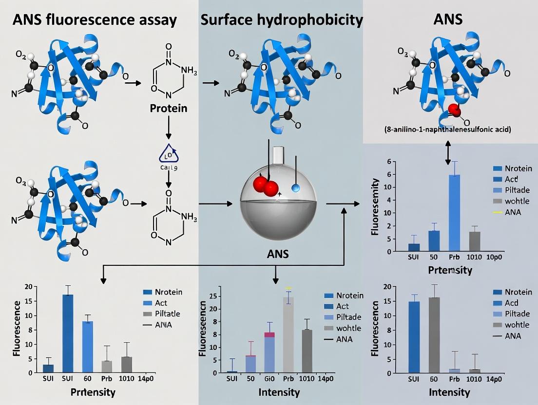
ANS Fluorescence Assay: A Complete Guide to Measuring Protein Surface Hydrophobicity for Researchers
This comprehensive guide details the ANS (1-anilinonaphthalene-8-sulfonate) fluorescence assay, a pivotal technique for quantifying protein surface hydrophobicity (PSH).
ANS Fluorescence Assay: A Complete Guide to Measuring Protein Surface Hydrophobicity for Researchers
Abstract
This comprehensive guide details the ANS (1-anilinonaphthalene-8-sulfonate) fluorescence assay, a pivotal technique for quantifying protein surface hydrophobicity (PSH). Targeted at researchers, scientists, and drug development professionals, the article covers the foundational principles of ANS-protein interaction and the critical role of PSH in protein function, stability, and aggregation. It provides a step-by-step methodological protocol with applications in biopharmaceutical characterization, including for monoclonal antibodies and biosimilars. The guide addresses common troubleshooting scenarios and optimization strategies for robust data, and critically evaluates the assay's validation, limitations, and comparison with complementary techniques like fluorescence spectroscopy and computational modeling. The conclusion synthesizes key takeaways and outlines the assay's implications for advancing protein engineering and therapeutic development.
Understanding ANS and Protein Surface Hydrophobicity: Principles and Biological Significance
What is Protein Surface Hydrophobicity (PSH) and Why Does It Matter?
Protein Surface Hydrophobicity (PSH) refers to the relative abundance of non-polar amino acid residues exposed on the surface of a protein's three-dimensional structure. These hydrophobic patches are critical for mediating interactions in aqueous biological environments. PSH is not a static property; it dynamically changes with protein folding, conformational changes, denaturation, and aggregation. It matters profoundly because it dictates key functional and pathological behaviors: it drives protein-protein interactions (e.g., antibody-antigen binding, enzyme-substrate complexes), influences protein stability and solubility, and is a primary factor in aberrant aggregation processes linked to diseases like Alzheimer's and Parkinson's. In biopharmaceuticals, PSH directly impacts the efficacy, stability, safety, and manufacturability of protein therapeutics, influencing aggregation propensity, immunogenicity, and viscosity.
Quantitative Data on PSH & Protein Behavior
Table 1: Correlation of PSH with Key Protein Properties
| Protein System | PSH Measurement (ANS Binding Affinity Kd, μM) | Observed Impact | Reference Context |
|---|---|---|---|
| Native vs. Heat-Denatured Lysozyme | Native: 15.2 ± 2.1; Denatured: 2.8 ± 0.4 | ~5.4x increase in affinity post-denaturation, indicating exposure of buried hydrophobic clusters. | Model for protein unfolding studies. |
| Therapeutic mAb: Stable vs. Stressed | Stable: 8.5 ± 1.3; Agitated: 4.1 ± 0.7 | 2.1x increase predicts aggregation onset under mechanical stress. | Biopharmaceutical formulation screening. |
| α-Synuclein (Parkinson's related) | Monomer: >50; Oligomer: 5.5 ± 1.2 | High affinity in oligomers correlates with membrane disruption & toxicity. | Neurodegenerative disease research. |
| Whey Protein Isolate (Food Science) | Native: 12.0; High-Pressure Processed: 6.5 | Increased PSH improves emulsification capacity and foam stability. | Food protein functionality. |
Table 2: ANS Fluorescence Response Parameters
| Parameter | Typical Range / Value | Significance |
|---|---|---|
| Excitation λ | 370 - 380 nm | ANS absorbance maximum. |
| Emission λ (in buffer) | ~520 nm | Weak fluorescence in aqueous medium. |
| Emission λ (bound to PSH) | 460 - 480 nm | Spectral blue shift indicates hydrophobic environment. |
| Fluorescence Intensity Increase | 10 to 200-fold | Proportional to accessible hydrophobic surface area. |
| Assay Temperature | 25°C (controlled) | Critical for reproducibility; PSH is temperature-sensitive. |
Experimental Protocols
Protocol 1: Standard ANS Fluorescence Assay for PSH Determination
This protocol is central to the thesis on ANS fluorescence for PSH research.
I. Principle The fluorescent dye 8-Anilino-1-naphthalenesulfonate (ANS) is virtually non-fluorescent in water but exhibits strong fluorescence with a blue-shifted emission maximum when bound to hydrophobic patches on proteins. The increase in fluorescence intensity is proportional to the protein's surface hydrophobicity.
II. Reagents & Materials
- Protein sample in appropriate buffer (e.g., PBS, Tris-HCl).
- ANS stock solution: 8.0 mM in distilled water or buffer. Store in the dark at 4°C.
- Assay buffer (identical to protein buffer).
- Black-walled, clear-bottom 96-well microplates or quartz cuvettes.
- Plate reader or spectrofluorometer with temperature control.
III. Procedure
- Sample Preparation: Dilute the protein sample to a series of concentrations (e.g., 0.05 to 0.5 mg/mL) in assay buffer. A blank (buffer only) is essential.
- ANS Addition: Add ANS stock to each protein sample and blank to achieve a final ANS concentration of 50 μM. Mix gently. Final sample volume: 200 μL in well or 2 mL in cuvette.
- Incubation: Incubate the mixture in the dark at controlled temperature (e.g., 25°C) for 10-15 minutes to allow binding equilibrium.
- Fluorescence Measurement:
- Mode: Fluorescence intensity.
- Excitation λ: 380 nm.
- Emission Scan: 400 - 600 nm OR fixed Emission λ: 470 nm.
- Bandwidths: Set to 5 nm for both excitation and emission.
- Read all samples and blanks.
- Data Analysis:
- Subtract the blank (ANS in buffer) fluorescence from all samples.
- Plot corrected fluorescence intensity (at 470 nm) vs. protein concentration. The initial slope of the curve is the PSH Index.
- For affinity determination: Perform titration of a fixed protein concentration with increasing ANS. Fit data (e.g., using Stern-Volmer or binding isotherm models) to calculate binding constant (Kd).
Protocol 2: Thermal Stress Assay to Monitor PSH Changes
I. Principle: To study how temperature-induced unfolding affects PSH, providing insights into protein stability.
II. Procedure:
- Prepare protein-ANS mixtures as in Protocol 1, step 2, in a thermally stable plate or cuvette.
- Place the sample in a fluorometer with a programmable temperature controller.
- Set a thermal ramp (e.g., from 20°C to 90°C at 1°C/min).
- Continuously monitor fluorescence intensity at 470 nm (ex 380 nm).
- Plot fluorescence vs. temperature. The inflection point (Tm) indicates the unfolding transition, and the magnitude of intensity increase reflects the extent of hydrophobic core exposure.
The Scientist's Toolkit: Key Research Reagent Solutions
| Item | Function in PSH Research |
|---|---|
| 8-Anilino-1-naphthalenesulfonate (ANS) | Primary fluorescent probe. Binds to accessible hydrophobic clusters, yielding enhanced, blue-shifted fluorescence. |
| 1,1'-Bi(4-anilino)naphthalene-5,5'-disulfonic acid (Bis-ANS) | Dimeric ANS analogue. Higher affinity for hydrophobic sites, used for more stable complexes or competitive binding studies. |
| Sypro Orange / Nile Red | Alternative hydrophobicity probes. Sypro Orange is a sensitive protein stain; Nile Red is excellent for lipids and molten globule states. |
| Size-Exclusion Chromatography (SEC) Columns | Aggregation analysis. Used in tandem with PSH assays to correlate hydrophobicity increase with oligomer/aggregate formation. |
| Dynamic Light Scattering (DLS) Instrument | Hydrodynamic size monitoring. Correlates changes in PSH with particle size distribution, crucial for aggregation studies. |
| Differential Scanning Calorimetry (DSC) | Thermodynamic stability. Provides complementary data on protein unfolding transitions observed in thermal PSH assays. |
Visualizations
ANS-PSH Binding and Fluorescence Response
Workflow: ANS Assay for PSH in Drug Development
PSH Role in Protein Aggregation Pathway
Application Notes on ANS in Protein Surface Hydrobicity Research
1-Anilinonaphthalene-8-sulfonate (ANS) is an amphipathic, environment-sensitive fluorescent probe central to protein surface hydrophobicity assays. Its utility stems from its unique photophysical properties, which change dramatically upon binding to hydrophobic surfaces, making it a vital tool in biophysical characterization and drug discovery.
1. Chemical Properties and Photophysical Mechanism
ANS is a naphthalene derivative with an anilino group and a sulfonate moiety. In aqueous solution, the molecule exists in a twisted conformation, leading to rapid non-radiative decay of its excited state and thus very low fluorescence quantum yield (~0.004) and a short fluorescence lifetime. Upon transfer to a non-polar environment or binding to a hydrophobic protein surface, several key changes occur:
- The molecule adopts a more planar conformation.
- The dielectric constant around the probe decreases.
- This restricts intramolecular rotation and solvation dynamics, significantly reducing non-radiative decay pathways. Consequently, bound ANS exhibits a large increase in fluorescence intensity (up to 100-200 fold), a substantial blue shift in its emission maximum (from ~515 nm to ~460-480 nm), and an increased fluorescence lifetime. The sulfonate group provides solubility in water and directs the probe to solvent-accessible hydrophobic patches on proteins.
Table 1: Photophysical Properties of ANS in Different Environments
| Property | Free in Aqueous Buffer | Bound to Hydrophobic Protein Surface |
|---|---|---|
| Quantum Yield | ~0.004 | 0.2 - 0.6 |
| Emission Max (λem) | ~515 nm | 460 - 480 nm |
| Fluorescence Intensity | Very Low | High (100-200x increase) |
| Lifetime | < 0.1 ns | 5 - 15 ns |
2. Key Protocols for ANS-Based Protein Hydrophobicity Assay
Protocol 1: Steady-State Titration for Binding Affinity & Hydrophobic Site Quantification
- Objective: Determine the dissociation constant (Kd) and the number of binding sites (n) for ANS on a target protein.
- Materials: Protein sample (in low-ionic strength buffer, e.g., 5-20 mM phosphate, pH 7.0), 1 mM ANS stock solution (in the same buffer or water), fluorescence spectrophotometer.
- Procedure:
- Prepare a 2 µM protein solution in a quartz cuvette.
- Set fluorometer excitation to 370-380 nm, and scan emission from 400-600 nm.
- Titrate ANS into the protein solution in small increments (e.g., 0.5, 1, 2, 5, 10 µL of 1 mM stock). Mix gently and incubate for 1-2 min.
- After each addition, record the emission spectrum. Monitor intensity at the peak (~470 nm).
- Perform a control titration of ANS into buffer alone to correct for background fluorescence.
- Correct for dilution and inner-filter effect if necessary.
- Analyze data by plotting corrected fluorescence intensity (F) vs. [ANS]. Fit to a binding isotherm (e.g., one-site specific binding model) to derive Kd and n.
- Interpretation: A lower Kd indicates higher affinity for hydrophobic surfaces. The parameter n provides an estimate of hydrophobic clusters accessible to ANS.
Protocol 2: Thermal or Chemical Denaturation Monitoring
- Objective: Monitor changes in surface hydrophobicity during protein unfolding/denaturation.
- Materials: Protein sample, ANS stock, fluorometer with temperature-controlled cuvette holder or plate reader.
- Procedure:
- Prepare a sample containing protein (e.g., 1 µM) and ANS (e.g., 20 µM) in appropriate buffer.
- Load into a quartz cuvette or multi-well plate.
- Set excitation to 380 nm, emission to 470 nm.
- For thermal denaturation: Ramp temperature from 20°C to 80°C at a rate of 1°C/min, recording fluorescence continuously. For chemical denaturation: Titrate in a denaturant (e.g., guanidine HCl or urea) stepwise, incubate, then measure.
- Plot fluorescence intensity versus temperature or [denaturant].
- Interpretation: A sigmoidal increase in ANS fluorescence indicates exposure of buried hydrophobic residues during unfolding. The mid-point of the transition corresponds to the melting temperature (Tm) or denaturant concentration at half-unfolding (C1/2).
3. Visualization of ANS Assay Workflow and Data Interpretation
Diagram 1: ANS Protein Hydrophobicity Assay General Workflow
Diagram 2: ANS Fluorescence Mechanism Upon Binding
4. The Scientist's Toolkit: Key Research Reagent Solutions
Table 2: Essential Materials for ANS Fluorescence Assays
| Item | Function & Importance |
|---|---|
| High-Purity ANS | Probe stock solution. Essential for reproducible fluorescence yields and avoiding contaminants. Use >95% purity, store desiccated, protected from light. |
| Ultra-Low Fluorescence Cuvettes/Plates | Sample containment for measurement. Must exhibit minimal background fluorescence at 370-500 nm to avoid signal interference. |
| Appropriate Protein Buffer | Sample environment. Low-ionic strength buffers (e.g., phosphate, Tris) without primary amines or detergents are critical to prevent artifacts and non-specific interactions. |
| Fluorometer with Peltier | Instrumentation. For precise thermal denaturation protocols, a temperature-controlled cuvette holder or plate reader is mandatory. |
| Chemical Denaturants (GdnHCl, Urea) | Unfolding agents. High-purity grades are necessary for chemical denaturation studies to ensure clean baselines and transitions. |
| Reference Fluorophore | Instrument calibration. A standard (e.g., quinine sulfate) is used to correct for instrument spectral sensitivity variations over time. |
Within the broader thesis on ANS fluorescence as a probe for protein surface hydrophobicity, this application note details the fundamental biophysical principles and standardized protocols. The fluorescence enhancement of 8-anilino-1-naphthalenesulfonic acid (ANS) upon binding to hydrophobic protein patches is a cornerstone technique for characterizing protein folding, aggregation, and ligand interactions in drug development.
Quantitative Binding & Fluorescence Parameters
The interaction between ANS and protein hydrophobic sites is characterized by measurable spectroscopic changes. The data below summarize key parameters from recent studies.
Table 1: Characteristic Fluorescence Parameters of ANS Upon Protein Binding
| Parameter | Free ANS in Aqueous Buffer | ANS Bound to Hydrophobic Protein Patches | Typical Measurement Conditions |
|---|---|---|---|
| Peak Emission Wavelength (λem max) | ~515 nm | 460 - 480 nm (Blue Shift) | Excitation: 370-380 nm |
| Fluorescence Quantum Yield (Φ) | ~0.004 | 0.2 - 0.6 (Up to 100-fold increase) | Reference: Quinine sulfate |
| Binding Constant (Kd) | Not Applicable | 10 - 500 µM (Protein-dependent) | Measured via titration |
| Fluorescence Lifetime | < 0.1 ns | 5 - 12 ns | Time-correlated single photon counting |
Table 2: Impact of Protein Conformational States on ANS Binding
| Protein State | Relative ANS Fluorescence Intensity | Observed Blue Shift (Δλ) | Inferred Hydrophobicity |
|---|---|---|---|
| Native (Compact) | Low to Moderate | Small (10-20 nm) | Buried / Minimal |
| Molten Globule / Partially Unfolded | Very High | Large (40-50 nm) | Transiently Exposed |
| Aggregated / Fibrillar | High | Moderate (30-40 nm) | Persistently Exposed |
| Fully Denatured (Unfolded) | Low | Minimal | Dispersed / No Patches |
Experimental Protocols
Protocol 1: Standard Titration for Binding Affinity (Kd) and Stoichiometry
Objective: Determine the dissociation constant (Kd) and number of binding sites (n) for ANS-protein interaction.
Materials:
- Protein sample in appropriate buffer (e.g., 20 mM phosphate, pH 7.4).
- Stock solution of ANS (e.g., 10 mM in buffer or methanol).
- Spectrofluorometer with temperature control.
- Quartz cuvette (sub-1 cm path length).
Procedure:
- Prepare a protein solution at a fixed concentration (typically 1-10 µM).
- In the cuvette, add 2 mL of the protein solution.
- Set the spectrofluorometer: Excitation (λex) = 380 nm, scan emission from 400 to 600 nm.
- Record the baseline spectrum of the protein alone.
- Titrate by adding small aliquots (2-10 µL) of ANS stock solution. Mix gently and incubate for 30-60 seconds.
- After each addition, record the fluorescence emission spectrum. Monitor the intensity at ~470 nm.
- Continue until no further increase in fluorescence is observed (signal saturation).
Data Analysis: Plot the corrected fluorescence intensity (F - F0) at 470 nm against the total ANS concentration. Fit the data to the following binding isotherm model: [ F = F{\text{max}} \cdot \frac{[ANS]}{Kd + [ANS]} ] where Fmax is the maximum fluorescence. For site number, use Scatchard or similar analysis if binding is not 1:1.
Protocol 2: Monitoring Protein Unfolding/Refolding Kinetics
Objective: Use ANS as a reporter to track changes in surface hydrophobicity during conformational transitions.
Materials:
- Native protein sample.
- Denaturant stock (e.g., 8 M Urea or 6 M Guanidine HCl).
- ANS stock solution.
- Stopped-flow attachment for spectrofluorometer or manual mixing with fast kinetics capability.
Procedure:
- Prepare two syringes/mixtures:
- Syringe A: Protein at 2x final concentration with ANS (typically 50-100 µM).
- Syringe B: Denaturant at 2x final concentration or refolding buffer.
- Load syringes into the stopped-flow instrument.
- Set λex = 380 nm and use a cutoff filter (e.g., 455 nm) or monochromator set to 470 nm to monitor emission.
- Rapidly mix equal volumes and trigger data acquisition.
- Record the fluorescence intensity change over time (milliseconds to minutes).
- For manual refolding, rapidly dilute denatured protein into refolding buffer containing ANS and monitor continuously.
Data Analysis: Fit the resulting kinetic trace to appropriate exponential models (single, double) to derive rate constants for the exposure/burial of hydrophobic clusters.
Visualizations
Diagram Title: ANS Binding and Fluorescence Enhancement Mechanism
Diagram Title: ANS Binding Affinity Titration Workflow
The Scientist's Toolkit: Essential Research Reagents & Materials
Table 3: Key Reagent Solutions for ANS-Protein Assays
| Item | Function & Description | Critical Notes |
|---|---|---|
| 8-Anilino-1-naphthalenesulfonate (ANS), ammonium salt | The fluorescent molecular probe. Its fluorescence is quenched in water but enhances in non-polar environments. | Prepare fresh stock in buffer or methanol. Protect from light. Concentration must be verified spectrophotometrically (ε~5000 M⁻¹cm⁻¹ at 350 nm). |
| High-Purity Protein Sample | The analyte of interest. Purity is critical to avoid spurious hydrophobic binding from contaminants. | Dialyze or desalt into a low-absorbance, non-fluorescent buffer (e.g., phosphate, Tris) before assay. |
| Reference Fluorophore (Quinine sulfate) | Used to determine the relative quantum yield of ANS-protein complexes. | Dissolve in 0.1 M H₂SO₄ (Φ=0.54 at 350 nm excitation). |
| Chemical Denaturants (Urea/Guanidine HCl) | Used to unfold protein and expose maximal hydrophobic surface for control experiments. | Use high-purity grade. Concentrate via weight. Avoid cyanate formation in urea solutions. |
| Low-Binding Microcentrifuge Tubes & Pipette Tips | To minimize loss of protein and ANS via adsorption to plastics. | Use polypropylene tubes. Consider pre-rinsing tips for very dilute samples. |
| Spectrofluorometer Cuvettes | Quartz cuvettes (path length ≤1 cm) for optimal signal in small volumes. | Meticulously clean to avoid fluorescent contaminants. Use dedicated cuvettes for dye studies. |
Protein Surface Hydrophobicity (PSH) is a critical physicochemical property that profoundly influences protein folding, conformational stability, macromolecular interactions, and aggregation propensity. Within the context of a broader thesis employing the ANS (1-Anilinonaphthalene-8-sulfonic acid) fluorescence assay for PSH quantification, this document delineates the application notes and protocols for investigating the biological and industrial implications of PSH. Understanding and modulating PSH is paramount for researchers, scientists, and drug development professionals working on protein therapeutics, enzyme engineering, and biomaterial design.
Quantitative Implications of PSH
The relationship between quantified PSH and key protein behaviors is summarized in the table below. Data is synthesized from recent studies on therapeutic monoclonal antibodies (mAbs), enzymes, and model proteins like bovine serum albumin (BSA) and lysozyme.
Table 1: Correlation of PSH with Protein Properties
| Protein System | PSH Measurement (ΔF/Relative Fluorescence) | Observed Impact on Stability (Tₘ/Tagg) | Aggregation Rate (kagg) | Functional Consequence |
|---|---|---|---|---|
| mAb (IgG1) at pH 5 | Low (Baseline = 100 A.U.) | High (Tₘ = 72°C) | Low (kagg < 0.01 hr⁻¹) | Maintains antigen binding (>95%) |
| mAb (IgG1) stressed (pH 3) | High (350% Increase) | Low (ΔTₘ = -12°C) | High (kagg > 0.05 hr⁻¹) | Loss of potency (40-60%) |
| Engineered Lipase (Variant A) | Moderate (150 A.U.) | Optimized (Tₘ = 65°C) | Low | High catalytic activity |
| Engineered Lipase (Variant B) | Very High (400 A.U.) | Low (Tₘ = 52°C) | Rapid (Visible precip.) | Loss of enzymatic function |
| BSU (in native state) | Reference (Set to 1.0) | -- | -- | -- |
| BSA (partially unfolded) | 3-5 fold increase | Decreased | Increased | Altered ligand binding |
Experimental Protocols
Protocol 1: ANS Fluorescence Assay for PSH Determination
- Objective: Quantify the relative surface hydrophobicity of a protein sample under native and stress-induced conditions.
- Materials: Protein sample (0.1-1.0 mg/mL in suitable buffer), 8 mM ANS stock solution in ethanol or DMSO, fluorescence spectrophotometer, microcuvettes, buffer (e.g., 20 mM phosphate, pH 7.0).
- Procedure:
- Prepare a series of protein solutions (2 mL) at a constant concentration.
- Add ANS dye from stock to each sample to a final concentration of 50 µM. Incubate in the dark for 10-15 minutes.
- Set fluorescence spectrophotometer: Excitation (λex) = 390 nm, Emission (λem) scan = 400-600 nm, Slit widths = 5 nm.
- Measure fluorescence intensity at the emission maximum (~470-480 nm).
- Run controls: Protein without ANS, ANS without protein, and buffer blanks.
- Data Analysis: Calculate ΔF = F(sample) - F(protein alone) - F(ANS alone). Report as relative fluorescence units (RFU) or normalized to a standard (e.g., BSA).
Protocol 2: Linking PSH to Thermal Stability via Intrinsic Fluorescence
- Objective: Determine the protein melting temperature (Tₘ) and correlate with PSH.
- Materials: Protein sample, fluorescence spectrophotometer with Peltier temperature controller, capillary cuvettes.
- Procedure:
- Load protein sample (devoid of external dyes) into the cuvette.
- Set λex = 280 nm (for Trp) and monitor λem = 320-350 nm (for Trp).
- Ramp temperature from 25°C to 95°C at a rate of 1°C/min.
- Plot fluorescence intensity or λem shift vs. temperature. Fit data to a sigmoidal curve.
- Derive Tₘ as the inflection point.
- Perform ANS assay (Protocol 1) on aliquots of the same protein pre-incubated at key temperatures. Correlate rising PSH with the onset of unfolding.
Protocol 3: Accelerated Stability and Aggregation Kinetics
- Objective: Monitor aggregation propensity under stress and quantify kinetics.
- Materials: Protein sample, thermal shaker or incubator, microcentrifuge tubes, dynamic light scattering (DLS) instrument or turbidity reader (A350).
- Procedure:
- Subject protein samples to stress condition (e.g., 40-45°C, agitation, low pH).
- At defined time intervals (0, 1, 2, 4, 7 days), withdraw aliquots.
- Analysis A (Turbidity): Measure absorbance at 350 nm (A350). Plot A350 vs. time.
- Analysis B (DLS): Measure hydrodynamic radius (Rh). Monitor increase in Rh or % polydispersity.
- Fit aggregation data to a kinetic model (e.g., first-order) to derive rate constant (kagg).
- Measure PSH via ANS on the initial protein sample and correlate with derived kagg.
Visualization of Workflows
Diagram 1: ANS Assay & PSH Correlation Workflow
Diagram 2: PSH Role in Protein Aggregation Pathway
The Scientist's Toolkit: Key Research Reagent Solutions
Table 2: Essential Materials for PSH and Stability Studies
| Item | Function & Importance |
|---|---|
| ANS Fluorescent Probe | Core Reagent. Binds dynamically to exposed hydrophobic clusters on protein surfaces; fluorescence enhancement provides a quantitative PSH index. |
| High-Purity Protein Standard (e.g., BSA) | Assay Control. Used to validate and normalize the ANS assay protocol across experiments and days. |
| Controlled-Environment Incubator/Shaker | Stress Induction. Enables precise application of thermal and agitation stresses for accelerated stability studies. |
| Fluorescence Spectrophotometer | Primary Detection. Must be sensitive and equipped with a temperature-controlled cuvette holder for both ANS (extrinsic) and Trp (intrinsic) fluorescence measurements. |
| Dynamic Light Scattering (DLS) Instrument | Aggregation Sizing. Quantifies aggregate size (hydrodynamic radius) and population distribution in real-time, complementing turbidity measurements. |
| Size-Exclusion Chromatography (SEC) Columns | Aggregate Quantification. Gold-standard for separating and quantifying soluble monomeric protein from higher-order aggregates post-stress. |
| Differential Scanning Calorimetry (DSC) | Stability Benchmarking. Provides direct, label-free measurement of thermal unfolding transitions (Tₘ, ΔH) to ground-truth PSH correlations. |
| Chemical Denaturants (GdnHCl, Urea) | Unfolding Titrants. Used to create controlled unfolded states for establishing PSH measurement ranges and validating assay sensitivity. |
The Historical Context and Evolution of the ANS Assay in Protein Science
Historical Context and Evolution
The 1-Anilino-8-Naphthalene Sulfonate (ANS) fluorescence assay represents a cornerstone technique in protein biophysics for probing surface hydrophobicity. Its development in the late 1960s and early 1970s, primarily by G. Weber and D. C. Turner, provided a simple, sensitive method to monitor protein conformational changes, folding/unfolding, and aggregation. Historically, ANS was first recognized for its "probe" properties due to its dramatic fluorescence enhancement (~100-200 fold) and blue spectral shift upon moving from an aqueous to a non-polar environment. This evolution mirrors the broader trajectory of protein science from static structural studies to dynamic, thermodynamic, and kinetic analyses. Its continued relevance in modern drug development lies in its ability to rapidly assess protein-ligand interactions, the stability of biologics, and the early stages of protein misfolding linked to diseases.
Application Notes
Core Principle and Quantitative Parameters
ANS binds to hydrophobic patches on protein surfaces or in molten globule states. The key quantitative parameters derived from the assay are:
- Fluorescence Intensity: Correlates with the number and accessibility of hydrophobic clusters.
- Emission λ_max (Wavelength of Maximum Emission): Shifts from ~515 nm in water to ~470-480 nm upon binding to hydrophobic sites, indicating the polarity of the binding pocket.
- Binding Constant (K_d): Measured through titration experiments.
- Quantum Yield: Increases substantially upon binding.
Table 1: Quantitative Spectral Characteristics of ANS
| Condition | Emission λ_max (nm) | Relative Fluorescence Intensity | Quantum Yield |
|---|---|---|---|
| In Aqueous Buffer | 515 - 520 | 1 (Baseline) | ~0.004 |
| Bound to Protein Hydrophobic Site | 470 - 480 | 100 - 200 | ~0.5 - 0.8 |
| In Pure Ethanol | 480 | ~50 | ~0.3 |
Modern Applications in Drug Development
- High-Throughput Screening (HTS): Used to identify small molecules that stabilize proteins or inhibit pathological aggregation.
- Biopharmaceutical Characterization: Monitoring aggregation propensity and conformational stability of monoclonal antibodies and other therapeutic proteins under stress (pH, temperature).
- Mechanistic Studies: Investigating membrane protein interactions and the formation of partially folded intermediates in neurodegenerative disease pathways.
Detailed Protocols
Protocol A: Basic ANS Binding Assay for Protein Hydrophobicity
Purpose: To determine relative surface hydrophobicity of a native or partially folded protein. Materials: See "The Scientist's Toolkit" below. Procedure:
- Prepare a 1 mM stock solution of ANS in high-purity water or buffer. Wrap in foil to protect from light. Store at 4°C for up to a week.
- Prepare protein samples in a suitable buffer (e.g., 20 mM phosphate, pH 7.0). A typical final protein concentration is 1-5 µM.
- In a cuvette or microplate well, mix:
- 1000 µL of protein solution
- 10 - 50 µL of ANS stock (Final ANS concentration 10 - 50 µM). Note: Optimize ANS:Protein molar ratio (typically 10:1 to 50:1).
- Incubate in the dark for 5-10 minutes at the experimental temperature (e.g., 25°C).
- Using a fluorometer, measure the fluorescence emission spectrum from 450 nm to 600 nm with an excitation wavelength of 350-380 nm.
- Run a blank containing ANS in buffer without protein. Subtract this spectrum from the sample spectrum.
Protocol B: ANS-Based Thermal Denaturation Assay
Purpose: To monitor the exposure of hydrophobic regions during thermal unfolding. Materials: As above, plus a fluorometer equipped with a Peltier temperature controller. Procedure:
- Prepare the protein-ANS mixture as in Protocol A, steps 1-4, directly in a thermally controlled cuvette.
- Set the fluorometer to record fluorescence intensity at 480 nm (emission) with excitation at 380 nm.
- Ramp the temperature from 20°C to 90°C at a constant rate (e.g., 1°C/min).
- Plot fluorescence intensity vs. temperature. The midpoint of the transition (Tm) and the shape of the curve provide information on protein stability and unfolding cooperativity.
Visualizations
Title: Basic ANS Fluorescence Assay Workflow
Title: ANS Fluorescence Response to Protein States
The Scientist's Toolkit
Table 2: Essential Research Reagent Solutions for ANS Assays
| Item | Function/Benefit | Typical Specification/Note |
|---|---|---|
| ANS (Ammonium Salt) | The fluorescent molecular probe. Exhibits environment-sensitive fluorescence. | High purity (>97%). Prepare fresh stock solutions. Light sensitive. |
| Buffer Components (e.g., PBS, Phosphate) | Maintain protein stability and physiological pH during assay. | Use high-purity reagents. Avoid amines (e.g., Tris) if exciting below 400 nm. |
| Reference Standard (e.g., Apomyoglobin) | Positive control for ANS binding in partially folded states. | Useful for method validation and inter-experiment comparison. |
| Denaturant (e.g., GdnHCl, Urea) | To unfold protein and create a positive control for maximal hydrophobic exposure. | Used in unfolding titrations to validate ANS response. |
| Plate Reader-Compatible Black Microplates | For high-throughput screening applications. | Low fluorescence background, non-binding surface. |
| Quartz or UV-Transparent Cuvettes | For standard fluorometry. | Required for excitation in the 350-380 nm range. |
Step-by-Step ANS Fluorescence Assay Protocol and Applications in Biopharma
Essential Reagents, Buffer Selection, and Instrumentation Setup
Within the thesis "Quantifying Conformational Changes in Therapeutic Proteins via ANS Fluorescence," the reliable measurement of surface hydrophobicity is critical. 1-Anilino-8-naphthalene sulfonate (ANS) fluorescence is a sensitive, solution-based technique used to probe protein folding, aggregation, and ligand binding. This protocol details the essential components for robust, reproducible ANS assays, emphasizing reagent purity, buffer compatibility, and instrument calibration to minimize artifacts.
The Scientist's Toolkit: Essential Research Reagent Solutions
| Item | Specification/Example | Function in ANS Assay |
|---|---|---|
| ANS Probe | 1-Anilinonaphthalene-8-sulfonate, ammonium salt (e.g., MilliporeSigma, >97% purity) | Environment-sensitive fluorescent dye; fluorescence increases & blueshifts in hydrophobic environments. |
| Protein Standard | Bovine Serum Albumin (fatty-acid free) or a well-characterized protein with known hydrophobic patches. | Positive control for assay validation and instrument calibration. |
| Buffer Salts | High-purity sodium phosphate, Tris-HCl, or citrate salts. | Maintains physiological pH and ionic strength. Choice affects protein stability and ANS background. |
| Chaotrope / Denaturant | Ultrapure Guanidine HCl or Urea. | Creates unfolded protein control for maximum hydrophobic exposure. |
| Surfactant / Quencher | Sodium dodecyl sulfate (SDS) or Acrylamide. | Control/validation agent; SDS exposes hydrophobic sites, acrylamide quenches fluorescence. |
| Filtration Units | 0.22 μm PVDF or cellulose acetate membrane filters. | Removes particulate matter and aggregates from buffers and ANS stock to reduce light scattering. |
| Spectroscopic Cuvettes | Quartz, fluorescence grade, 10 mm path length, low fluorescence background. | Holds sample for measurement; material and quality critically affect signal-to-noise. |
| pH Meter & Standard Buffers | Calibrated pH meter with temperature compensation. | Ensures precise and reproducible buffer pH, a critical parameter for protein-dye interaction. |
Detailed Protocols
Protocol 3.1: Preparation of ANS Stock Solution and Working Solutions
Objective: To prepare a stable, concentrated ANS stock and serial dilutions for assay titration.
- Weighing: Weigh 7.92 mg of ANS ammonium salt (Molecular Weight: 396.47 g/mol) into a 1.5 mL amber microcentrifuge tube. This yields a 20 mM stock.
- Dissolution: Add 1.0 mL of target assay buffer (e.g., 20 mM sodium phosphate, pH 7.0). Vortex vigorously for 30 seconds.
- Filtration: Filter the solution through a 0.22 μm syringe filter into a new amber vial to remove undissolved dye aggregates.
- Aliquoting & Storage: Aliquot into small volumes (e.g., 50 μL). Wrap in foil. Store at -20°C for up to 3 months. Avoid repeated freeze-thaw cycles.
- Working Solution: Thaw an aliquot and dilute in assay buffer to a 200 μM working solution on the day of the experiment.
Protocol 3.2: Standard ANS Fluorescence Titration Assay
Objective: To determine the optimal ANS:protein molar ratio and measure the increase in fluorescence intensity (FI) upon binding.
- Instrument Setup: Configure fluorometer (see Section 4). Set excitation to 370 nm, emission scan from 400 to 600 nm. Use 5 nm slits for both.
- Background Measurement: In a quartz cuvette, add 1495 μL of assay buffer and 5 μL of buffer (no dye). Measure and save spectrum as buffer blank.
- Free ANS Baseline: To the same cuvette, add 5 μL of 200 μM ANS working solution (final [ANS] = 50 μM). Mix gently by inversion. Measure and save spectrum as free dye baseline.
- Protein Titration: To the cuvette containing free ANS, sequentially add small volumes (e.g., 2-10 μL) of a concentrated protein stock. Mix gently and incubate for 1 minute at constant temperature (e.g., 25°C) before each measurement.
- Data Processing: Subtract the buffer blank from all spectra. Plot the fluorescence intensity at λmax (typically ~470-480 nm for bound ANS) versus the protein concentration or ANS:protein ratio. Fit the binding curve to determine apparent Kd.
Protocol 3.3: Thermal Denaturation Monitored by ANS Fluorescence
Objective: To monitor the exposure of hydrophobic surfaces as a function of temperature.
- Sample Preparation: Prepare a solution containing target protein and ANS at the optimal ratio determined in Protocol 3.2. Include a protein-free ANS control.
- Instrument Setup: Use a fluorometer equipped with a Peltier temperature controller. Set excitation to 370 nm, emission to 480 nm, and monitor intensity over time.
- Temperature Ramp: Set a linear temperature ramp from 20°C to 90°C at a rate of 1°C/min. Record fluorescence intensity and temperature simultaneously.
- Data Analysis: Plot FI vs. Temperature. The inflection point of the sigmoidal curve, determined by first derivative analysis, is reported as the melting temperature (Tm) for hydrophobic exposure.
Instrumentation Setup & Validation
Fluorometer Configuration:
- Light Source: Xenon arc lamp preferred for stable output across UV-Vis range.
- Monochromators/Gratings: Set to 370 nm (Ex) and 475 nm (Em) for peak measurements, or scan emission.
- Detector: Photomultiplier Tube (PMT) voltage should be set to auto or manually adjusted to avoid signal saturation (typically between 500-700 V). Use the same voltage for an entire experiment set.
- Cuvette Holder: Thermostatted holder is essential. Allow 5-10 minutes for temperature equilibration before measurement.
- Validation: Daily, perform a Raman scan of distilled water (Ex 350 nm, Em scan 365-450 nm). The peak should be near 397 nm. The peak intensity should be consistent day-to-day (<5% CV).
Quantitative Data & Buffer Selection Guidelines
Table 1: Effect of Common Buffer Components on ANS Fluorescence Signal
| Buffer Component | Typical Concentration | Effect on Free ANS FI (λem ~515 nm) | Effect on Protein-Bound ANS FI (λem ~475 nm) | Recommendation |
|---|---|---|---|---|
| NaCl / KCl | 0 - 150 mM | Slight decrease (~10%) | Moderate increase (~20-30%) up to 100 mM | Use to modulate ionic strength; maintain consistency. |
| MgCl2 / CaCl2 | 1 - 10 mM | Can cause precipitation | May enhance or quench based on protein | Avoid unless biologically required; filter carefully. |
| DTT / TCEP | 1 - 5 mM | Negligible | Negligible on FI; critical for reducing disulfide bonds. | Use fresh; TCEP is more stable and does not absorb at 280 nm. |
| Glycerol | 5 - 10% (v/v) | Significant increase (artifactual) | Significant increase (artifactual) | AVOID. Creates hydrophobic microenvironments. |
| Polysorbate 20/80 | 0.01 - 0.05% | Large increase (micelle formation) | Large increase (competes with protein) | AVOID. Use only in necessary controls for formulation studies. |
Table 2: Optimal Instrument Parameters for ANS Assay (Typical Setup)
| Parameter | Setting | Rationale |
|---|---|---|
| Excitation Wavelength | 370 - 380 nm | Near ANS absorption maximum, minimizes protein UV absorption. |
| Emission Scan Range | 400 - 600 nm | Captures full spectral shift from free (~515 nm) to bound (~475 nm) ANS. |
| Slit Widths (Ex/Em) | 5 nm / 5 nm | Balances signal intensity with spectral resolution. Can be reduced to 3 nm for high-concentration samples. |
| Integration Time / Scan Speed | 0.5 - 1 sec per nm (scan), 1 sec (point) | Ensures adequate signal averaging. Faster scans may reduce resolution. |
| Temperature | 25°C (or physiologically relevant) | Controlled temperature is critical for reproducibility of binding equilibria. |
Visualized Workflows and Pathways
Title: ANS Fluorescence Titration Experimental Workflow
Title: ANS Assay Detects Protein Unfolding via Hydrophobic Exposure
Within the context of an ANS (8-anilino-1-naphthalenesulfonic acid) fluorescence assay for protein surface hydrophobicity research, sample preparation is the critical first determinant of data reliability. The fluorescent quantum yield of ANS is exquisitely sensitive to the protein's conformational state, which is directly influenced by its concentration and the chemical composition of the surrounding buffer. This document details the application notes and protocols for optimizing these parameters to ensure consistent, interpretable results for researchers and drug development professionals.
The Impact of Protein Concentration and Buffer
ANS binds to solvent-accessible hydrophobic clusters on protein surfaces. Aggregation, misfolding, or unintended interactions induced by suboptimal concentration or buffer conditions can artificially alter the number and accessibility of these clusters, leading to erroneous hydrophobicity measurements.
Key Considerations:
- Protein Concentration: Must be within a range that minimizes intermolecular aggregation while providing a sufficient signal-to-noise ratio. Too high a concentration promotes aggregation; too low yields weak fluorescence.
- Buffer Components: Ions, pH, and additives can stabilize or destabilize native conformation, modulate ANS affinity, and directly quench fluorescence. Compatibility is paramount.
Table 1: Effect of Common Buffer Components on ANS Fluorescence Signal
| Buffer Component | Typical Concentration | Effect on Protein | Effect on ANS Fluorescence | Recommendation for ANS Assay |
|---|---|---|---|---|
| Sodium Chloride (NaCl) | 0-500 mM | Can modulate solubility & aggregation. | Minimal direct effect. | Use ≤150 mM to prevent salting-out. |
| Imidazole | 0-250 mM | Common eluent in His-tag purification. | Can significantly quench fluorescence. | Must dialyze out; keep ≤20 mM in final assay. |
| Glycerol | 0-20% (v/v) | Stabilizes protein structure. | Increases background fluorescence. | Limit to ≤5% or match concentration in all blanks. |
| DTT / β-Mercaptoethanol | 1-10 mM | Prevents disulfide bond formation. | Reduces ANS fluorescence intensity. | Use at minimal necessary concentration (e.g., 1 mM). |
| Detergents (e.g., Triton X-100) | > CMC | Solubilizes aggregates. | Abolishes ANS binding and signal. | AVOID in assay buffer. |
| HEPES, Phosphate, Tris | 10-50 mM | Standard buffering agents. | Generally inert. Slight pH-dependent variance. | Preferred. Maintain consistent pH ±0.1. |
Table 2: Optimized Protein Concentration Ranges for ANS Assay
| Protein Size (kDa) | Recommended Concentration Range (µM) | Rationale |
|---|---|---|
| < 20 | 5 - 15 µM | Smaller proteins have fewer binding sites; need higher conc. for detectable signal. |
| 20 - 60 | 2 - 10 µM | Standard range balancing signal and aggregation risk. |
| > 60 | 1 - 5 µM | Larger proteins have more sites; lower conc. minimizes aggregation and inner filter effects. |
| Aggregation-Prone | 0.5 - 2 µM (with verification via DLS/SEC) | Ultra-low concentration to maintain monodispersity is critical. |
Detailed Experimental Protocols
Protocol 1: Buffer Exchange and Optimization for ANS Assay
Objective: To transfer purified protein into an ANS-compatible assay buffer. Materials: Protein sample, dialysis tubing (or centrifugal filters), ANS assay buffer (e.g., 20 mM phosphate, pH 7.4, 50 mM NaCl), storage buffer.
- Choose Assay Buffer: Select a low-fluorescence buffer (e.g., phosphate, HEPES). Avoid imidazole, detergents, and high concentrations of reducing agents.
- Perform Buffer Exchange:
- Dialysis: Dialyze protein against ≥500x volume of assay buffer at 4°C for 4-6 hours. Change buffer and dialyze overnight.
- Centrifugal Filtration: Use a MWCO filter concentrator. Concentrate sample, dilute with assay buffer to original volume, and repeat 3x.
- Clarify Sample: Centrifuge the buffer-exchanged sample at 14,000 x g for 10 minutes at 4°C to remove any aggregates.
- Determine Concentration: Measure protein concentration using a suitable method (A280, BCA) after buffer exchange.
Protocol 2: Determining Optimal Protein Concentration for ANS Binding
Objective: To identify the protein concentration that yields a strong, linear ANS fluorescence response without aggregation. Materials: Buffer-exchanged protein stock, ANS stock solution (e.g., 2 mM in assay buffer or DMSO), assay buffer, fluorometer.
- Prepare a dilution series of the protein in assay buffer. Use the range suggested in Table 2 as a starting point (e.g., 1, 2, 5, 10, 15 µM).
- Prepare a master mix of ANS at a final concentration of 50-100 µM (from stock) in assay buffer.
- Mix 490 µL of each protein dilution with 10 µL of ANS stock (or vice versa for scaling) in a cuvette. Prepare a blank (assay buffer + ANS).
- Incubate in the dark for 5-10 minutes.
- Measure fluorescence (λex ~370-380 nm, λem ~470-480 nm, bandwidth 5 nm).
- Plot Net Fluorescence Intensity (Protein+ANS blank - ANS-only blank) vs. Protein Concentration. The optimal range is within the linear region before the curve plateaus, indicating saturation or aggregation.
Protocol 3: Assessing Buffer Compatibility via ANS Fluorescence Titration
Objective: To evaluate the impact of a specific buffer component on protein surface hydrophobicity. Materials: Protein in base buffer, concentrated stock of test component (e.g., 1M NaCl, 500 mM Imidazole), ANS stock, fluorometer.
- Prepare the protein at its optimal concentration (from Protocol 2) in the base assay buffer.
- Prepare a series of samples where the concentration of the test component varies (e.g., 0, 25, 50, 100, 250 mM NaCl), while keeping protein and ANS concentrations constant.
- Add ANS, incubate, and measure fluorescence as in Protocol 2.
- Plot Net Fluorescence Intensity vs. Concentration of Test Component. A sharp decrease may indicate conformational change or quenching. A increase may suggest exposure of hydrophobic patches.
Visualizations
Workflow for Optimizing Protein Samples for ANS Assay
Consequences of Poor Sample Prep on ANS Assay Results
The Scientist's Toolkit: Research Reagent Solutions
Table 3: Essential Materials for Sample Preparation in ANS Assays
| Item | Function in Sample Prep | Key Consideration |
|---|---|---|
| Low-Fluorescence Assay Buffer (e.g., PBS, HEPES) | Provides a stable, inert chemical environment for the protein and ANS. | Must be filtered (0.22 µm) and degassed to reduce light scattering and artifacts. |
| Dialysis Tubing/Cassettes (Appropriate MWCO) | Removes incompatible small molecules (imidazole, DTT) via equilibrium dialysis. | Pre-soak according to manufacturer instructions to remove preservatives. |
| Centrifugal Filter Concentrators (MWCO) | For rapid buffer exchange and precise protein concentration adjustment. | Choose MWCO 3-5x smaller than protein size. Do not over-concentrate to dryness. |
| High-Purity ANS (≥98%) | The fluorescent molecular probe for hydrophobic surface characterization. | Prepare fresh stock in appropriate solvent (buffer/DMSO); store in dark, -20°C. |
| Compatible Reducing Agent (e.g., TCEP) | Maintains cysteine residues in reduced state with minimal fluorescence quenching. | More stable and less quenching than DTT/β-mercaptoethanol at low concentrations. |
| Dynamic Light Scattering (DLS) Instrument | Validates monodispersity and detects aggregates after sample preparation. | Essential quality control step before performing the ANS assay on a new sample. |
Within the broader thesis investigating protein surface hydrophobicity using ANS fluorescence, the precise titration and incubation of 1-anilino-8-naphthalene sulfonate (ANS) is a critical methodological component. ANS is an amphipathic fluorescent probe whose quantum yield increases dramatically upon binding to hydrophobic protein surfaces. The protocol detailed herein provides a standardized, reliable method for determining the optimal ANS-to-protein molar ratio and incubation conditions to ensure reproducible quantification of surface hydrophobicity, a key parameter in protein folding, stability, and interaction studies relevant to biopharmaceutical development.
Key Research Reagent Solutions
The following table lists essential materials and their functions for the ANS fluorescence assay.
| Reagent/Material | Function in Assay | Notes for Preparation |
|---|---|---|
| 1-anilino-8-naphthalene sulfonate (ANS), ammonium salt | Fluorescent molecular probe. Binds to accessible hydrophobic clusters on the protein surface. | Prepare a stock solution (e.g., 5-10 mM) in buffer or purified water. Store in the dark at 4°C. |
| Purified Target Protein | The analyte whose surface hydrophobicity is being quantified. | Dialyze extensively against the assay buffer to remove interfering small molecules. Determine accurate concentration (A280 or BCA assay). |
| Assay Buffer (e.g., 10-50 mM phosphate, pH 7.0-7.4) | Provides a consistent chemical environment. Must be free of amines (e.g., Tris) that can quench ANS fluorescence. | Filter through 0.22 µm membrane to minimize light scatter. |
| Fluorometer/Spectrofluorometer | Instrument to measure fluorescence intensity. | Equipped with a thermostatted cuvette holder. Standard settings: λex = 370-380 nm, λem = 470-480 nm. |
| Quartz or UV-transparent Microcuvette | Holds the sample for fluorescence measurement. | Low fluorescence background is essential. |
Detailed Titration and Incubation Protocol
Preliminary ANS Stock Solution Standardization
Note: ANS concentration must be verified spectroscopically prior to use.
- Dilute an aliquot of the ANS stock solution in assay buffer to an approximate concentration of 10 µM.
- Measure the absorbance at 350 nm.
- Calculate the exact concentration using the molar extinction coefficient (ε) of ~5,000 M⁻¹cm⁻¹.
Primary Titration: Determining Optimal ANS:Protein Molar Ratio
The goal is to identify the ANS concentration that saturates available hydrophobic sites without causing non-specific aggregation or inner-filter effects.
Procedure:
- Prepare a master solution of your target protein in assay buffer at a concentration 2x your final assay concentration (e.g., 2 µM for a final of 1 µM).
- Prepare a series of ANS dilutions in assay buffer to cover a range of final concentrations from 0 to 100 µM in the cuvette.
- In a 96-well plate or series of microcentrifuge tubes, mix equal volumes (e.g., 250 µL) of the 2x protein solution and each ANS dilution. This yields a constant final protein concentration (e.g., 1 µM) and a gradient of ANS concentrations.
- Incubate mixtures in the dark at the desired assay temperature (e.g., 25°C) for 15 minutes.
- Transfer each mixture to the fluorometer cuvette and measure fluorescence intensity (FI) at λem 470 nm (with λex 370 nm). Perform triplicate measurements.
- Include controls: ANS in buffer alone (background fluorescence) and protein in buffer alone (autofluorescence).
Data Analysis:
- Subtract the average fluorescence of the appropriate controls (ANS-only at each concentration + protein-only) from each sample measurement.
- Plot corrected fluorescence intensity versus the final ANS concentration (or ANS:Protein molar ratio).
- Identify the inflection point or plateau where further ANS addition does not significantly increase fluorescence. This indicates saturation of available hydrophobic sites. The ANS concentration at this point is the optimal molar excess for your protein under these conditions.
Table 1: Example Titration Data (Hypothetical Protein, 1 µM final)
| Final ANS Conc. (µM) | ANS:Protein Ratio | Corrected FI (a.u.) | Notes |
|---|---|---|---|
| 0 | 0:1 | 0 | Baseline |
| 5 | 5:1 | 1250 ± 85 | Linear increase |
| 10 | 10:1 | 2450 ± 110 | Linear increase |
| 20 | 20:1 | 4200 ± 150 | Near saturation |
| 40 | 40:1 | 4800 ± 130 | Saturation plateau |
| 60 | 60:1 | 4850 ± 140 | Plateau |
| 80 | 80:1 | 4900 ± 135 | Plateau; possible slight inner-filter effect |
| 100 | 100:1 | 4850 ± 155 | Plateau |
Conclusion from Example: An optimal ANS:Protein molar ratio of 40:1 is selected for subsequent assays.
Incubation Parameter Optimization
Objective: To define the necessary time and temperature for equilibrium binding before measurement.
Time-Course Experiment:
- Prepare your protein-ANS mixture at the optimal molar ratio determined in Section 3.2.
- Immediately after mixing, transfer to the cuvette (pre-equilibrated at assay temperature) and start time-course measurement.
- Record fluorescence intensity every 30 seconds for 20-30 minutes.
- Plot FI vs. time. The time required for the signal to stabilize is the minimum required incubation time.
Temperature Considerations:
- Standard Assay: 25°C is common for stability studies.
- Thermal Denaturation Studies: Temperature is the variable. Crucial: After each temperature increment, allow ample equilibration time (e.g., 5-10 min) for both the sample and the fluorometer cuvette holder before measuring.
- For all assays, once the sample is at the target temperature, a consistent incubation period (e.g., 3-5 minutes) after mixing ANS and protein is recommended before measurement.
Finalized Standard Operating Protocol (SOP)
Based on the titration and incubation optimization results, the standard protocol is defined.
Procedure:
- Turn on the fluorometer and set excitation to 370 nm and emission to 470 nm. Allow lamp to warm up for 15 minutes.
- Set the cuvette holder temperature to the desired assay temperature (e.g., 25.0°C).
- Prepare the ANS Working Solution in assay buffer at a concentration that, when mixed 1:1 with the protein solution, yields the optimal final ratio (e.g., 80 µM ANS for a 40:1 ratio with 1 µM final protein).
- Prepare the Protein Solution in assay buffer at 2x the final desired concentration (e.g., 2 µM).
- In a low-binding microcentrifuge tube, mix 250 µL of Protein Solution with 250 µL of ANS Working Solution by gentle pipetting. Start a timer.
- Incubate the mixture in the dark at the assay temperature for exactly 15 minutes.
- Transfer the mixture to a clean quartz cuvette and place it in the fluorometer.
- Measure the fluorescence intensity. Perform three independent replicates.
Calculations:
- Average the replicate readings for the sample (F_sample).
- Prepare and measure controls: ANS-only (FANS) and protein-only (Fprotein) at the same concentrations/dilutions.
- Calculate the corrected fluorescence: Fcorrected = Fsample - FANS - Fprotein.
- Report F_corrected as a direct measure of relative surface hydrophobicity. For comparative studies, results may be normalized to the sample with the lowest hydrophobicity (set to 1) or expressed as a percentage of a maximum value.
Diagram 1: ANS Assay Development Workflow (82 chars)
Diagram 2: ANS Binding & Fluorescence Signal Mechanism (77 chars)
Within the context of investigating protein surface hydrophobicity using the ANS (1-anilinonaphthalene-8-sulfonate) fluorescence assay, precise fluorescence measurement is paramount. This protocol details the critical steps of excitation/emission wavelength selection and robust data acquisition, which are fundamental for generating reliable, quantitative data on protein conformational changes, aggregation, and ligand binding.
Core Principles: ANS Photophysics and Wavelength Selection
ANS is an environmentally sensitive extrinsic fluorophore. In aqueous solution, it exhibits weak fluorescence with an emission maximum (~515 nm). Upon binding to hydrophobic patches on a protein surface, its fluorescence intensity increases significantly, and its emission spectrum blue-shifts to ~470-490 nm. Proper wavelength selection captures this shift and intensity change.
Table 1: Recommended Wavelength Parameters for ANS-Protein Assay
| Parameter | Value/Range | Rationale |
|---|---|---|
| Excitation (Ex) | 350 - 380 nm | Near the ANS absorbance maximum; minimizes direct protein fluorescence. |
| Emission Scan Range | 400 - 600 nm | Captures the full spectral shift from bound and unbound ANS. |
| Emission Max (Bound ANS) | 470 - 490 nm | Primary data point for hydrophobicity quantification. |
| Slit Widths (Ex/Em) | 5 nm / 5 nm | Balanced to provide sufficient signal while maintaining spectral resolution. Adjust based on instrument and sample. |
| Integration Time | 0.1 - 1.0 sec | Optimize for signal-to-noise; avoid photobleaching. |
Detailed Experimental Protocol
Materials and Reagent Setup
- Protein Sample: Purified protein in suitable buffer (e.g., phosphate, Tris). Determine concentration spectrophotometrically.
- ANS Stock Solution: 5-10 mM ANS in high-purity water or buffer. Aliquot and store in the dark at -20°C. Warm to room temperature and vortex before use.
- Assay Buffer: Identical to protein buffer, without protein.
- Cuvettes: Use quartz cuvettes for UV excitation (350-380 nm). Plastic or glass cuvettes are not suitable.
- Spectrofluorometer: Instrument capable of scanning emission spectra with temperature control.
Step-by-Step Procedure for Spectral Acquisition
A. Instrument Preparation and Blank Measurement
- Power on the spectrofluorometer and associated temperature controller. Allow lamps to warm up for 15-30 minutes.
- Set the instrument parameters based on Table 1: Ex = 370 nm, Em scan from 400 to 600 nm, slit widths = 5/5 nm, medium scan speed.
- Prepare a blank sample: Add 2 mL of assay buffer and 10-20 µL of ANS stock solution to a final ANS concentration of 10-50 µM in the cuvette. Mix gently by inversion.
- Place the blank in the sample compartment, equilibrate to desired temperature (e.g., 25°C) for 2-3 minutes.
- Acquire the emission spectrum of the blank. Save this spectrum; it will be used for background subtraction.
B. Protein Sample Measurement
- Prepare the sample: To 2 mL of protein solution in a cuvette, add the same volume of ANS stock as used for the blank. Final protein concentration should be determined empirically but often ranges from 0.05 to 1 mg/mL. The ANS:protein molar ratio is critical; a common starting point is 50:1 to 200:1.
- Mix gently and incubate in the dark for 1-5 minutes to allow ANS binding to reach equilibrium.
- Place the sample in the spectrometer. Ensure consistent cuvette orientation.
- Acquire the emission spectrum using the identical parameters used for the blank.
- Repeat: Perform measurements in triplicate for each protein condition (e.g., native, denatured, with/without ligand).
C. Data Processing
- Subtract the blank spectrum from each sample spectrum.
- Plot corrected fluorescence intensity (y-axis) vs. wavelength (x-axis).
- Record key quantitative parameters:
- λmax (nm): The emission wavelength at maximum intensity.
- Fmax (a.u.): The fluorescence intensity at λmax.
- Spectral Center of Mass: Calculated as ∑(Ii * λi) / ∑Ii, providing a weighted average emission wavelength.
Data Acquisition Best Practices & Analysis
- Inner Filter Effect Correction: For samples with high absorbance at excitation or emission wavelengths (>0.1), apply correction: Fcorr = Fobs * antilog[(Aex + Aem)/2], where A are absorbances at the relevant wavelengths.
- Signal Averaging: Use multiple scans to improve signal-to-noise ratio.
- Baseline Stability: Monitor baseline drift by periodically measuring a blank or reference standard.
- Quenching Controls: Include controls with known quenchers (e.g., acrylamide) to confirm ANS is in a bound state.
Table 2: Key Data Outputs for Hydrophobicity Analysis
| Output Metric | Definition | Interpretation in Hydrophobicity Assay |
|---|---|---|
| Blue Shift (Δλmax) | λmax(blank) - λmax(sample) | Indicates the degree of ANS entry into a hydrophobic environment. Larger shift = more non-polar binding pocket. |
| Fluorescence Enhancement | Fmax(sample) / Fmax(blank) | Reflects the number of binding sites and/or the quantum yield increase upon binding. |
| Integrated Area | Area under curve (400-600 nm) | A holistic measure of total fluorescence change, combining intensity and shift effects. |
Visualization of Workflow and Data Interpretation
Diagram 1: ANS Fluorescence Assay Workflow
Diagram 2: Spectral Interpretation of Protein States
The Scientist's Toolkit: Essential Research Reagents & Materials
Table 3: Key Research Reagent Solutions for ANS Assay
| Item | Function/Description | Critical Notes |
|---|---|---|
| ANS (Ammonium Salt) | Environmentally sensitive fluorescent probe. Binds to hydrophobic protein surfaces. | High-purity grade (>97%). Prepare fresh stock solutions or freeze aliquots. Light-sensitive. |
| Protein of Interest | Target macromolecule for surface hydrophobicity analysis. | Must be highly purified, in a known, non-fluorescent buffer. Concentration accurately determined. |
| Assay Buffer (e.g., PBS, Tris-HCl) | Provides consistent ionic strength and pH for binding. | Must be free of fluorescent contaminants and detergents unless being studied. |
| Chemical Denaturant (e.g., Guanidine HCl, Urea) | Positive control. Maximally exposes hydrophobic residues. | Use high-purity, freshly prepared solutions. Determine final concentration in sample. |
| Reference Fluorophore (e.g., Quinine Sulfate) | Instrument performance validation and cross-day calibration. | Standard for quantum yield and wavelength accuracy checks. |
| Quartz Cuvettes (Semi-micro) | Holds sample for measurement. Transparent to UV/Vis light. | Meticulously clean with detergent, rinsed with ethanol/water. Handle by top edges only. |
This application note is framed within a broader thesis investigating protein surface hydrophobicity using the 8-Anilino-1-naphthalenesulfonic acid (ANS) fluorescence assay. Accurate data analysis of fluorescence intensity and spectral shifts is critical for quantifying hydrophobicity changes, which correlate with protein folding, aggregation, and ligand binding—key parameters in biophysical characterization and drug development.
Table 1: Key Fluorescence Parameters for ANS-Protein Binding Analysis
| Parameter | Description | Typical Range/Value | Significance in Hydrophobicity Assay |
|---|---|---|---|
| λ_max (nm) | Wavelength of maximum emission intensity. | 470-520 nm (bound ANS) | Blue shift indicates binding to hydrophobic pockets. |
| F_max (a.u.) | Maximum fluorescence intensity. | Variable; sample-dependent. | Quantifies amount of ANS bound to hydrophobic sites. |
| Spectral Shift (Δλ) | Difference in λ_max vs. free ANS in buffer (~515 nm). | 0 to ~45 nm | Magnitude correlates with hydrophobicity of binding site. |
| Binding Constant (K_d) | Equilibrium dissociation constant. | µM to mM range | Affinity of ANS for hydrophobic protein surfaces. |
| Quantum Yield (Φ) | Efficiency of photon emission. | Increases upon binding. | Enhanced upon transfer to non-polar environment. |
Table 2: Data Analysis Outputs for Hypothetical Proteins
| Protein Sample | Condition | λ_max (nm) | F_max (a.u.) | Δλ (nm) | Relative Hydrophobicity (F_max norm.) |
|---|---|---|---|---|---|
| Native State | 25°C, pH 7.4 | 472 | 15000 | 43 | 1.00 |
| Heat-Denatured | 60°C, pH 7.4 | 485 | 22000 | 30 | 1.47 |
| Ligand-Bound | +10 µM Drug | 469 | 10500 | 46 | 0.70 |
| Aggregating | Shaken, 48h | 490 | 35000 | 25 | 2.33 |
Detailed Experimental Protocols
Protocol 1: Steady-State ANS Fluorescence Assay for Protein Hydrophobicity
Objective: To measure changes in ANS fluorescence emission spectrum upon binding to protein hydrophobic surfaces. Materials: See The Scientist's Toolkit below. Procedure:
- Sample Preparation:
- Prepare protein solution in appropriate buffer (e.g., 20 mM phosphate, pH 7.0). Typical protein concentration: 0.1-1.0 mg/mL.
- Prepare ANS stock solution (e.g., 5 mM in buffer or water). Protect from light.
- Prepare assay mixture: Combine protein sample with ANS to a final ANS concentration of 50-200 µM. Incubate in the dark for 3-5 minutes.
- Prepare a blank containing ANS in buffer only (no protein).
- Spectrofluorometer Setup:
- Set excitation wavelength to 370-380 nm.
- Configure emission scan from 400 nm to 600 nm.
- Set appropriate slit widths (e.g., 5 nm/5 nm) and scan speed.
- Maintain constant temperature (e.g., 25°C) using a Peltier cuvette holder.
- Data Acquisition:
- Place blank in cuvette and acquire baseline emission spectrum.
- Replace with sample and acquire full emission spectrum.
- Perform replicates (n≥3).
- Initial Data Processing:
- Subtract the blank spectrum from the sample spectrum.
- Identify the wavelength of maximum fluorescence intensity (λ_max).
- Record the intensity at λmax (Fmax).
Protocol 2: Data Analysis for Spectral Shifts and Intensity
Objective: To calculate quantitative parameters from raw fluorescence spectra. Procedure:
- Spectral Centroid (Mean Emission Wavelength) Calculation:
- For advanced analysis, calculate the centroid (first moment) of the spectrum to detect subtle shifts.
- Use formula:
<λ> = Σ (λ_i * I_i) / Σ I_i, where λi is wavelength and Ii is intensity.
- Determining Spectral Shift (Δλ):
- Δλ = λmax (free ANS) - λmax (sample). Free ANS λ_max is typically 515 nm in aqueous buffer.
- A positive Δλ indicates a blue shift (hydrophobic binding).
- Normalizing Intensity Data:
- Normalize F_max values to a control sample (e.g., native protein) for comparative studies:
F_norm = F_max(sample) / F_max(control).
- Normalize F_max values to a control sample (e.g., native protein) for comparative studies:
- Binding Isotherm Analysis (for Kd):
- Titrate a fixed protein concentration with increasing ANS.
- Plot ΔF (Fmax - Finitial) vs. [ANS].
- Fit data to a suitable binding model (e.g., one-site specific binding) to derive Kd.
Mandatory Visualizations
Diagram Title: ANS Assay Experimental and Analysis Workflow
Diagram Title: Relationship Between Protein Change and ANS Signal
The Scientist's Toolkit
Table 3: Essential Research Reagent Solutions for ANS Assay
| Item | Function/Benefit | Example/Note |
|---|---|---|
| 8-Anilino-1-naphthalenesulfonic acid (ANS) | Fluorescent probe. Non-polar environment increases quantum yield & causes blue shift. | Magnesium salt often used for solubility. Prepare fresh or store aliquots at -20°C protected from light. |
| High-Purity Buffer Salts | Maintain protein stability and consistent ionic environment. | e.g., Phosphate, Tris, HEPES. Avoid amines or components that fluoresce near 370-520 nm. |
| Reference Fluorophore | Instrument performance validation (wavelength, intensity). | e.g., Quinine sulfate in 0.1 M H₂SO₄ (λex=350 nm, λem=450 nm). |
| Protein Standard (Positive Control) | Assay validation. A known hydrophobic protein under denaturing conditions. | e.g., Bovine Serum Albumin (BSA) in native and urea-denatured states. |
| Temperature Control System | Ensures reproducible binding kinetics and stability. | Peltier-controlled cuvette holder is essential for thermal denaturation studies. |
| Quartz or UV-Transparent Cuvettes | Minimal autofluorescence and high transmission at low UV wavelengths. | Use 10 mm pathlength, ensure proper cleaning to avoid contaminant fluorescence. |
| Data Analysis Software | For spectral processing, peak fitting, and binding isotherm analysis. | e.g., Origin, GraphPad Prism, Python (with NumPy/SciPy), or fluorometer vendor software. |
1. Introduction Within the broader research thesis on ANS fluorescence as a probe for protein surface hydrophobicity, this application note details its utility in two critical areas of biopharmaceutical development: the characterization of monoclonal antibody (mAb) conformational stability and the demonstration of biosimilarity. The hydrophobic dye 8-Anilino-1-naphthalenesulfonic acid (ANS) binds to solvent-exposed hydrophobic clusters, which become exposed upon thermal or chemical stress. The resulting increase in fluorescence intensity and blue shift in emission maximum provides a sensitive, rapid, and low-sample-consumption method to monitor unfolding intermediates and compare higher-order structures.
2. Key Quantitative Data Summary
Table 1: Representative ANS Fluorescence Data for mAb Conformational Stability Under Thermal Stress
| mAb Sample | Midpoint Unfolding Temp (Tm) (°C) | Onset Unfolding Temp (Tonset) (°C) | Maximum Fluorescence Intensity (A.U.) | Δ Emission λmax (nm vs. Native) |
|---|---|---|---|---|
| Reference mAb | 67.5 ± 0.3 | 62.1 ± 0.4 | 850 ± 25 | 0 (Baseline) |
| Stressed mAb* | 64.2 ± 0.5 | 58.7 ± 0.6 | 1050 ± 40 | +15 |
| Biosimilar A | 67.3 ± 0.4 | 61.8 ± 0.5 | 830 ± 30 | -1 |
| Biosimilar B | 66.0 ± 0.6 | 60.5 ± 0.5 | 920 ± 35 | +5 |
*Stressed at 40°C for 14 days.
Table 2: ANS Binding Data for Biosimilarity Assessment
| Analytical Parameter | Acceptance Criterion | Reference mAb | Biosimilar Candidate | Conclusion |
|---|---|---|---|---|
| Tm by ANS (℃) | ±1.0°C | 71.2 | 71.5 | Pass |
| Relative Fluorescence Gain at Tm | ±15% | 100% | 95% | Pass |
| Chemical Denaturation EC50 (GdnHCl, M) | ±0.2 M | 1.65 | 1.70 | Pass |
3. Detailed Experimental Protocols
Protocol 1: ANS Fluorescence Thermal Melt for Conformational Stability Objective: Determine the thermal unfolding profile of a mAb.
- Sample Preparation: Dialyze mAb samples into a formulation buffer (e.g., PBS, pH 7.4). Centrifuge at 14,000 x g for 10 min to remove aggregates.
- ANS Solution: Prepare a 500 µM stock of ANS in the same buffer. Protect from light.
- Labeling: Combine mAb (final concentration 0.1 - 0.5 mg/mL) with ANS (final concentration 50 µM). Incubate in the dark at 4°C for 30 min.
- Instrument Setup: Use a fluorometer equipped with a thermal Peltier. Set excitation to 380 nm, emission scan from 400 to 600 nm, or monitor at 480 nm.
- Run Parameters: Load 100 µL of sample in a quartz cuvette or microplate. Temperature gradient: 25°C to 95°C, with a ramp rate of 1°C/min. Record fluorescence at 480 nm continuously.
- Data Analysis: Plot fluorescence intensity vs. temperature. Fit data to a sigmoidal Boltzmann equation to determine Tm (inflection point) and Tonset.
Protocol 2: ANS-Based Biosimilarity Assessment via Chemical Denaturation Objective: Compare the structural resilience of a biosimilar to its reference product.
- Denaturant Series: Prepare a gradient of Guanidine Hydrochloride (GdnHCl) from 0 M to 3 M in formulation buffer.
- Equilibration: Mix mAb (reference and biosimilar) at 0.2 mg/mL with each denaturant concentration. Incubate at 25°C for 1 hour.
- ANS Addition: Add ANS to each sample to a final concentration of 50 µM. Incubate in the dark for 15 min.
- Measurement: Record fluorescence emission spectra (ex 380 nm, em 400-600 nm) at a constant 25°C.
- Analysis: Plot the fluorescence intensity at 480 nm vs. GdnHCl concentration. Calculate the denaturant concentration at half-maximal unfolding (EC50). Compare profiles of reference and biosimilar.
4. The Scientist's Toolkit: Essential Research Reagents
Table 3: Key Reagent Solutions for ANS Fluorescence Assays
| Reagent/Material | Function & Rationale |
|---|---|
| 8-Anilino-1-naphthalenesulfonic acid (ANS) | Hydrophobic fluorescent probe; binds to exposed protein hydrophobic patches, signal increases upon unfolding. |
| Monoclonal Antibody (Reference & Test) | The protein analyte of interest for stability or biosimilarity studies. |
| Phosphate Buffered Saline (PBS) pH 7.4 | Common physiological formulation buffer for mAbs, ensuring relevant solution conditions. |
| Guanidine Hydrochloride (GdnHCl) | Chemical denaturant used to perturb protein conformation and probe unfolding energetics. |
| Disposable Size-Exclusion Spin Columns | For rapid buffer exchange to ensure uniform sample buffer composition. |
| Black/Wall, Clear-Bottom 96- or 384-Well Plates | Optically suitable plates for high-throughput fluorescence measurements with minimal crosstalk. |
| Quartz Cuvettes (Sub-micro volume) | For high-sensitivity fluorescence measurements in a cuvette-based fluorometer. |
5. Workflow and Data Interpretation Diagrams
Diagram Title: Workflow for ANS-Based mAb Stability & Biosimilarity Analysis
Diagram Title: ANS Signal Mechanism Upon Protein Unfolding
1. Introduction & Thesis Context Within the broader thesis on ANS (1-Anilinonaphthalene-8-sulfonate) fluorescence as a probe for protein surface hydrophobicity, this document establishes its critical application in monitoring dynamic protein states. ANS fluorescence assays provide a sensitive, solution-based method to detect transient exposure of hydrophobic patches—a common feature in protein unfolding, aggregation, and conformational shifts upon ligand binding. These notes detail standardized protocols and data interpretation for these key biophysical events.
2. Key Research Reagent Solutions
| Reagent/Material | Function & Rationale |
|---|---|
| ANS (Na Salt) | The fluorophore probe. Binds to solvent-accessible hydrophobic protein patches, resulting in a blue shift and large increase in fluorescence intensity. |
| Purified Target Protein | Protein of interest (>95% purity recommended) in a suitable, well-characterized buffer to minimize artifacts. |
| Chemical Denaturants (e.g., Urea, GdnHCl) | Used to induce controlled, reversible unfolding in equilibrium unfolding studies. |
| Aggregation Inducers (e.g., Agitated Incubation, Heat) | Stress conditions to promote protein aggregation and expose hydrophobic interfaces. |
| Candidate Ligands/Compounds | Small molecules or other binding partners to test for conformational stabilization or changes. |
| Low-Binding Microplates/Tubes | To minimize nonspecific adsorption of protein and probe, ensuring accurate signal measurement. |
| Plate Reader or Spectrofluorometer | Instrument capable of measuring fluorescence intensity with appropriate filters/excitation (∼370 nm) and emission (∼480 nm) for ANS. |
3. Experimental Protocols
Protocol 3.1: Standard ANS Binding Assay for Baseline Hydrophobicity Objective: Establish the intrinsic surface hydrophobicity of the native protein state.
- Prepare a 10 mM ANS stock solution in buffer (e.g., 20 mM phosphate, pH 7.0). Protect from light.
- Prepare a dilution series of your purified protein (e.g., 0.5 to 10 µM) in assay buffer.
- In a black 96-well plate or cuvette, mix 100 µL of protein solution with 5 µL of ANS stock (final [ANS] typically 200-500 µM). Include controls: ANS + buffer only (background) and protein without ANS.
- Incubate in the dark for 5-15 minutes at constant temperature (e.g., 25°C).
- Measure fluorescence (Ex: 370-380 nm, Em: 470-480 nm, bandwidth 5-10 nm). Subtract the ANS+buffer control.
- Plot corrected fluorescence intensity vs. protein concentration to determine the binding profile.
Protocol 3.2: Monitoring Chemical-Induced Protein Unfolding Objective: Track the unfolding transition and identify intermediate states.
- Prepare a master solution of native protein (e.g., 2 µM final) and ANS (e.g., 300 µM final) in assay buffer.
- Prepare a series of denaturant solutions (e.g., 0 to 8 M Urea) in the same buffer.
- Mix equal volumes of the protein-ANS master solution with each denaturant solution in the plate/cuvette. Final protein concentration 1 µM.
- Equilibrate for 30 minutes at constant temperature.
- Measure fluorescence intensity and emission wavelength maximum (λmax) for each sample.
- Plot both Fluorescence Intensity and λmax against denaturant concentration. A cooperative increase in intensity with a blue shift indicates hydrophobic exposure during unfolding.
Protocol 3.3: Real-Time Monitoring of Protein Aggregation Objective: Detect early aggregation events via hydrophobic patch exposure.
- Prepare a protein solution (e.g., 5-10 µM) with ANS (300 µM) in a low-binding microplate.
- Place the plate in a temperature-controlled plate reader.
- Initiate aggregation by either: a) applying constant agitation, or b) rapidly raising the temperature above the protein's melting point.
- Continuously monitor ANS fluorescence (Ex 370, Em 480) and light scattering (Ex 340, Em 340) every 2-5 minutes.
- Correlate the kinetic trace of ANS fluorescence increase with the light scattering signal. ANS increase often precedes large aggregate formation detected by scattering.
Protocol 3.4: Detecting Ligand-Induced Conformational Changes Objective: Assess if ligand binding alters protein surface hydrophobicity.
- Prepare a fixed concentration of protein-ANS complex (e.g., 1 µM protein, 300 µM ANS).
- Titrate in increasing concentrations of the candidate ligand. Include a vehicle control.
- After each addition, incubate 5 min and measure fluorescence intensity and λmax.
- Plot the change in fluorescence (ΔF) or λmax versus ligand concentration. A decrease in ANS fluorescence suggests ligand binding buries hydrophobic patches, while an increase suggests induced exposure.
4. Data Presentation & Interpretation
Table 1: Summary of ANS Fluorescence Signatures for Protein Events
| Protein Event | Typical ANS Fluorescence Change | Emission λmax Shift | Interpretation |
|---|---|---|---|
| Native State (Compact) | Low Baseline | ~500-520 nm | Limited hydrophobic exposure. |
| Molten Globule/Unfolding Intermediate | Strong Increase | Blue Shift (~470-490 nm) | Substantial, solvent-accessible hydrophobic core exposure. |
| Full Unfolding (Denatured) | Decrease from peak | Red Shift (>520 nm) | Hydrophobic residues fully solvated, ANS displaced. |
| Aggregation | Sustained Increase | Slight Blue Shift or None | Hydrophobic interfaces exposed for protein-protein association. |
| Stabilizing Ligand Binding | Decrease | Red Shift | Ligand binding buries or shields hydrophobic patches. |
Table 2: Example Quantitative Data from a Model Unfolding Experiment (Lysozyme + GdnHCl)
| [GdnHCl] (M) | Fluorescence Intensity (a.u.) | Emission λmax (nm) | Apparent State |
|---|---|---|---|
| 0.0 | 150 ± 10 | 510 ± 2 | Native |
| 1.5 | 1050 ± 45 | 480 ± 1 | Intermediate |
| 3.0 | 2200 ± 120 | 472 ± 1 | Maximally Exposed |
| 5.0 | 450 ± 25 | 525 ± 3 | Unfolded |
5. Diagrams
Diagram 1: Pathways of Protein Unfolding & Aggregation
Diagram 2: Generic ANS Assay Workflow
Solving Common ANS Assay Problems: A Troubleshooting and Optimization Handbook
Addressing Low Signal-to-Noise Ratio and High Background Fluorescence
Within the context of a broader thesis on the use of 8-Anilinonaphthalene-1-sulfonic acid (ANS) fluorescence assays for quantifying protein surface hydrophobicity, addressing low signal-to-noise ratio (SNR) and high background fluorescence is a critical methodological challenge. These issues can obscure true binding events, lead to inaccurate quantification, and compromise the reproducibility of research with implications for drug development, particularly in understanding protein-ligand interactions and aggregation-prone regions. This application note details current strategies and protocols for mitigating these artifacts to obtain robust, high-fidelity data.
The primary sources of noise and background in ANS assays are summarized in the table below.
Table 1: Common Sources of Noise & Background in ANS Fluorescence Assays
| Source | Typical Impact on Signal | Quantitative Mitigation Target |
|---|---|---|
| Unbound/Free ANS in Solution | High background fluorescence at ~515 nm. | Reduce free [ANS] to < 2% of total. |
| Buffer/Reagent Impurities | Scattering, unwanted fluorescence. | Use ultra-pure water (18.2 MΩ·cm), HPLC-grade buffers. |
| Protein Aggregation | Non-specific ANS binding, increased scattering. | Maintain protein monomericity (e.g., via SEC, DLS). |
| Inner Filter Effects | Signal attenuation at high [ANS] or [Protein]. | Keep Absorbance at λex (370-380 nm) < 0.1. |
| Photobleaching | Signal decay over time. | Limit exposure; use stable light sources. |
| Cuvette/Plate Material | Background fluorescence & scattering. | Use quartz cuvettes or low-fluorescence plates. |
Table 2: Effect of Optimization on Assay Parameters (Representative Data)
| Condition | SNR (Typical) | Background Fluorescence (a.u.) | Z'-Factor (for HTS) |
|---|---|---|---|
| Unoptimized Assay | 3:1 - 5:1 | 500 - 1000 | < 0.2 |
| After Optimization | 15:1 - 50:1 | 50 - 150 | > 0.5 |
Detailed Experimental Protocols
Protocol 1: Purification and Preparation of ANS Stock Solution
Objective: To minimize inherent fluorescent impurities.
- Dissolve ANS dye in spectroscopic-grade methanol or ethanol to a concentration of 10 mM.
- Filter the solution through a 0.22 µm PTFE syringe filter.
- Determine the exact concentration spectrophotometrically using the molar extinction coefficient (ε) of 5000 M⁻¹cm⁻¹ at 350 nm in methanol.
- Aliquot, shield from light, and store at -20°C for up to 3 months. Avoid repeated freeze-thaw cycles.
Protocol 2: Optimized Protein Preparation and Assay Buffer Formulation
Objective: To reduce scattering and non-specific binding.
- Protein Purification: Perform size-exclusion chromatography (SEC) as a final polishing step using a buffer containing 20 mM phosphate, 150 mM NaCl, pH 7.4. Filter the protein sample (0.1 µm centrifugal filter).
- Buffer Preparation: Use high-purity salts (e.g., ≥99.5% purity). Prepare buffer with Milli-Q or equivalent water (18.2 MΩ·cm resistivity). Degas and filter (0.22 µm) before use.
- Sample Clarification: Centrifuge all samples (protein, ANS, buffer) at 16,000 x g for 10 minutes at assay temperature immediately before use to remove micro-aggregates.
Protocol 3: Titration Protocol with Background Subtraction and Inner Filter Effect Correction
Objective: To accurately measure specific ANS-protein binding fluorescence.
- Prepare a master mix of protein at 2x the desired final concentration in assay buffer. A typical starting [Protein] is 1-5 µM.
- Prepare a dilution series of purified ANS in the same buffer, typically from 0.5 to 100 µM.
- In a low-fluorescence 96- or 384-well plate (or quartz cuvette), mix equal volumes of protein master mix and ANS dilution to achieve the desired final concentrations. Include triplicates.
- Critical Controls: Include wells containing (a) ANS only (at each concentration) and (b) protein only.
- Incubate in the dark for 15-30 minutes at constant temperature (e.g., 25°C).
- Measurement: Using a plate reader or fluorometer, excite at 370-380 nm (5 nm bandwidth) and record emission spectra from 400-600 nm or a single emission point at 470-480 nm (10-20 nm bandwidth).
- Data Correction:
- Background Subtraction: Corrected Signal = (Protein + ANS sample) - (ANS only control at same [ANS]) - (Protein only control).
- Inner Filter Effect Correction: Apply the formula: Fcorr = Fobs * antilog[(Aex + Aem)/2], where Aex and Aem are the absorbance values of the sample at excitation and emission wavelengths, respectively.
Protocol 4: Determining Binding Affinity (Kd) from Corrected Data
- Plot corrected fluorescence intensity (at 470 nm) vs. total ANS concentration.
- Fit the data to a one-site specific binding model using nonlinear regression analysis (e.g., in GraphPad Prism): F = Fmax * [ANS] / (Kd + [ANS]) + Background Where F is corrected fluorescence, Fmax is maximum fluorescence at saturation, and Kd is the dissociation constant.
Visualizations
Title: Optimized ANS Assay Workflow
Title: Data Correction Pathway for ANS Assays
The Scientist's Toolkit: Research Reagent Solutions
Table 3: Essential Materials for High-SNR ANS Assays
| Item | Specification/Example | Critical Function |
|---|---|---|
| ANS Probe | Purified ≥95% (HPLC), e.g., MilliporeSigma A1028 | High-purity dye minimizes fluorescent contaminants. |
| Protein Purification Resin | Size-exclusion resin (e.g., Superdex 75) | Removes protein aggregates that cause non-specific binding. |
| Buffer Filtration Kit | 0.22 µm PES or PVDF membrane filters | Removes particulate matter that causes light scattering. |
| Water Purification System | Milli-Q or equivalent (18.2 MΩ·cm) | Eliminates ionic/organic fluorophores from water. |
| Microplate/Cuvette | Black quartz microplate or quartz cuvette | Minimizes autofluorescence and maximizes light transmission. |
| Spectrophotometer | Nanodrop or cuvette-based UV-Vis | Accurately measures sample absorbance for inner filter correction. |
| Fluorometer | Instrument with temperature control & low-stray light | Ensures stable, precise measurement of weak signals. |
| Data Analysis Software | Prism, Origin, or custom Python/R scripts | Enables robust nonlinear fitting for Kd determination. |
Within the broader thesis investigating ANS (1-anilinonaphthalene-8-sulfonate) fluorescence as a sensitive probe for protein surface hydrophobicity, a universal challenge is the determination of the optimal ANS:protein molar ratio. This ratio is not a fixed value but varies significantly across protein systems due to differences in size, structure, solvent-exposed hydrophobic patches, and aggregation state. This application note provides a standardized, systematic protocol for determining this critical parameter, ensuring accurate and reproducible measurements of surface hydrophobicity for diverse proteins, from globular monomers to complex multi-subunit assemblies.
Core Principle and Rationale
ANS is a hydrophobic fluorescent dye whose quantum yield increases dramatically upon binding to non-polar protein surfaces. Insufficient dye leads to incomplete reporting of hydrophobic sites, while excess dye results in non-specific aggregation, self-quenching, and high background fluorescence, distorting the signal. The optimal molar ratio saturates available binding sites without causing these artifacts, enabling valid comparisons between different protein systems.
Systematic Protocol for Determining Optimal ANS:Protein Ratio
Materials & Reagent Solutions
Table 1: Research Reagent Solutions Toolkit
| Item | Specification/Formula | Function |
|---|---|---|
| ANS Stock Solution | 8 mM in distilled water or DMSO (store at 4°C in the dark) | Fluorescent molecular probe for hydrophobic surfaces. |
| Protein Sample Buffer | e.g., 20 mM phosphate, 50 mM Tris-HCl, pH 7.4 | Provides consistent, non-interfering ionic environment for protein and ANS. |
| Reference Protein | Bovine Serum Albumin (BSA), 2 mg/mL in buffer | Positive control with known ANS-binding behavior. |
| Buffer Blank | Identical to protein sample buffer without protein | For background fluorescence subtraction. |
| Microplate or Cuvettes | Low-binding, non-fluorescent (e.g., black polystyrene) | Vessel for fluorescence measurement, minimizing signal loss. |
| Fluorimeter/Plate Reader | Capable of λ~ex=370-380 nm, λ~em=470-480 nm | Instrument for detecting ANS fluorescence emission. |
Step-by-Step Procedure
- Sample Preparation: Prepare a series of protein solutions in the desired buffer at a constant, low concentration (typically 0.05-0.2 mg/mL) to avoid inner-filter effects. A final volume of 2 mL (cuvette) or 200 µL (microplate well) is standard.
- ANS Titration: To each protein sample, add small aliquots of the ANS stock solution. Create a series covering a wide range of ANS:protein molar ratios. A typical range is 0:1 to 100:1, with more points in the lower range (e.g., 0, 0.5:1, 1:1, 2:1, 5:1, 10:1, 20:1, 50:1, 100:1).
- Incubation: Mix gently and incubate all samples in the dark at a constant temperature (e.g., 25°C) for 15 minutes to allow binding equilibrium.
- Fluorescence Measurement: Measure fluorescence intensity (FI) at λ~ex=380 nm and λ~em=480 nm. Always include a buffer + ANS blank for each ANS concentration to account for background.
- Data Analysis: For each protein sample, plot Net Fluorescence Intensity (Sample FI - Corresponding Blank FI) versus the ANS:Protein Molar Ratio.
- Determination of Optimum: Identify the point where the increase in fluorescence intensity plateaus. The molar ratio at the onset of this plateau, before any decline or excessive scatter in data points, is considered optimal. A sharp plateau indicates specific binding, while a gradual increase suggests non-specific interactions.
Example Data from Model Proteins
Table 2: Optimal ANS:Protein Molar Ratios for Representative Protein Systems
| Protein System | Structural Characteristics | Approx. Molecular Weight (kDa) | Typical Optimal ANS:Protein Molar Ratio (Range) | Key Consideration |
|---|---|---|---|---|
| Bovine Serum Albumin (BSA) | Monomer, multiple hydrophobic pockets | 66.5 | 10:1 to 20:1 | Well-characterized, often used as a standard. |
| β-Lactoglobulin | Dimer, hydrophobic calyx | 18.4 (monomer) | 5:1 to 10:1 | Ratio is often given per monomer. |
| Lysozyme | Compact, globular, low surface hydrophobicity | 14.3 | 50:1 to 100:1 | High ratio needed due to few binding sites. |
| Caseins (e.g., β-Casein) | Unstructured, open conformation | 24 | 2:1 to 5:1 | Highly accessible hydrophobic clusters. |
| Heat-Denatured Proteins | Unfolded, aggregated | Variable | 1:1 to 3:1 | Massive hydrophobic surface exposure; prone to aggregation at high ANS. |
| Monoclonal Antibody (IgG1) | Large, multi-domain | 150 | 15:1 to 30:1 | Ratio depends on structural integrity and formulation. |
Advanced Protocol: Multi-Parameter Optimization Workflow
For novel or complex systems (e.g., membrane proteins in detergent, protein-drug complexes), a multi-step optimization is recommended.
Title: Workflow for Multi-Parameter ANS Assay Optimization
Data Interpretation and Troubleshooting
Table 3: Common Fluorescence Profile Interpretations
| Observed Curve Profile | Likely Interpretation | Recommended Action |
|---|---|---|
| Sharp rise, clear plateau | Ideal specific binding. | Optimal ratio is at plateau onset. |
| Gradual, linear increase | Non-specific or continuous binding. | Use lowest ratio giving reproducible signal; consider alternative probe (e.g., bis-ANS). |
| Rise then sharp decline | Dye aggregation or protein precipitation at high [ANS]. | Optimal ratio is just before the decline. |
| No significant increase | Very low surface hydrophobicity or inactive dye. | Confirm protein integrity and ANS stock activity with a BSA control. |
Integration into Broader Hydrophobicity Research
The optimized ratio is the foundational step for subsequent experiments within the thesis framework, such as measuring hydrophobicity changes under denaturing conditions, probing ligand-binding interactions, or comparing protein variants.
Title: Role of Ratio Optimization in Broader Research
Mitigating Inner Filter Effects and Other Spectral Artefacts
Application Notes: For ANS Fluorescence Assay in Protein Hydrophobicity Research
In the quantitative assessment of protein surface hydrophobicity using 8-anilino-1-naphthalenesulfonic acid (ANS) fluorescence, spectral artefacts pose a significant threat to data accuracy. This document details protocols for identifying and correcting for Inner Filter Effects (IFE) and other common artefacts, ensuring reliable correlation between fluorescence intensity and hydrophobic site availability—a critical parameter in drug development for understanding protein-ligand interactions, aggregation propensity, and stability.
Understanding and Quantifying Inner Filter Effects (IFE)
IFE occur when the absorbance of the sample at the excitation or emission wavelengths is sufficiently high to attenuate the observed fluorescence signal. In ANS assays, high protein or ANS concentrations can lead to significant absorption. The primary correction is applied using the following relationship:
[ F{corr} = F{obs} \times antilog\left(\frac{A{ex} + A{em}}{2}\right) ]
Where ( F{corr} ) is the corrected fluorescence, ( F{obs} ) is the observed fluorescence, and ( A{ex} ) and ( A{em} ) are the absorbance values at the excitation and emission wavelengths, respectively.
Table 1: Impact and Correction of IFE Across Typical ANS Assay Conditions
| Sample Condition | [ANS] (μM) | [Protein] (mg/mL) | A280 | A372 (Ex) | A480 (Em) | Observed F (a.u.) | Corrected F (a.u.) | % Error Uncorrected |
|---|---|---|---|---|---|---|---|---|
| Low Conc. Control | 50 | 0.05 | 0.03 | 0.02 | 0.01 | 15,200 | 15,280 | +0.5% |
| Standard Assay | 250 | 0.5 | 0.25 | 0.15 | 0.05 | 84,500 | 92,100 | +9.0% |
| High Conc. (Risk) | 500 | 2.0 | 1.10 | 0.65 | 0.12 | 112,000 | 158,000 | +41.1% |
Protocol 1.1: Direct Absorbance Measurement for IFE Correction
- Prepare Samples: Prepare your ANS-protein samples as per standard assay protocol (e.g., in 20 mM phosphate buffer, pH 7.0).
- Measure Absorbance: Prior to fluorescence measurement, use a UV-Vis spectrophotometer to obtain the absorbance spectrum (250-500 nm) of each sample. Use a matched buffer blank.
- Extract Key Values: Record the absorbance values at the excitation wavelength (typically 372-375 nm) and at the emission maximum (typically 480-485 nm).
- Apply Correction: For each sample, apply the formula above using the extracted ( A{ex} ) and ( A{em} ) values to calculate ( F_{corr} ).
- Validation: Ensure the absorbance at the excitation wavelength in the sample cuvette is <0.1 for minimal IFE. If >0.1, correction is mandatory.
Mitigating Other Spectral Artefacts
2.1 Light Scattering (Rayleigh & Raman) Scattering from aggregates or particulate matter can artificially inflate the signal. Raman scatter from solvent has a characteristic wavelength shift.
- Protocol: Always run a blank containing buffer + ANS (no protein). Subtract this blank spectrum from all sample spectra. Use ultrapure, filtered buffers. Centrifuge protein stocks prior to assay.
2.2 Concentration-Dependent Aggregation of ANS At high concentrations (>300 μM), ANS can form excimers or aggregates, leading to redshifted emission and non-linear response.
- Protocol: Perform an ANS titration (e.g., 10-400 μM) at a fixed, low protein concentration to establish the linear range for your system. Stay within this range for quantitative assays.
2.3 Protein Autofluorescence & Background Tryptophan/Tyrosine fluorescence can contribute to background.
- Protocol: Excite at 372 nm, not at 280 nm, to minimize direct protein excitation. Verify the absence of peaks at ~340 nm (Trp) in the final corrected spectrum.
2.4 Photobleaching ANS can photobleach under prolonged illumination.
- Protocol: Use minimal slit widths and fast scan speeds. Use a shutter to block the beam between readings. Use the same integration time for all samples.
Experimental Workflow for Artefact-Free ANS Assay
Diagram 1: Workflow for Corrected ANS Fluorescence Assay
The Scientist's Toolkit: Key Research Reagent Solutions
Table 2: Essential Materials for Robust ANS Assay
| Item | Function & Rationale |
|---|---|
| High-Purity ANS (>98%) | Minimizes fluorescent impurities that contribute to background noise. |
| Ultra-Pure Water (e.g., Milli-Q) | Reduces Raman scattering and particulate-induced light scattering. |
| Low-Autofluorescence Buffers | Phosphate or HEPES buffers prepared from high-grade salts minimize background. |
| UV-Transparent Microplates/Cuvettes | Ensure high transmission at low wavelengths (e.g., 372 nm excitation). |
| Spectrophotometer with Stirred Cuvette | For accurate pre-fluorescence absorbance measurement (A372, A480). |
| Fluorometer with Monochromators | Provides precise wavelength selection for both excitation and emission, crucial for scatter discrimination. |
| 0.02 μm Anotop or Similar Syringe Filters | For definitive removal of particulates from buffer and sample solutions. |
| Concentrated Protein Stock Solution | Allows working in low-volume, low-absorbance regime to inherently minimize IFE. |
Integrated Correction Protocol
Protocol 2: Comprehensive ANS Assay with Built-In Artefact Mitigation
- Prepare Solutions: Filter all buffers through a 0.02 μm filter. Prepare a concentrated protein stock. Prepare ANS stock (e.g., 10 mM in buffer).
- Sample Dilution: Dilute protein stock into a series of concentrations in buffer. Add ANS from stock to a final, predetermined optimal concentration (e.g., 200 μM). Include a blank (buffer + ANS).
- Absorbance Scan: Immediately after mixing, measure absorbance of each sample from 350 to 500 nm. Record A372 and A480.
- Fluorescence Measurement: In a fluorometer, excite at 372 nm (slit 2-5 nm), scan emission from 400 to 600 nm (slit 5 nm). Use fast scan speed. Measure blank identically.
- Data Processing: a. Subtract the blank emission spectrum from each sample spectrum. b. Apply the IFE correction formula using the measured A372 and A480. c. Extract parameters from the corrected spectrum: peak intensity (Fmax), emission maximum (λmax), and area under the curve (AUC).
Diagram 2: Decision Path for Spectral Artefact Mitigation
Application Notes
Within the context of an ANS (1-anilinonaphthalene-8-sulfonic acid) fluorescence assay for protein surface hydrophobicity research, buffer composition is a critical, yet often overlooked, variable. Inaccurate results and poor reproducibility frequently stem from inadequate control of pH, ionic strength, and contaminating detergents. This document outlines their effects and provides protocols for systematic troubleshooting.
1. pH Effects ANS fluorescence is highly sensitive to pH, which alters the probe's anionic charge state and the ionization states of protein surface residues. A shift in pH can cause conformational changes in the protein, altering ANS binding sites and quantum yield.
Table 1: Effect of pH on ANS Fluorescence Intensity with a Model Protein (e.g., BSA)
| pH | Relative Fluorescence Intensity (λem 470 nm) | Probable Cause |
|---|---|---|
| 4.0 | 15 | Protein may be near/isoelectric point; ANS in protonated form. |
| 7.0 | 100 (Reference) | Optimal for native conformation and ANS binding. |
| 9.0 | 65 | Protein may undergo alkaline-induced unfolding/structural shift. |
2. Ionic Strength Effects Salt concentration influences electrostatic shielding. Moderate ionic strength can enhance ANS binding by screening repulsive charges, while high concentrations can promote non-specific aggregation or "salting-out" of the probe.
Table 2: Effect of NaCl Concentration on ANS-Protein Complex Signal
| [NaCl] (mM) | Fluorescence Intensity (%) | Observed λem max shift | Interpretation |
|---|---|---|---|
| 0 | 85 | None | Possible electrostatic repulsion limiting access. |
| 50 | 100 | None | Optimal screening of surface charges. |
| 500 | 120 | +5 nm (e.g., 470→475 nm) | Increased hydrophobic aggregation/ANS stacking. |
| 1000 | 60 | +10 nm | Probe & protein precipitation; signal artifacts. |
3. Detergent Interference Trace detergents (e.g., SDS, Triton X-100, Tween) from labware or protein purification are potent interferents. They form micelles that sequester ANS, producing high background fluorescence and outcompeting protein binding sites.
Table 3: Detergent Interference in ANS Assays
| Detergent (at Critical Micelle Concentration) | Apparent "Protein" Fluorescence (Background) | Effect on Protein+ANS Signal |
|---|---|---|
| SDS (0.1%) | Very High | >90% Suppression |
| Triton X-100 (0.01%) | High | ~75% Suppression |
| Tween-20 (0.01%) | Moderate | ~50% Suppression |
Experimental Protocols
Protocol 1: Systematic pH Titration for ANS Assay Optimization Objective: To determine the optimal pH for ANS binding to your target protein. Materials: Protein sample, ANS stock solution (e.g., 10 mM in methanol), 20 mM buffer series (Citrate-phosphate for pH 4-7, Tris-HCl for pH 7-9, Glycine-NaOH for pH 9-10), fluorometer. Procedure:
- Prepare 1 mL samples containing a fixed concentration of protein (e.g., 1 µM) and ANS (e.g., 50 µM) in each buffer pH.
- Incubate in the dark for 15 minutes at constant temperature (e.g., 25°C).
- Measure fluorescence emission spectra from 400-600 nm with excitation at 350-380 nm.
- Plot fluorescence intensity at λem,max versus pH. The optimal pH yields the highest signal-to-noise ratio without protein precipitation.
Protocol 2: Ionic Strength Screening Objective: To assess the impact of salt on ANS-protein interaction. Materials: Protein in low-salt buffer (e.g., 5 mM Tris, pH 7.4), 4M NaCl stock, ANS stock. Procedure:
- Prepare a master mix of protein and ANS at desired concentrations.
- Aliquot the master mix into tubes. Add small volumes of NaCl stock to achieve final concentrations from 0 to 1000 mM.
- After incubation, measure fluorescence intensity and note any visual precipitation.
- Analyze both intensity and any spectral shift (blue shift suggests hydrophobic environment; red shift suggests more polar or stacked ANS).
Protocol 3: Detergent Contamination Test & Decontamination Objective: To test for and eliminate detergent interference. Materials: Assay buffer, protein sample, ANS, fluorometer, activated charcoal (Norit A), 1M HCl. Procedure for Testing:
- Run an ANS assay with buffer only (no protein). A significant fluorescence signal indicates detergent or contaminant presence. Procedure for Decontamination (Buffer/Protein):
- Buffer Treatment: Add 1-2% (w/v) activated charcoal to buffer, stir for 1 hour at 4°C, and filter through a 0.22 µm membrane.
- Protein Sample Caution: Charcoal treatment can adsorb protein. As an alternative, use detergent-removal resins (e.g., Bio-Beads SM-2) per manufacturer protocol, or ensure protein is purified via detergent-free methods (e.g., acetone precipitation).
Mandatory Visualizations
Diagram Title: Buffer Effects on ANS Assay Signal Flow
Diagram Title: ANS Assay Troubleshooting Workflow
The Scientist's Toolkit: Key Research Reagent Solutions
| Item | Function / Role in Troubleshooting |
|---|---|
| High-Purity ANS Dye | Minimizes intrinsic fluorescent contaminants; prepare fresh stock in anhydrous methanol. |
| Detergent-Free Buffers | Use buffers prepared from high-purity salts and water (HPLC/spectroscopic grade). |
| Activated Charcoal (Norit A) | Removes trace organic contaminants and detergents from buffers via adsorption. |
| Bio-Beads SM-2 Resin | Specifically removes detergents from protein samples without denaturation. |
| Reference Protein (e.g., BSA) | Provides a standardized control to validate assay performance under new buffer conditions. |
| Concentrated Salt Stocks | For precise, reproducible modulation of ionic strength without diluting protein/ANS. |
| pH Calibration Standards | Ensures accuracy of pH meter readings across the relevant range (pH 4-10). |
| Low-Binding/Glass Labware | Prevents adsorption of ANS/protein and leaching of contaminants from plasticware. |
Best Practices for Handling Photo-bleaching and Sample Degradation
Within the context of a broader thesis on the ANS fluorescence assay for protein surface hydrophobicity research, managing photo-bleaching and sample degradation is critical. These phenomena directly compromise data reproducibility and quantitative accuracy, particularly in long-term or high-intensity studies common in drug development.
Key Challenges & Quantitative Impact
The following table summarizes primary factors and their measured impact on ANS fluorescence assays.
Table 1: Factors Affecting ANS Fluorescence Signal Integrity
| Factor | Typical Effect on Signal | Approximate Rate of Signal Loss* | Primary Influence |
|---|---|---|---|
| High-Intensity Excitation | Permanent fluorophore destruction | 5-20% per minute (continuous) | Photo-bleaching |
| Prolonged Exposure to Ambient Light | Gradual signal decay | 1-5% per hour | Photo-degradation |
| Elevated Temperature (>4°C) | Increased bleaching & aggregation | 2-10% per hour (at 25°C) | Thermal degradation |
| Repeated Freeze-Thaw Cycles | Protein aggregation & ANS binding loss | 5-15% per cycle | Structural degradation |
| Presence of Reactive Oxygen Species | Enhanced fluorophore oxidation | Varies widely with contaminants | Chemical degradation |
*Rates are highly dependent on specific experimental conditions (e.g., light source intensity, sample composition).
Detailed Experimental Protocols
Protocol 1: Minimizing Photo-bleaching in ANS Kinetic Assays
Objective: To measure the time-dependent exposure of hydrophobic protein surfaces while minimizing ANS photo-bleaching.
Materials:
- Protein sample in appropriate buffer.
- 8-Anilino-1-naphthalenesulfonate (ANS) stock solution (e.g., 10 mM in water or buffer).
- Spectrofluorometer with temperature control and shutter.
- Quartz cuvette (low fluorescence grade).
- Aluminum foil or light-blocking cuvette holder.
Methodology:
- Sample Preparation: Incubate protein with ANS at the desired molar ratio (e.g., 50:1 ANS:protein) in the dark for 15-30 minutes at the assay temperature. Wrap tubes in foil.
- Instrument Setup:
- Set excitation to 350-380 nm (ANS-specific), emission to 450-500 nm.
- Set instrument shutter to open only during data acquisition.
- Use the smallest slit widths that provide adequate signal (typically 2.5-5 nm).
- Set temperature control to desired level (e.g., 25°C).
- Data Acquisition:
- Place sample in cuvette, cover the holder.
- Program a kinetic scan with minimal interval time (e.g., 1 sec) but use intermittent data collection. For example, acquire a 0.1 sec reading every 10 seconds, keeping the shutter closed between readings.
- Limit total exposure time. Perform a control with ANS-buffer alone to subtract baseline bleaching.
- Data Analysis: Correct the fluorescence intensity versus time curve using the bleaching rate obtained from the buffer control.
Protocol 2: Assessing and Mitigating Sample Degradation
Objective: To evaluate the stability of the protein-ANS complex over time and under storage conditions.
Materials:
- Prepared protein-ANS complexes.
- Spectrofluorometer.
- Centrifugal filters (e.g., 10 kDa MWCO).
- Dynamic Light Scattering (DLS) instrument or native-PAGE equipment.
Methodology – Stability Assessment:
- Time-Course Measurement: Prepare a master mix of protein-ANS complex. Aliquot into multiple low-binding microcentrifuge tubes. Wrap in foil.
- Store aliquots under different conditions: on ice (4°C), at room temperature (25°C), and frozen (-80°C) for a subset.
- At set time points (0, 1, 2, 4, 8, 24 hours), rapidly analyze one aliquot from each condition using a pre-set, short fluorescence scan.
- Aggregation Check: At the final time point, centrifuge samples (e.g., 16,000 x g, 15 min) or pass through a 0.1 µm filter. Measure fluorescence of the supernatant/filtrate. A significant drop indicates complex aggregation/precipitation.
- Structural Integrity: Analyze parallel samples via DLS (for hydrodynamic radius) or native-PAGE to monitor oligomeric state changes.
Signaling Pathways and Workflow Diagrams
Title: Molecular Pathway of ANS Photo-bleaching
Title: Workflow for Sample Degradation Assessment
The Scientist's Toolkit: Key Research Reagent Solutions
Table 2: Essential Materials for ANS Assay Integrity
| Item | Function & Rationale |
|---|---|
| 8-Anilino-1-naphthalenesulfonate (ANS), High-Purity Grade | The extrinsic fluorophore; high purity reduces background fluorescence and spurious signals from contaminants. |
| Low-Binding Microcentrifuge Tubes & Plates | Minimizes adsorption of protein and ANS to plastic surfaces, ensuring accurate concentration. |
| Oxygen Scavenging System (e.g., Glucose Oxidase/Catalase) | Reduces dissolved oxygen, mitigating ROS formation and oxidative damage during long experiments. |
| Anti-fade Reagents (e.g., Trolox, Ascorbic Acid) | Neutralizes free radicals generated during excitation, specifically reducing photo-bleaching rates. |
| Spectrophotometric Grade, Aprotic Solvents (DMSO) | For preparing stable, concentrated ANS stock solutions, preventing hydrolysis. |
| Size-Exclusion Spin Columns or Dialysis Cassettes | For rapid buffer exchange to remove unbound ANS after incubation, reducing background signal. |
| Quartz Cuvettes (Stopped-Flow Compatible) | For kinetic studies; quartz withstands rapid temperature changes and has optimal UV transmission. |
| Programmable Spectrofluorometer with Shutter | Allows automated, intermittent data collection, keeping the sample in the dark between readings. |
Within the broader thesis research on utilizing 8-anilino-1-naphthalenesulfonic acid (ANS) fluorescence to probe protein surface hydrophobicity, a significant challenge arises when applying the assay to aggregation-prone proteins. Such proteins often undergo rapid conformational changes and self-association, which can confound standard assay protocols and lead to inconsistent or misleading hydrophobicity readings. This application note details a systematic optimization of the ANS assay for a model aggregation-prone protein, "Protein X," a therapeutic antibody fragment with a known propensity for aggregation under physiological pH and mild thermal stress.
The Challenge with Protein X
Standard ANS assays (10 µM ANS, 5 µM protein in 20 mM phosphate buffer, pH 7.4) yielded highly variable fluorescence intensity (FI) readings (coefficient of variation >25% across replicates) and a non-linear protein concentration-FI relationship. Dynamic light scattering confirmed the formation of soluble oligomers during the assay timeframe, which non-specifically bound ANS and created artefactual signals.
Optimization Strategy & Results
The optimization focused on four key parameters to minimize aggregation while maintaining protein structural integrity for a valid surface hydrophobicity measurement.
Table 1: Summary of Optimization Parameters and Quantitative Outcomes
| Parameter Tested | Standard Condition | Optimized Condition | Key Outcome (Fluorescence Intensity @ 470 nm) | Aggregation State (DLS Hydrodynamic Radius) |
|---|---|---|---|---|
| Incubation Temperature | 25°C | 4°C | FI increased by 15%; CV reduced to 8% | Reduced from 12 nm (oligomers) to 5 nm (monomer) |
| ANS:Protein Molar Ratio | 2:1 | 10:1 | FI signal enhanced 3-fold; saturation achieved | No significant change from monomeric state at 4°C |
| Buffer Additive | None | 150 mM NaCl | FI stabilized, minimal temporal decay (<5% over 30 min) | Stabilized monomeric radius at 5 nm for >1 hour |
| Incubation Time | Immediate reading | 5 min post-mixing | Consistent, maximized FI (peak signal) | Stable monomeric population |
| Final Protocol Result | N/A | Combined optimized conditions | FI: 850 ± 45 AU (Mean ± SD, n=6) | Rh: 5.1 ± 0.3 nm (Monomeric) |
Detailed Experimental Protocols
Protocol 1: Optimized ANS Assay for Aggregation-Prone Proteins
Objective: To measure the relative surface hydrophobicity of Protein X under conditions that suppress transient aggregation. Materials: See "Research Reagent Solutions" below. Procedure:
- Prepare assay buffer: 20 mM sodium phosphate, 150 mM NaCl, pH 7.4. Filter through a 0.22 µm membrane.
- Prepare a 500 µM ANS stock solution in the same buffer. Protect from light.
- Dilute Protein X to 5 µM in chilled (4°C) assay buffer. Keep on ice.
- In a low-binding, black-walled 96-well plate, mix 100 µL of 5 µM protein with 10 µL of 500 µM ANS stock (final [ANS]=45.5 µM, ~10:1 molar ratio).
- Incubate the plate in the dark at 4°C for 5 minutes.
- Measure fluorescence immediately using a plate reader with excitation at 380 nm and emission scan from 450-600 nm. Record peak intensity at ~470 nm.
- Include controls: ANS alone in buffer, buffer alone, and a non-aggregating reference protein (e.g., lysozyme).
Protocol 2: Validation via Dynamic Light Scattering (DLS)
Objective: To confirm the monomeric state of the protein during the ANS assay. Procedure:
- Using the same mixture from Protocol 1, Step 4, transfer 50 µL to a micro-cuvette or DLS plate.
- Immediately place the sample in a DLS instrument pre-equilibrated to 4°C.
- Measure the hydrodynamic radius (Rh) using a 90° scattering angle. Perform a minimum of 10 acquisitions.
- Analyze the correlation function using a cumulants model. The primary peak should correspond to the expected Rh of monomeric Protein X (~5 nm).
Visualizing the Optimization Workflow & Impact
Title: ANS Assay Optimization Workflow for Aggregation-Prone Protein
Title: Specific vs Non-Specific ANS Binding Pathways
The Scientist's Toolkit: Research Reagent Solutions
Table 2: Essential Materials for the Optimized ANS Assay
| Item | Function & Rationale | Example (Supplier Agnostic) |
|---|---|---|
| High-Purity ANS | The fluorescent probe. Batch-to-batch consistency is critical for reproducible fluorescence quantum yield. | 8-Anilino-1-naphthalenesulfonic acid, ammonium salt, >98% purity. |
| Low-Binding Microplates | Minimizes adsorptive loss of aggregation-prone proteins and the ANS probe to plastic surfaces. | Black-walled, clear-bottom 96-well plates with polymer surface treatment. |
| Precision Buffer Components | To prepare the optimized assay buffer with controlled ionic strength and pH, filtering is essential. | Sodium phosphate dibasic, Sodium chloride, 0.22 µm syringe filters. |
| Temperature-Controlled Plate Reader | Enables incubation and reading at 4°C, a key factor in suppressing aggregation during measurement. | Multimode reader with cooled chamber and kinetic capability. |
| Dynamic Light Scattering (DLS) Instrument | The critical validation tool for confirming the monomeric state of the protein under assay conditions. | Nano-particle analyzer with temperature control down to 4°C. |
| Aggregation-Prone Target Protein | The subject of the study, requiring careful handling. | Purified protein aliquots, flash-frozen, stored at -80°C. |
This case study, within the wider thesis on ANS fluorescence, demonstrates that a mechanistic understanding of a protein's colloidal stability is prerequisite to a valid hydrophobicity assay. For aggregation-prone proteins like Protein X, simply following a standard protocol is insufficient. The optimized method—centered on low temperature (4°C), a high ANS:Protein ratio (10:1), inclusion of stabilizing salt (150 mM NaCl), and a controlled incubation time (5 min)—successfully decoupled specific surface binding from non-specific aggregation binding. This approach transformed the ANS assay from an unreliable measurement into a robust, quantitative tool for tracking conformational changes in Protein X under various formulation stresses, providing critical data for its development as a biotherapeutic.
Validating ANS Assay Data: Comparison with Alternative Hydrophobicity Methods
Application Notes
Within the broader thesis investigating protein surface hydrophobicity using 8-Anilino-1-naphthalenesulfonic acid (ANS) fluorescence, rigorous assay validation is paramount. This document details the core parameters of reproducibility, sensitivity, and linearity, ensuring data reliability for research and early-stage biotherapeutic characterization (e.g., assessing aggregation propensity, stability under stress).
1. Reproducibility (Precision) Reproducibility confirms the assay's reliability across different runs, days, and analysts. For ANS assays, this is challenged by ANS photobleaching and protein-adsorption to surfaces.
- Intra-assay Precision: Measured as repeatability (multiple replicates within a plate).
- Inter-assay Precision: Measured as intermediate precision (across different days, analysts).
- Key Metric: Percent Coefficient of Variation (%CV) of the fluorescence intensity at a defined protein/ANS condition.
Table 1: Representative Reproducibility Data for ANS Assay
| Precision Type | Sample | Mean Fluorescence Intensity (RFU) | Standard Deviation (SD) | %CV | Acceptance Criterion Met (Y/N) |
|---|---|---|---|---|---|
| Intra-assay (n=12) | Native Protein (Control) | 15,250 | 610 | 4.0% | Y (≤5%) |
| Intra-assay (n=12) | Heat-Denatured Protein | 45,500 | 2,275 | 5.0% | Y (≤5%) |
| Inter-assay (n=18, 3 days) | Native Protein (Control) | 14,900 | 1,192 | 8.0% | N (≤7%) |
| Inter-assay (n=18, 3 days) | Heat-Denatured Protein | 44,100 | 3,528 | 8.0% | N (≤7%) |
Data indicates inter-assay variability requires protocol optimization, likely in ANS handling or plate reader calibration.
Protocol 1: Determining Reproducibility
- Objective: Determine intra- and inter-assay precision of ANS fluorescence measurement.
- Materials: Protein sample (1 mg/mL in suitable buffer), ANS stock solution (10 mM in distilled water), assay buffer (e.g., 20 mM phosphate, pH 7.0), black 96-well microplate, fluorescence plate reader.
- Procedure:
- Prepare an ANS working solution (e.g., 200 µM) in assay buffer, protected from light.
- For intra-assay precision: In one plate, prepare 12 replicate wells each containing 95 µL of protein sample and 95 µL of ANS working solution. Include 12 replicate wells of buffer-only (blank) and ANS-only controls.
- Incubate in the dark for 10-15 minutes.
- Read fluorescence (λex = 370-380 nm, λem = 470-520 nm).
- For inter-assay precision: Repeat steps 2-4 on three separate days, with two different analysts preparing stocks, using the same instrument.
- Subtract the appropriate blank (buffer + ANS) from sample readings.
- Calculate mean, SD, and %CV for each sample set.
2. Sensitivity Sensitivity defines the lowest detectable change in protein hydrophobic surface area. It is assessed via the Limit of Detection (LoD) and Limit of Quantification (LoQ) for the fluorescence signal change upon denaturation.
Table 2: Sensitivity Parameters for ANS Assay
| Parameter | Calculation Method | Result | Interpretation |
|---|---|---|---|
| Limit of Detection (LoD) | 3.3 * σ / S | 1.8% denatured protein | Minimal detectable level of hydrophobic exposure. |
| Limit of Quantification (LoQ) | 10 * σ / S | 5.5% denatured protein | Reliable quantitative measurement threshold. |
| Assay Dynamic Range | -- | 5.5% - 100% denatured protein | Range over which the assay provides quantitative data. |
σ = SD of the response (blank); S = Slope of the denaturation standard curve.
Protocol 2: Determining Sensitivity (LoD/LoQ)
- Objective: Establish the minimum detectable and quantifiable increase in ANS fluorescence.
- Materials: As in Protocol 1. A "denatured standard" (e.g., fully heat-denatured protein) is required.
- Procedure:
- Prepare a series of samples containing fixed total protein concentration but with increasing proportions of denatured protein (0%, 1%, 5%, 10%, 25%, 50%, 100%) mixed with native protein.
- Incubate each sample with ANS in 8 replicates.
- Measure fluorescence as in Protocol 1.
- Plot mean fluorescence intensity (Y) vs. % denatured protein (X).
- Perform linear regression on the low-concentration points (e.g., 0-10%).
- LoD = 3.3 * (SD of 0% denatured sample) / Slope.
- LoQ = 10 * (SD of 0% denatured sample) / Slope.
3. Linearity Linearity assesses the ability of the assay to produce results directly proportional to the amount of hydrophobic surface present. It is tested across a range of protein concentrations or denaturation states.
Table 3: Linearity Data for ANS Assay Across Protein States
| Sample Condition | Concentration Range Tested | R² Value | Linearity Acceptance Met (Y/N) |
|---|---|---|---|
| Native Protein | 0.025 - 0.5 mg/mL | 0.991 | Y (≥0.98) |
| Heat-Denatured Protein | 0.025 - 0.5 mg/mL | 0.998 | Y (≥0.98) |
| Chemical Denaturant Titration | 0 - 4 M GdnHCl | 0.975 (Sigmoidal) | N (for linearity) |
Data confirms linear response for fixed-state protein, while denaturation is a non-linear transition.
Protocol 3: Establishing Assay Linearity
- Objective: Verify the linear relationship between fluorescence signal and protein concentration at defined states.
- Materials: As in Protocol 1.
- Procedure:
- Prepare a two-fold serial dilution of protein sample in assay buffer, covering the expected relevant range (e.g., 0.025 to 0.5 mg/mL).
- For each dilution, prepare a separate set of samples in the native state and the fully denatured state (e.g., heat-treated).
- Mix each protein dilution 1:1 with ANS working solution.
- Incubate and read fluorescence as before.
- Plot net fluorescence intensity (blank-corrected) versus protein concentration for both native and denatured series.
- Perform linear regression analysis. The assay demonstrates acceptable linearity if R² ≥ 0.98 across the specified range.
Visualization
Title: Assay Validation Parameter Workflow and Decision Logic
Title: ANS Fluorescence Mechanism for Detecting Hydrophobic Patches (HPs)
The Scientist's Toolkit: Key Research Reagent Solutions
| Item | Function in ANS Hydrophobicity Assay |
|---|---|
| 8-Anilino-1-naphthalenesulfonic acid (ANS) Magnesium Salt | The extrinsic fluorescent probe. Its fluorescence increases dramatically (~100-fold) and blue-shifts upon binding to hydrophobic protein surfaces. Magnesium salt enhances solubility. |
| Black, Flat-Bottom 96- or 384-Well Microplates | Minimizes optical crosstalk and background light scattering during fluorescence measurement, maximizing signal-to-noise ratio. |
| Fluorescence Plate Reader with Temperature Control | Enables high-throughput, consistent measurement of ANS fluorescence (λex ~370-380 nm, λem ~470-520 nm). Temperature control is critical for stability studies. |
| Chemical Denaturants (GdnHCl, Urea) | Used to create a standard curve of hydrophobic exposure or to stress proteins in a controlled manner to assess stability profiles. |
| Standardized Buffers (e.g., Phosphate, Tris) | Consistent ionic strength and pH (typically 7.0-8.0) are crucial for reproducible ANS binding kinetics and protein stability. |
| Reference Protein Standards (e.g., Native/Denatured BSA) | Provide a system suitability control to validate assay performance (sensitivity, reproducibility) across different experimental runs. |
| Low-Binding Microcentrifuge Tubes & Pipette Tips | Reduces loss of protein, especially aggregates or hydrophobic species, via adsorption to plastic surfaces. |
Cross-Validation with Other Probe-Based Assays (e.g., Bis-ANS, SYPRO Orange)
Within the broader thesis on the ANS fluorescence assay for quantifying protein surface hydrophobicity, cross-validation with complementary probe-based assays is critical. ANS (1-anilinonaphthalene-8-sulfonate) provides valuable data, but its specificity for hydrophobic clusters and sensitivity to experimental conditions necessitate verification. Employing orthogonal methods like Bis-ANS and SYPRO Orange enhances the robustness of conclusions regarding protein conformational changes, aggregation propensity, and stability—key parameters in drug development.
Core Principles of Complementary Probes
While ANS binds to solvent-exposed hydrophobic patches, other probes have distinct mechanisms:
- Bis-ANS: A dimer of ANS with significantly higher quantum yield upon binding. It is more sensitive and binds with higher affinity to aggregation-prone intermediates and molten globule states.
- SYPRO Orange: A hydrophobic dye that fluoresces strongly in nonpolar environments. It is primarily used in protein thermal shift assays, detecting unfolding as hydrophobic cores become exposed to the dye.
Quantitative Data Comparison
Table 1: Key Characteristics of Hydrophobicity Probes
| Probe | Primary Excitation/Emission (nm) | Binding Target | Key Application | Relative Sensitivity to ANS |
|---|---|---|---|---|
| ANS | 370 / 480 | Surface hydrophobic clusters | Equilibrium binding, folding kinetics | Baseline (1x) |
| Bis-ANS | 385 / 500 | Hydrophobic clusters & molten globules | Aggregation intermediates, high-affinity sites | 10-100x higher |
| SYPRO Orange | 470 / 570 | Buried hydrophobic regions (upon unfolding) | Thermal shift assays (TSA), stability screening | Context-dependent |
Table 2: Example Cross-Validation Data for Model Protein (Lysozyme) under Denaturation
| Condition | ANS Fluorescence Intensity (a.u.) | Bis-ANS Fluorescence Intensity (a.u.) | SYPRO Orange Tm (°C) | Interpretation |
|---|---|---|---|---|
| Native (pH 7.0) | 100 ± 5 | 150 ± 10 | 72.5 ± 0.3 | Stable, compact structure. |
| Partial Denaturation (2 M GdnHCl) | 450 ± 20 | 5200 ± 250 | 58.1 ± 0.5 | Exposure of hydrophobic clusters; Bis-ANS shows extreme sensitivity. |
| Acidic Molten Globule (pH 2.0) | 300 ± 15 | 3800 ± 200 | 51.4 ± 0.6 | Formation of molten globule state; high Bis-ANS binding is diagnostic. |
Detailed Experimental Protocols
Protocol 1: Bis-ANS Binding Assay for High-Affinity Sites
Objective: To validate and extend ANS findings by identifying high-affinity hydrophobic sites associated with intermediate states. Reagents: Protein sample, Bis-ANS stock solution (in DMSO or buffer), assay buffer (e.g., 20 mM phosphate, pH 7.4). Procedure:
- Prepare a 1 mM Bis-ANS stock solution in DMSO. Protect from light.
- In a black 96-well plate or quartz cuvette, mix protein (0.1-1 µM final concentration) with assay buffer.
- Titrate Bis-ANS from 0 to 50 µM final concentration. Keep final DMSO < 1%.
- Incubate in the dark for 5-10 minutes at constant temperature (e.g., 25°C).
- Measure fluorescence (λex = 385 nm, λem = 500 nm).
- Plot fluorescence intensity vs. [Bis-ANS]. Fit data to a quadratic binding model or Scatchard plot to determine binding affinity (Kd) and stoichiometry (n).
Protocol 2: SYPRO Orange Thermal Shift Assay (TSA)
Objective: To correlate surface hydrophobicity (ANS) with global thermal stability and unfolding transitions. Reagents: Protein sample, SYPRO Orange dye (5000x stock in DMSO), compatible buffer. Procedure:
- Prepare a 1x SYPRO Orange working solution by diluting the stock in buffer.
- In a real-time PCR tube or plate, mix protein (0.1-2 mg/mL, 50 µL final volume) with 1x SYPRO Orange dye (5-10 µL of 1x solution).
- Seal the plate. Centrifuge briefly to remove bubbles.
- Load plate into a real-time PCR instrument with fluorescence detection.
- Run a thermal ramp from 25°C to 95°C at a rate of 0.5-1°C/min, monitoring fluorescence in the ROX or HEX channel (λex/~470 nm, λem/~570 nm).
- Analyze the resulting melt curve. The first derivative (dF/dT) identifies the melting temperature (Tm), the point of maximum unfolding rate.
Experimental Workflow & Data Integration
Workflow for Probe Cross-Validation
Probe Sensitivity to Protein States
The Scientist's Toolkit: Research Reagent Solutions
Table 3: Essential Materials for Cross-Validation Assays
| Item | Function & Key Characteristics | Example Vendor/Cat. No. (Illustrative) |
|---|---|---|
| ANS (Ammonium Salt) | Primary probe for surface hydrophobicity. Highly soluble in aqueous buffers. | Sigma-Aldrich, A1028 |
| Bis-ANS | High-sensitivity probe for hydrophobic intermediates and aggregates. Stock in DMSO. | Thermo Fisher Scientific, B153 |
| SYPRO Orange Dye (5000x) | Environment-sensitive dye for thermal shift assays. Compatible with RT-PCR systems. | Thermo Fisher Scientific, S6650 |
| Black 96-/384-Well Plates | Low background fluorescence for plate-reader-based assays. | Corning, 3915 |
| Real-Time PCR Instrument | Precise thermal control and fluorescence detection for TSA. | Applied Biosystems QuantStudio |
| Spectrofluorometer | High-sensitivity scanning for wavelength-specific measurements. | Horiba Fluorolog |
| DMSO (Anhydrous) | Solvent for dye stock solutions. | Sigma-Aldrich, 276855 |
| Standard Assay Buffer | Provides consistent pH and ionic strength (e.g., PBS, phosphate). | N/A (Lab prepared) |
| Chemical Denaturants | To induce controlled unfolding (e.g., GdnHCl, Urea). | Sigma-Aldrich, G4505 |
| Data Analysis Software | For curve fitting (Kd, Tm) and statistical comparison. | GraphPad Prism, OriginLab |
Introduction Within the context of a broader thesis investigating the ANS fluorescence assay for protein surface hydrophobicity, it is crucial to compare this spectroscopic technique with established chromatographic methods. Hydrophobic Interaction Chromatography (HIC) stands as a primary orthogonal technique that directly exploits hydrophobicity for separation, providing complementary and often more scalable data. These Application Notes detail the principles, protocols, and comparative analysis of HIC relative to the ANS assay.
Principles and Comparative Framework The ANS (1-Anilinonaphthalene-8-sulfonate) fluorescence assay provides a rapid, solution-based measurement of solvent-accessible hydrophobic patches on protein surfaces, reporting changes via fluorescence intensity shifts. In contrast, HIC separates biomolecules based on the differential interaction of their hydrophobic surfaces with a weakly hydrophobic stationary phase under high-salt conditions. The key distinction lies in ANS measuring a potential for interaction in solution, while HIC measures an actual binding event under specific chromatographic conditions. This makes HIC highly relevant for predicting protein behavior in purification, stability, and aggregation.
Key Data Comparison
Table 1: Comparison of ANS Fluorescence Assay and Hydrophobic Interaction Chromatography (HIC)
| Parameter | ANS Fluorescence Assay | Hydrophobic Interaction Chromatography (HIC) |
|---|---|---|
| Measurement Principle | Fluorescence enhancement of dye upon binding to hydrophobic patches. | Differential adsorption/desorption on hydrophobic resin. |
| Output Metric | Relative Fluorescence Units (RFU), λmax shift. | Retention time (tR), capacity factor (k'). |
| Sample Throughput | High (plate-based). | Low to medium (column-based). |
| Sample Consumption | Low (µg scale). | Moderate to high (mg scale for analytical columns). |
| Key Strengths | Fast, sensitive to subtle changes, high-throughput. | Direct functional separation, scalable, orthogonal. |
| Key Limitations | Dye-specific artifacts, qualitative without careful controls. | Non-physiological salt conditions, method development time. |
| Primary Application | Initial screening, stability studies, conformational change. | Purification process development, aggregation analysis. |
Table 2: Correlation of ANS and HIC Data for Model Proteins (Hypothetical Dataset)
| Protein / Condition | ANS Fluorescence Intensity (% of Native) | HIC Retention Time (min) | Inferred Hydrophobicity Trend |
|---|---|---|---|
| Native State Protein A | 100 | 15.2 | Baseline |
| Heat-Denatured Protein A | 320 | 22.5 | Increased |
| Native State Protein B | 75 | 10.1 | Lower than A |
| Formulation w/ Stabilizer | 90 | 14.0 | Slight reduction |
Experimental Protocols
Protocol 1: Standard ANS Fluorescence Assay Objective: To determine relative surface hydrophobicity (S0) of proteins in solution.
- Prepare a 10 mM ANS stock solution in suitable buffer (e.g., 20 mM phosphate, pH 7.0).
- Dilute protein samples to a consistent concentration (e.g., 0.1 mg/mL) in the same buffer.
- In a black 96-well plate, mix 190 µL of protein sample with 10 µL of ANS stock (final [ANS] ~500 µM).
- Incubate in the dark for 5-10 minutes.
- Measure fluorescence using a plate reader (λex = 370 nm, λem = 470-480 nm). Subtract background fluorescence from a well containing ANS and buffer only.
- Plot fluorescence intensity versus protein concentration. The initial slope is proportional to S0.
Protocol 2: Analytical HIC for Hydrophobicity Assessment Objective: To characterize protein hydrophobicity via chromatographic retention.
- Column: Use an analytical HIC column (e.g., polypropyl or phenyl-based, 4.6 x 100 mm).
- Mobile Phase A (Elution): 20 mM sodium phosphate, pH 7.0.
- Mobile Phase B (Binding): 2.0 M ammonium sulfate in 20 mM sodium phosphate, pH 7.0.
- System Equilibration: Equilibrate column at 1.0 mL/min with 90% B for at least 10 column volumes.
- Sample Preparation: Dialyze or dilute protein sample into 90% B. Filter (0.22 µm) prior to injection.
- Gradient Elution: Inject 20-50 µg of protein. Run a descending linear gradient from 90% B to 0% B over 25 minutes. Monitor absorbance at 280 nm.
- Data Analysis: Record retention time (tR). The capacity factor k' = (tR - t0)/t0, where t0 is the column void time, serves as a quantitative hydrophobicity index.
Workflow and Relationship Diagram
Diagram Title: Complementary Hydrophobicity Analysis Workflow
The Scientist's Toolkit: Essential Research Reagent Solutions
Table 3: Key Materials for Combined Hydrophobicity Studies
| Item | Function / Description |
|---|---|
| ANS Fluorescent Dye | Polarity-sensitive probe; fluorescence increases in hydrophobic environments. |
| HIC Analytical Column (e.g., Butyl, Phenyl, Octyl) | Stationary phase with immobilized hydrophobic ligands for selectivity tuning. |
| Ammonium Sulfate, USP Grade | High-salinity salt for promoting hydrophobic interactions in HIC binding buffer. |
| Size-Exclusion Desalting Columns | For rapid buffer exchange of protein samples into HIC starting buffer. |
| Black 96-Well Microplates | For optimal fluorescence signal measurement with minimal crosstalk. |
| HPLC/UPLC System with UV/Vis Detector | For precise, reproducible HIC method execution and data collection. |
| Protein Stability Buffers (e.g., with Sucrose, Arginine) | To modulate and study protein hydrophobicity under different conditions. |
Within the broader thesis investigating 1-anilino-8-naphthalene sulfonate (ANS) fluorescence as a probe for protein surface hydrophobicity, a critical challenge lies in data validation and interpretation. ANS binding is influenced by multiple factors, including local dielectric constant, probe accessibility, and dynamic protein conformational states. Therefore, correlating ANS fluorescence metrics—such as emission maximum shift (λmax), intensity fold-change, and binding affinity (Kd)—with data from orthogonal biophysical techniques is essential. This application note provides detailed protocols and frameworks for integrating Differential Scanning Fluorimetry (DSF), Differential Scanning Calorimetry (DSC), and Spectroscopy (UV-Vis, CD) with ANS assays. This multi-technique approach validates hydrophobicity measurements and provides a more comprehensive understanding of protein stability, folding, and ligand interactions relevant to drug development.
Quantitative Data Correlation Table
Table 1: Correlative Signatures from ANS and Complementary Biophysical Techniques
| Protein State / Perturbation | ANS Fluorescence Signature | DSF (Tm Shift) | DSC (ΔH, Tm) | Far-UV CD | Correlation Insight |
|---|---|---|---|---|---|
| Native Folded | Moderate intensity, ~480-490 nm λmax | Distinct, sharp Tm | High ΔH, single cooperative transition | Characteristic secondary structure minima | Baseline established for native surface hydrophobicity. |
| Thermal Unfolding (Mid-point) | Large intensity increase, λmax red-shift to ~500+ nm | Tm value recorded | Tm value recorded; ΔH decrease | Loss of ellipticity signal | ANS spike correlates with loss of secondary structure (CD) and peak heat capacity (DSC/DSF). |
| Chemical Denaturation (e.g., Urea) | Gradual increase in intensity, red shift | Decrease in Tm | Lower ΔH, broader transition | Gradual loss of structure | ANS data tracks with unfolding mid-point (Cm) from CD & DSC. |
| Ligand Binding (Stabilizing) | Decrease in intensity, blue shift (~470 nm) | ΔTm increase (+2 to +10°C) | Increase in Tm & possibly ΔH | Possible subtle changes | Reduced ANS signal correlates with thermal stabilization (DSF/DSC), suggesting binding buries hydrophobic patches. |
| Aggregation/Amyloid Formation | Extreme intensity increase, large blue shift (~460-470 nm) | Often irreversible, high fluorescence | Irreversible, non-two-state transition | β-sheet increase | ANS binds exposed β-sheet grooves; correlates with CD β-signal and loss of reversible thermal transition. |
| Molten Globule State | High intensity, blue shift (~470 nm) | Broad, low-temperature transition | Broad, low-enthalpy transition | Native-like secondary structure | High ANS with retained CD structure confirms loose tertiary fold with exposed hydrophobicity. |
Experimental Protocols
Protocol 1: Integrated ANS-DSF Thermal Melt
Objective: To simultaneously monitor thermal stability and hydrophobic exposure.
- Sample Preparation: Prepare protein samples (2-5 µM) in desired buffer. Add ANS at a molar ratio of 50:1 (ANS:Protein) and SYPRO Orange dye (as per DSF kit) at a 5X final concentration. Include controls (protein+ANS, protein+SYPRO, buffer+dyes).
- Plate Setup: Load 20 µL of each sample/control into a transparent 96-well PCR plate in triplicate. Seal plate optically.
- Run Simultaneous Detection:
- DSF Channel: Use a real-time PCR instrument with a FRET channel (ex: 470±10 nm, em: 570±10 nm) for SYPRO Orange.
- ANS Channel: Use a high-resolution melt instrument or a plate reader capable of thermal ramping with a filter set for ANS (ex: 360-380 nm, em: 460-520 nm). Note: Not all instruments allow dual-color ramping; a sequential run may be required.
- Thermal Ramp: Ramp from 20°C to 95°C at a rate of 1°C/min with fluorescence reading at each interval.
- Data Analysis:
- Plot fluorescence vs. temperature for both dyes.
- Fit sigmoidal curves to determine Tm from each dye.
- Compare Tm(ANS) and Tm(SYPRO). A lower Tm(ANS) suggests ANS binding precedes large-scale unfolding.
- Analyze the maximum fluorescence intensity (Fmax) of ANS as a direct metric of hydrophobic exposure during unfolding.
Protocol 2: ANS Titration with DSC Validation
Objective: To correlate ANS binding affinity with thermodynamic stability parameters.
- ANS Binding Affinity (Kd):
- Titrate ANS (0-500 µM) into a fixed protein concentration (2 µM) in buffer at 25°C.
- Measure fluorescence intensity (ex 380 nm, em 470 nm) after each addition. Correct for inner filter effect.
- Fit corrected data to a one-site binding model to derive Kd and ΔFmax.
- DSC Validation of Stabilization:
- Prepare three samples: Apo protein, Protein + ANS at Kd concentration, Protein + ANS at saturating concentration.
- Dialyze all samples identically against reference buffer.
- Load samples into a high-sensitivity DSC instrument. Scan from 20°C to 100°C at a rate of 1°C/min.
- Analyze thermograms using a non-two-state model if necessary. Determine Tm, calorimetric enthalpy (ΔHcal), and van't Hoff enthalpy.
- Correlation: A decreased ANS Kd (tighter binding) coupled with an increased Tm and/or ΔH from DSC indicates ANS binding stabilizes the native state. A unchanged Tm but altered ΔH suggests binding to an unfolded/molten state.
Protocol 3: Spectroscopy Triangulation (ANS, UV-Vis, CD)
Objective: To correlate hydrophobicity changes with secondary/tertiary structural alterations.
- Sample Series: Create a series of identical protein samples under varying conditions (e.g., pH gradient, denaturant gradient, or with/without ligand).
- Parallel Measurements:
- ANS Scan: To each sample, add ANS (final 50 µM). Incubate 5 min. Record emission spectra (400-600 nm, ex 380 nm).
- Intrinsic Fluorescence (Tryptophan): Using the same protein samples without ANS, record emission spectra (300-400 nm, ex 295 nm). Monitor λmax shift.
- Far-UV CD: Using protein samples (without ANS) in a 1 mm pathlength cuvette, record spectra from 260-200 nm. Calculate mean residue ellipticity.
- UV-Vis Absorbance: Scan from 240-350 nm to check for aggregation (light scattering) or tyrosine changes.
- Data Integration: Plot ANS λmax & intensity, Trp λmax, and CD signal at 222 nm vs. the perturbation variable (e.g., [Denaturant]). Coincident transitions indicate global unfolding. Decoupled transitions (e.g., ANS change without CD change) indicate a molten globule or local hydrophobic collapse.
Visualization Diagrams
Diagram Title: Multi-Technique Correlation Workflow
Diagram Title: ANS Response & Correlation Logic
The Scientist's Toolkit: Key Research Reagent Solutions
Table 2: Essential Materials for Integrated ANS-Biophysical Studies
| Item | Function & Relevance |
|---|---|
| Ultrapure ANS (8-Anilino-1-naphthalenesulfonate) | The core fluorescent probe. High purity is critical for reproducible Kd measurements and avoiding background fluorescence. |
| SYPRO Orange Protein Gel Stain | The standard dye for DSF. Its environmental sensitivity differs from ANS, providing complementary unfolding data. |
| Standardized Unfolding Controls (e.g., Lysozyme, BSA) | Well-characterized proteins for validating instrument performance and experimental protocols across techniques. |
| High-Stability Buffer Kits (e.g., Hampton Research) | For screening pH and buffer conditions with minimal artifact in DSC and DSF, ensuring ANS signals are protein-specific. |
| Precision Denaturants (Ultrapure Urea, GdnHCl) | For creating controlled unfolding gradients to correlate ANS, CD, and DSC transition midpoints (Cm). |
| Sealed DSC Cells & Compatible Plates | Essential consumables for obtaining high-quality, artifact-free thermodynamic data for correlation with ANS Kd. |
| Chemical Chaperones / Known Ligands (e.g., ATP, Small Molecules) | Positive controls for observing ANS signal decrease coupled with thermal stabilization (ΔTm in DSF/DSC). |
The 8-Anilino-1-naphthalenesulfonic acid (ANS) fluorescence assay is a widely used technique in protein science for estimating surface hydrophobicity. The hydrophobic anilinonaphthalene moiety of ANS binds to accessible apolar regions on proteins, resulting in a significant increase in fluorescence quantum yield and a blue shift in emission maxima. While invaluable, its interpretation requires careful consideration of its inherent limitations within the broader context of protein biophysics and drug development.
What the ANS Assay CAN Tell You
- Relative Hydrophobic Exposure: It provides a semi-quantitative measure of relative changes in solvent-accessible hydrophobic patches. An increase in fluorescence intensity often correlates with increased hydrophobic surface area.
- Conformational Changes: It can detect ligand-induced, denaturation-induced, or mutation-induced conformational alterations that affect hydrophobic surface topology.
- Aggregation Propensity: Early-stage aggregation, often driven by hydrophobic interactions, can be monitored via ANS fluorescence changes.
- Molten Globule States: ANS is a classic probe for identifying molten globule intermediates, which have a loosely packed, hydrophobic core.
Table 1: Typical ANS Fluorescence Responses to Protein States
| Protein State | Typical ANS Fluorescence Intensity | Typical Emission Max (λmax) Shift |
|---|---|---|
| Native (folded) | Low to Moderate | ~480-520 nm |
| Unfolded (Denatured) | Low (hydrophobic residues dispersed/solvated) | ~515-520 nm |
| Molten Globule | Very High | Strong blue shift (~470-480 nm) |
| Aggregating/Partially Unfolded | High (increasing over time) | Blue shift (~475-490 nm) |
| Ligand-Bound (if binding site is hydrophobic) | May Increase or Decrease | May blue shift |
Critical Limitations: What the ANS Assay CANNOT Tell You
- Absolute Hydrophobicity: It does not provide an absolute, quantitative measure of surface hydrophobicity (e.g., in Ų). Data is always comparative.
- Specific Binding Site Mapping: ANS binds promiscuously to any accessible hydrophobic patch. It cannot pinpoint the exact location or number of binding sites without complementary techniques like X-ray crystallography or NMR.
- Distinction Between Static and Dynamic Exposure: It cannot easily differentiate between a permanently exposed hydrophobic cluster and one that is transiently exposed due to protein dynamics.
- Probe-Independent Measurement: The measurement is probe-dependent. ANS itself is amphiphilic and can influence protein conformation, especially at high ratios. Other probes (e.g., bis-ANS, SYPRO Orange) may give different results.
- Interference from Non-Protein Factors: Fluorescence is affected by buffer conditions, ionic strength, pH, and the presence of detergents or other fluorophores. Results can be confounded by intrinsic protein fluorescence (Trp).
- Silent Changes: Conformational changes that do not alter the topography or accessibility of hydrophobic surfaces will be "invisible" to ANS.
Detailed Experimental Protocol: ANS Binding Assay
Objective: To measure the change in protein surface hydrophobicity upon thermal denaturation.
Research Reagent Solutions:
| Item | Function & Specification |
|---|---|
| Pure Target Protein | Analyte. >95% purity recommended to avoid artifacts. |
| ANS, Ammonium Salt | Fluorescent probe. Prepare fresh 5 mM stock in buffer or water, protect from light. |
| Assay Buffer | e.g., 20 mM phosphate, pH 7.4. Must be free of amines (e.g., Tris) that can quench ANS fluorescence. |
| Microplate or Cuvette | Compatible with fluorimeter. Black-walled plates reduce cross-talk. |
| Fluorescence Spectrophotometer | Capable of scanning emission spectra (excitation ~370-380 nm). |
| Thermal Control Block | For temperature-dependent studies. |
Methodology:
- Sample Preparation:
- Prepare protein samples in assay buffer at a final concentration typically between 0.5-5 µM.
- Prepare an ANS working solution from stock to achieve a final concentration of 50-100 µM in the assay.
- Pre-mix protein and ANS in a 1:1 ratio (e.g., 100 µL protein + 100 µL ANS) in a microplate well. Include controls: Protein only, ANS only, Buffer only.
- Incubate in the dark at room temperature for 5-15 minutes.
Fluorescence Measurement:
- Set fluorimeter parameters: Excitation wavelength = 380 nm. Emission scan range = 400-600 nm. Use appropriate slit widths (e.g., 5 nm).
- For thermal denaturation: Set the thermal controller to ramp from 20°C to 80°C at a rate of 1°C/min, with a 1-minute equilibration and fluorescence read at each temperature interval.
- Record the fluorescence emission spectrum for each sample and control.
Data Analysis:
- Subtract the signal from the ANS-only control from the protein-ANS sample.
- Plot Fluorescence Intensity at λmax vs. Temperature.
- Determine the apparent melting temperature (Tm) from the inflection point of the sigmoidal curve.
- Alternatively, report the integrated fluorescence area under the curve (AUC) between 450-550 nm for comparative studies.
Complementary Techniques & Data Integration Flowchart
Diagram Title: Integrating ANS Assay Data with Complementary Techniques
The ANS assay is a powerful, rapid, and sensitive tool for probing conformational states and relative hydrophobic surface changes. However, its findings must be framed by its limitations as an indirect, probe-dependent method. For critical applications in structural biology and drug development—such as characterizing biotherapeutic aggregation or identifying cryptic binding pockets—data from ANS assays should be validated and enriched by orthogonal biophysical techniques. Used judiciously within this integrated framework, it remains a cornerstone in the protein scientist's toolkit.
Integrating ANS Data with Computational Modeling of Protein Surfaces
This Application Note is situated within a broader thesis investigating the utility of the 8-Anilino-1-naphthalenesulfonic acid (ANS) fluorescence assay for probing protein surface hydrophobicity. The core hypothesis is that quantitative ANS fluorescence data—characterized by emission intensity and spectral shifts—can be effectively integrated with computational models of protein surfaces to predict and validate hydrophobic patches, ligand-binding sites, and aggregation-prone regions. This synergy between wet-lab biophysics and in silico analysis provides a powerful framework for researchers in structural biology and drug development.
Core Quantitative Data from ANS-Protein Binding
The interaction between ANS and a target protein yields key photophysical parameters. These metrics serve as the essential quantitative bridge to computational models.
Table 1: Key Quantitative Parameters from ANS Fluorescence Assays
| Parameter | Symbol | Typical Range for Bound ANS | Interpretation for Surface Modeling |
|---|---|---|---|
| Fluorescence Intensity | F_max | 10-1000 fold increase vs. free ANS | Correlates with the size/accessibility of the hydrophobic cluster. |
| Emission λ_max | λ_em | 460-520 nm | Lower λ_em (~470-480 nm) indicates a more apolar, buried environment. |
| Binding Constant | K_d | 1-500 µM | Affinity of ANS for the hydrophobic site(s). |
| Spectral Blue Shift | Δλ | 20-60 nm (vs. free ANS in water, ~515 nm) | Magnitude indicates the hydrophobicity of the binding pocket. |
Table 2: Correlation of ANS Data with Computed Surface Properties
| ANS Experimental Readout | Corresponding Computational Descriptor | Typical Correlation Method |
|---|---|---|
| Fluorescence Intensity at λ_max | Total Solvent-Exposed Non-polar Surface Area (NPSA) | Linear Regression |
| Emission λ_max (Blue Shift) | Average Local Hydrophobicity / Hydropathy Index | Non-linear Fitting |
| Number of Binding Sites (n) | Count of Topographical Hydrophobic Patches | Cluster Analysis |
| ANS-derived K_d | Computed Binding Free Energy (ΔG) from Docking | Scoring Function Validation |
Experimental Protocols
Protocol A: Standard ANS Fluorescence Titration for Protein Surface Mapping
Purpose: To obtain binding affinity (K_d), stoichiometry (n), and fluorescence enhancement data for integration with computational models.
Materials:
- Purified target protein in suitable buffer (e.g., 20 mM phosphate, pH 7.4).
- ANS stock solution (e.g., 5 mM in buffer or methanol). Protect from light.
- Fluorescence spectrophotometer with thermostatic control.
- Quartz cuvette (path length 1 cm).
Procedure:
- Prepare a protein solution at a fixed concentration (typically 1-5 µM) in 2 mL of buffer.
- Prepare a series of ANS working solutions from the stock.
- Set fluorometer parameters: Excitation λ = 370-380 nm, Emission scan = 400-600 nm, slit widths 5 nm.
- Titrate ANS into the protein solution in incremental steps (e.g., 0.5, 1, 2, 5, 10, 20, 50 µL of ANS stock). Mix gently and incubate for 1-2 min before each scan.
- Record the full emission spectrum after each addition.
- Data Analysis:
- Plot fluorescence intensity at λmax (e.g., 470 nm) vs. ANS concentration.
- Fit data to a standard binding isotherm (e.g., quadratic equation for 1:1 binding or Scatchard plot for multiple sites) to derive Kd and n.
- Plot λ_em shift vs. ANS concentration.
Protocol B: Competitive Displacement Assay for Binding Site Validation
Purpose: To validate if a computational predicted ligand-binding site coincides with the ANS-binding hydrophobic patch.
Materials:
- Protein-ANS complex from Protocol A (at ~K_d ANS concentration).
- Putative competitive ligand (small molecule drug candidate).
Procedure:
- Prepare a sample with protein and ANS at a concentration giving ~80% of maximal fluorescence.
- Titrate increasing concentrations of the competitive ligand into the complex.
- Monitor the decrease in ANS fluorescence intensity at λ_max.
- A significant decrease (quenching) indicates the ligand displaces ANS, confirming overlap between the computational binding pocket and the experimentally mapped hydrophobic patch.
Integration Workflow & Computational Protocols
Protocol C: Computational Pipeline for Surface Hydrophobicity Analysis
Purpose: To generate computational models of protein surface hydrophobicity for direct comparison with ANS fluorescence data.
Materials/Software:
- Protein Data Bank (PDB) file of the target protein.
- Molecular visualization software (PyMOL, UCSF Chimera).
- Computational tools: PDB2PQR, APBS (for electrostatic maps), CASTp (for pocket detection), AutoDock/Vina (for ANS docking).
Procedure:
- Structure Preparation: Obtain the 3D structure (PDB ID). Remove water and heteroatoms. Add missing hydrogen atoms and assign charges using PDB2PQR.
- Surface Property Calculation:
- Calculate the solvent-accessible surface area (SASA) using tools like DSSP or NACCESS.
- Map hydrophobicity scales (e.g., Kyte-Doolittle) onto the molecular surface.
- Pocket Detection: Use a cavity detection algorithm (e.g., in CASTp, PyMOL, or SiteMap) to identify potential hydrophobic pockets. Record their volume, depth, and surface composition (% non-polar residues).
- Molecular Docking of ANS: Dock the ANS probe into the identified pockets using flexible-ligand docking (AutoDock Vina). Cluster results and analyze the top-scoring poses for binding energy and orientation.
- Data Integration & Correlation: Correlate the characteristics of the top computational pockets (size, hydrophobicity score) with experimental ANS parameters (Kd, Δλ, Fmax) from Table 2.
Diagram Title: Computational & Experimental Data Integration Pipeline
Diagram Title: ANS Binding Informs Computational Surface Models
The Scientist's Toolkit: Key Research Reagent Solutions
Table 3: Essential Materials for ANS-Protein Surface Studies
| Item | Function & Role in Integration |
|---|---|
| 8-Anilino-1-naphthalenesulfonic acid (ANS), magnesium salt | The extrinsic fluorescent probe. Its quantum yield increases and emission blueshifts upon binding to hydrophobic protein surfaces. |
| High-Purity Target Protein (>95%) | Essential for reproducible fluorescence measurements and accurate correlation with computational models derived from a single conformational state. |
| ANS Fluorescence Assay Kit (Commercial) | Provides optimized buffers, controls, and protocols for standardized data collection, ensuring consistency for comparative analysis. |
| Molecular Modeling Suite (e.g., Schrodinger, MOE) | Software platform for performing the computational pipeline: surface mapping, pocket detection, and molecular docking of the ANS molecule. |
| Hydrophobicity Scale Datafile (e.g., KD, Wimley-White) | Digital dataset used by software to map theoretical hydrophobicity values onto protein residues, generating a computational surface for comparison. |
| Titration-Compatible Cuvette | Allows for sequential addition of ANS or competitor ligand while monitoring fluorescence in real-time, crucial for binding isotherms. |
Within the broader thesis on ANS fluorescence assay for protein surface hydrophobicity (PSH) research, selecting an appropriate analytical method is critical for data validity and project relevance. This framework guides researchers through method selection based on project-specific parameters.
Comparative Analysis of Core PSH Methods
| Method | Principle | Key Metric | Dynamic Range | Sample Throughput | Key Advantage | Primary Limitation |
|---|---|---|---|---|---|---|
| Steady-State ANS Fluorescence | Binding of extrinsic fluorophore (ANS) to hydrophobic patches. | Fluorescence Intensity (λex~370-380 nm, λem~470-480 nm). | ~10 nM - 100 µM protein. | High (plate reader compatible). | Simple, rapid, high throughput. | Semi-quantitative; sensitive to environmental factors. |
| Tryptophan Fluorescence Quenching | Quenching of intrinsic Trp fluorescence by soluble quenchers (e.g., acrylamide). | Stern-Volmer Constant (Ksv). | ~1 µM - 50 µM protein. | Medium. | Probes intrinsic hydrophobicity; label-free. | Only for proteins with Trp; complex data analysis. |
| Hydrophobic Interaction Chromatography (HIC) | Differential retention on a hydrophobic stationary phase. | Retention Time / Salt Concentration for Elution. | Broad. | Low to Medium. | Robust, separates conformational variants. | Non-native conditions (high salt); low resolution for subtle changes. |
| Fluorescence Dye-Based Thermal Shift (e.g., SYPRO Orange) | Dye binding to exposed hydrophobic regions upon protein unfolding. | Melting Temperature (Tm). | ~0.1 - 10 µM protein. | Very High (384-well format). | Assesses stability & hydrophobicity in one assay. | Measures hydrophobicity only in unfolded state. |
| Two-Phase Partitioning | Distribution equilibrium between aqueous and hydrophobic phases. | Partition Coefficient (K). | - | Low. | Direct measure of surface hydrophobicity. | Labor-intensive; difficult to miniaturize. |
Experimental Protocols
Protocol 1: Standard Steady-State ANS Fluorescence Assay Objective: Quantify relative PSH of native protein samples.
- Reagent Preparation: Prepare ANS stock solution (e.g., 5 mM in methanol or DMSO). Prepare protein samples in desired buffer (e.g., 20 mM phosphate, pH 7.0). Note: Avoid amines (e.g., Tris) as they quench ANS fluorescence.
- Sample Mixing: In a black 96- or 384-well plate, mix protein sample (final concentration 1-10 µM) with ANS (final concentration 50-200 µM). Final volume typically 100-200 µL. Include controls: ANS alone (no protein) and protein alone (no ANS).
- Incubation: Incubate mixture in the dark at 25°C for 10-15 minutes for binding equilibrium.
- Fluorescence Measurement: Using a plate reader, measure fluorescence intensity with excitation at 370-380 nm and emission at 470-480 nm. Set bandwidths to 5-10 nm.
- Data Analysis: Subtract the signal of ANS alone from the sample signals. Plot corrected fluorescence intensity vs. protein concentration or condition.
Protocol 2: Tryptophan Quenching with Acrylamide Objective: Determine surface accessibility of hydrophobic Trp residues.
- Sample Preparation: Prepare a 4-5 µM protein solution in appropriate degassed buffer.
- Titration: Prepare a series of acrylamide stock solutions (0.1M to 5M). Add small aliquots to the protein sample, mixing gently. Keep protein concentration constant.
- Intrinsic Fluorescence Measurement: After each addition, measure Trp fluorescence (λex = 295 nm, λem = 300-400 nm scan). Correct for dilution and inner filter effect using the formula: Fcorr = Fobs * antilog[(Aex + Aem)/2], where A is absorbance.
- Stern-Volmer Analysis: Plot F0/F vs. [Acrylamide], where F0 is initial fluorescence and F is fluorescence after quenching. Fit data to the Stern-Volmer equation: F0/F = 1 + Ksv[Q], where Ksv is the quenching constant.
Mandatory Visualization
PSH Method Selection Decision Tree
ANS Fluorescence Assay Workflow
The Scientist's Toolkit: Key Research Reagent Solutions
| Reagent / Material | Function / Role in PSH Analysis |
|---|---|
| 8-Anilino-1-naphthalenesulfonate (ANS) | Extrinsic fluorescent probe; binds to hydrophobic protein surface patches, exhibiting a strong fluorescence increase. |
| SYPRO Orange Dye | Environment-sensitive dye used in thermal shift assays; binds hydrophobic regions exposed during thermal denaturation. |
| Acrylamide | Neutral, water-soluble quencher; dynamically quenches tryptophan fluorescence, revealing residue accessibility. |
| HIC Resin (e.g., Butyl/Phenyl Sepharose) | Hydrophobic stationary phase for HIC; separates proteins based on surface hydrophobicity under high-salt conditions. |
| Black/Clear Bottom 384-Well Plates | Microplate format for high-throughput fluorescence and thermal shift assays, minimizing cross-talk. |
| Microplate Reader with Thermal Control | Instrument for measuring fluorescence intensity and polarization across sample arrays with temperature ramping capability. |
| Size-Exclusion Chromatography (SEC) Columns | For protein purification and ensuring monomeric/oligomeric state consistency prior to PSH analysis. |
Conclusion
The ANS fluorescence assay remains an indispensable, accessible, and information-rich tool for probing protein surface hydrophobicity, a key determinant of macromolecular behavior. By mastering its foundational principles, rigorous methodology, and optimization strategies outlined here, researchers can generate robust, interpretable data critical for understanding protein stability, interactions, and aggregation pathways. While the assay has limitations, its value is maximized when used as part of an orthogonal analytical strategy, validated against techniques like HIC and computational analysis. Future directions point toward high-throughput adaptation for biopharmaceutical screening, real-time monitoring of protein processing, and enhanced probe development for specific hydrophobic sub-classes. For drug development professionals, leveraging the ANS assay effectively can de-risk candidate selection, guide formulation development, and ensure the quality of biologic therapeutics, directly impacting the advancement of biomedical and clinical research.