Anfinsen's Dogma Revisited: The Guiding Principle of Protein Folding in Modern Drug Discovery
This article provides a comprehensive exploration of Anfinsen's dogma, the foundational principle that a protein's amino acid sequence uniquely determines its native three-dimensional structure.
Anfinsen's Dogma Revisited: The Guiding Principle of Protein Folding in Modern Drug Discovery
Abstract
This article provides a comprehensive exploration of Anfinsen's dogma, the foundational principle that a protein's amino acid sequence uniquely determines its native three-dimensional structure. Tailored for researchers, scientists, and drug development professionals, we examine the dogma's historical context and core tenets, assess its validation through modern computational and experimental methodologies like AlphaFold and cryo-EM, and address its limitations in understanding complex folding phenomena such as chaperone-assisted folding and misfolded disease states. We further detail its critical applications and troubleshooting in protein engineering, therapeutic design (e.g., for neurodegenerative diseases and cancer), and biologics manufacturing. Finally, we compare Anfinsen's central framework with competing paradigms, synthesizing its enduring legacy and future implications for predicting protein behavior, combating protein-misfolding diseases, and designing novel biologics.
What is Anfinsen's Dogma? The Foundational Principle of Protein Folding
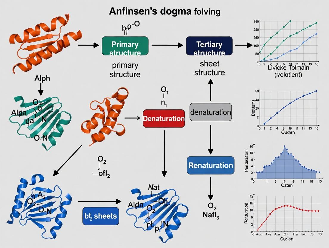
The 1972 Nobel Prize in Chemistry awarded to Christian B. Anfinsen stands as a foundational pillar in molecular biology. His work on Ribonuclease A (RNase A) crystallized the principle now known as Anfinsen's Dogma: the primary amino acid sequence of a protein uniquely determines its three-dimensional, native, and functional conformation under a given set of physiological conditions. This in-depth guide examines the historical experiment that led to this insight, its technical execution, and its enduring legacy in protein folding research and therapeutic development.
The Core Experiment: RNase A Denaturation and Refolding
Experimental Objective
To demonstrate that all information required for a protein to achieve its native, biologically active structure is encoded in its amino acid sequence.
Key Methodology & Protocol
Materials:
- Protein: Bovine pancreatic Ribonuclease A (124 amino acids, 4 disulfide bonds).
- Denaturant: 8M Urea or 6M Guanidine Hydrochloride.
- Reducing Agent: β-mercaptoethanol to reduce disulfide bonds.
- Oxidizing Environment: Atmospheric oxygen in a diluted, buffered solution.
- Assay Buffer: For activity measurement (e.g., using RNA or cyclic cytidine monophosphate as substrate).
Detailed Protocol:
- Native Purification: Isolate and purify RNase A to homogeneity.
- Complete Denaturation & Reduction:
- Prepare a solution of RNase A (~1 mg/mL) in 8M urea (or 6M GuHCl) containing 0.1M β-mercaptoethanol.
- Incubate at room temperature for several hours (or at elevated temperature, e.g., 37°C, for 1-2 hours).
- This step unfolds the polypeptide chain and reduces the four native disulfide bonds (Cys26-Cys84, Cys40-Cys95, Cys58-Cys110, Cys65-Cys72) to free sulfhydryl groups.
- Renaturation Initiation:
- Dilute the denatured/reduced mixture 100-fold into a chilled, aerated, mild buffer (e.g., 0.1M Tris-HCl, pH 8.0).
- This rapid dilution decreases the concentration of denaturant and reductant, allowing refolding and oxidation.
- Oxidation & Folding:
- Allow the diluted solution to stand at room temperature for several hours. Disulfide bonds reform spontaneously in the presence of atmospheric oxygen.
- Activity Recovery Measurement:
- At time intervals, aliquot the refolding solution.
- Measure enzymatic activity using a spectrophotometric assay (e.g., hydrolysis of cCMP, monitoring increase in absorbance at 296 nm).
- Compare activity to a native RNase A control and a fully denatured/reduced control.
- Variants: Experiments were performed with scrambled disulfides (allowing non-native pairing) followed by exposure to a trace of reductant to permit disulfide isomerization to the native state.
Table 1: Key Quantitative Outcomes from RNase A Refolding Experiments
| Parameter | Native RNase A (Control) | Denatured/Reduced RNase A | Refolded RNase A (After Oxidation) | Notes |
|---|---|---|---|---|
| Specific Activity | 100% | <1% | ~95-100% | Recovery of catalytic function. |
| Disulfide Bonds | 4 (Native pairs) | 0 (Reduced) | 4 (Re-formed as native pairs) | Verified by peptide mapping. |
| Refolding Yield | N/A | N/A | >90% | Dependent on exact conditions (pH, temperature, dilution rate). |
| Key Observation | Fully active, folded. | Unfolded, inactive. | Regains native structure & function. | Proves sequence encodes fold. |
Table 2: Modern Validation & Extensions of Anfinsen's Principle
| Aspect | Classic View (Anfinsen) | Modern Understanding (Post-RNase A) | Relevance to Drug Development |
|---|---|---|---|
| Folding Driver | Thermodynamic control; global free energy minimum. | Kinetic pathways and intermediate states are critical; some proteins require chaperones. | Misfolding diseases (e.g., Alzheimer's, ALS); chaperones as therapeutic targets. |
| Disulfide Bond Role | Formed post-folding to stabilize native structure. | Can guide and stabilize folding intermediates. | Critical for recombinant antibody and protein therapeutic production. |
| In Vitro Refolding | Spontaneous for many small proteins like RNase A. | Often inefficient for large, complex proteins; requires optimized redox buffers. | Major challenge in industrial biopharmaceutical manufacturing. |
Visualizing the Experimental Workflow and Dogma
Diagram 1: RNase A Refolding Proof-of-Principle Workflow
Diagram 2: Modern View of Anfinsen's Dogma in Context
The Scientist's Toolkit: Key Research Reagent Solutions
Table 3: Essential Materials for Protein Folding/Refolding Studies (Inspired by RNase A Experiments)
| Reagent/Material | Function in Folding Research | Modern Example/Note |
|---|---|---|
| Chaotropic Denaturants (Urea, GuHCl) | Disrupt hydrogen bonding & hydrophobic interactions to unfold proteins. Critical for establishing unfolding baselines. | High-purity, enzyme-grade to avoid cyanate contamination (for urea). |
| Reducing Agents (β-mercaptoethanol, DTT, TCEP) | Reduce disulfide bonds to free thiols. Essential for studying disulfide-coupled folding. | TCEP is more stable and effective at lower pH than DTT. |
| Redox Buffering Systems | Control the thiol-disulfide exchange equilibrium during refolding. | Glutathione (GSH/GSSG) or cysteine/cystine systems are standard. |
| Spectroscopic Probes (Intrinsic Fluorescence, CD, NMR) | Monitor changes in secondary/tertiary structure in real-time during folding/unfolding transitions. | Stopped-flow devices coupled to fluorometers enable millisecond resolution. |
| Analytical Chromatography (SEC, RP-HPLC) | Separate and quantify folded monomers, aggregates, and misfolded intermediates. | SEC-MALS (Multi-Angle Light Scattering) determines oligomeric state. |
| Chaperone Proteins (GroEL/ES, DnaK, etc.) | Assist in vivo folding by preventing aggregation or providing folding compartments. Used in vitro to study assisted folding mechanisms. | Key targets for understanding protein homeostasis networks. |
| Activity Assay Reagents | Measure functional recovery as the ultimate proof of correct folding. | For RNase A: cCMP or small RNA substrates. |
Legacy and Impact on Modern Drug Development
The RNase A experiment transcended a single finding. It established the conceptual framework for:
- Recombinant Protein Therapeutics: The premise that a protein expressed in a heterologous system (e.g., E. coli, CHO cells) can fold into its active form is rooted in Anfinsen's Dogma. In vitro refolding protocols are critical for drugs like insulin and some antibodies.
- Misfolding Disease Research: Diseases like Alzheimer's, Parkinson's, and cystic fibrosis are viewed through the lens of protein folding gone awry, where the polypeptide chain fails to reach or maintain its native state.
- Computational Drug Design & De Novo Protein Engineering: Predicting protein structure from sequence (AlphaFold) and designing novel functional proteins rely on the fundamental principle that sequence dictates fold.
- Biopharmaceutical Formulation: Stabilizing the native state of protein drugs against aggregation and denaturation during storage and delivery is a direct application of folding thermodynamics.
While subsequent research has introduced complexity—kinetic traps, chaperone requirements, and conformational diseases—the core insight from the RNase A experiment remains unchallenged: the linear amino acid sequence is the intrinsic blueprint for the three-dimensional architecture of life's molecular machines.
The foundational principle of structural biology, enshrined in Christian Anfinsen's Nobel Prize-winning work, posits that a protein's amino acid sequence uniquely determines its three-dimensional native conformation, which in turn dictates its biological function. This "thermodynamic hypothesis" established the paradigm of spontaneous, reversible folding driven by the search for a global free energy minimum. While revolutionary, contemporary research reveals a more nuanced landscape where chaperones, environmental factors, and kinetic traps influence the folding landscape. This whitepores the core tenet through the lens of modern protein folding research and its critical implications for therapeutic intervention.
Quantitative Foundations: Energy Landscapes and Folding Kinetics
The folding process is governed by a complex energy landscape. Key quantitative metrics are summarized below.
Table 1: Key Quantitative Metrics in Protein Folding Research
| Metric | Description | Typical Range/Value | Experimental Method |
|---|---|---|---|
| ΔGfolding | Free energy change of folding (Native vs. Unfolded) | -5 to -15 kcal/mol | Thermal or Chemical Denaturation |
| Tm | Melting Temperature (50% unfolded) | 40°C to 80°C | Differential Scanning Calorimetry (DSC) |
| Cm | Denaturant Concentration at midpoint of unfolding | 3-8 M Urea; 1.5-4 M GdnHCl | Equilibrium Denaturation |
| Folding Rate (kf) | Rate constant for folding | μs to seconds | Stopped-Flow Fluorescence |
| Unfolding Rate (ku) | Rate constant for unfolding | s-1 to hr-1 | Stopped-Flow or Temperature Jump |
| Φ-value | Fraction of native contacts in transition state (0-1) | 0 (unfolded-like) to 1 (native-like) | Protein Engineering & Kinetics |
Table 2: Impact of Sequence Mutations on Stability (ΔΔG) and Kinetics
| Mutation Type | Typical ΔΔG (kcal/mol) | Effect on Folding Rate (kf) | Common Functional Consequence |
|---|---|---|---|
| Core Hydrophobic to Polar | +1.5 to +4.0 (Destabilizing) | Decrease by 10-1000x | Loss of function, Aggregation |
| Surface Polar to Hydrophobic | +0.5 to +2.0 | Variable | Potential Misfolding, Altered Interactions |
| Salt Bridge Removal | +0.5 to +3.0 | Mild decrease | Reduced specificity, Altered allostery |
| Proline in Flexible Loop | Variable (often neutral) | Minimal | Altered conformational dynamics |
| Glycine to Alanine (in turn) | +0.5 to +2.0 (Destabilizing) | Decrease | Impaired loop formation, Slowed folding |
Core Experimental Methodologies
Determining Structure from Sequence: X-ray Crystallography Protocol
- Objective: Obtain atomic-resolution 3D structure of a purified protein.
- Key Steps:
- Cloning & Expression: Gene of interest is cloned into an expression vector (e.g., pET) and expressed in a host (E. coli, insect, mammalian cells).
- Purification: Affinity (e.g., His-tag), ion-exchange, and size-exclusion chromatography yield >95% pure, monodisperse protein.
- Crystallization: Using vapor diffusion (hanging/sitting drop), protein is mixed with precipitant solutions to form ordered crystals. Screening robots test 100s-1000s of conditions.
- Data Collection: Crystal is flash-cooled with liquid N2 and exposed to high-intensity X-rays at a synchrotron. Diffraction patterns are collected.
- Phasing & Model Building: Phase problem is solved via molecular replacement (using homologous structure) or experimental phasing (e.g., SAD with selenomethionine). An atomic model is built into the electron density map (e.g., using Coot).
- Refinement & Validation: Model is refined (e.g., with PHENIX.refine) against diffraction data. Stereochemical quality is validated (e.g., with MolProbity).
Probing Folding Kinetics: Stopped-Flow Fluorescence Protocol
- Objective: Measure the rate of folding/unfolding on millisecond-to-second timescales.
- Key Steps:
- Sample Preparation: Purified protein is labeled with an environment-sensitive fluorophore (e.g., Tryptophan intrinsic, or extrinsic ANS) or engineered to have a FRET pair.
- Initiation: Two syringes are loaded: one with native protein in buffer, the other with a high concentration of denaturant (for unfolding) or buffer alone (for refolding of denatured protein). Syringes are rapidly mixed (dead time ~1 ms).
- Detection: Fluorescence emission at a specific wavelength is monitored continuously over time via a photomultiplier tube.
- Data Analysis: The resulting trace (fluorescence intensity vs. time) is fit to single or multi-exponential equations to extract observed rate constants (kobs). Chevron plots (log(kobs) vs. [denaturant]) are constructed to derive intrinsic folding (kf) and unfolding (ku) rates.
The Scientist's Toolkit: Key Research Reagent Solutions
Table 3: Essential Reagents for Protein Folding & Structure Research
| Reagent / Material | Function & Rationale |
|---|---|
| Urea & Guanidine HCl | Chemical denaturants. Used to unfold proteins reversibly for equilibrium and kinetic folding studies. |
| ANS (1-Anilinonaphthalene-8-sulfonate) | Hydrophobic dye. Binds to exposed hydrophobic patches in molten globule/folding intermediates, used as a fluorescent probe. |
| DTT (Dithiothreitol) / TCEP | Reducing agents. Break disulfide bonds to study unfolded state or prevent non-native crosslinking. |
| HEPES / Tris Buffers | Maintain constant pH during experiments, critical as folding can be pH-sensitive. |
| Size Exclusion Chromatography Resins (e.g., Superdex) | Separate folded monomers from aggregates or oligomers, assessing folding quality and monodispersity. |
| Crystallization Screens (e.g., JC SG, Morpheus) | Pre-formulated sparse matrix screens of precipitant, salt, and buffer conditions to identify initial crystallization hits. |
| Cryoprotectants (e.g., Glycerol, Ethylene Glycol) | Prevent ice crystal formation during flash-cooling of protein crystals for X-ray data collection. |
| Thermostable DNA Polymerases (e.g., Phusion) | For high-fidelity PCR in site-directed mutagenesis to create sequence variants for Φ-value analysis. |
Visualizing the Paradigm: Pathways and Workflows
Protein Folding Central Dogma
Experimental Workflow for Folding Studies
Folding Energy Landscape Schematic
Contemporary Challenges and Therapeutic Implications
The core tenet directly underpins rational drug design and the understanding of disease. Misfolding, leading to aggregation (e.g., amyloid-β in Alzheimer's, α-synuclein in Parkinson's), is a failure of the sequence-to-structure pathway. Conversely, targeting specific protein structures (e.g., kinase ATP pockets, protease active sites) remains the cornerstone of small-molecule drug development. Emerging fields like cryo-electron microscopy (cryo-EM) provide high-resolution structures of previously intractable targets (e.g., membrane proteins), while AI/ML systems like AlphaFold2 and RoseTTAFold have revolutionized structure prediction from sequence alone. These advances validate Anfinsen's dogma at scale but also highlight the ongoing challenge of predicting functional dynamics and allosteric regulation from static structure alone. The next frontier lies in integrating sequence-structure predictions with folding kinetics, conformational ensembles, and the cellular environment to achieve a truly predictive understanding of protein function.
The Thermodynamic Hypothesis, central to Anfinsen's dogma, posits that the native, functional conformation of a protein is the one in which its Gibbs free energy is at a global minimum under physiological conditions. Anfinsen's seminal ribonuclease A experiments demonstrated that the information required for folding is encoded entirely within the protein's amino acid sequence. This established the foundational principle that the native state is both thermodynamically stable and kinetically accessible. Modern protein folding research continues to test and refine this hypothesis, particularly in the context of complex, multi-domain proteins and the role of cellular machinery like chaperones.
Theoretical Foundations: Energy Landscapes and Folding Funnels
The global free energy minimum concept is best visualized through the energy landscape theory. A protein's conformational space is not a flat plain but a rugged funnel. The broad top represents the vast ensemble of unfolded states with high entropy and energy. The steepness of the funnel walls corresponds to the folding rate, while the ruggedness represents kinetic traps (e.g., misfolded states). The narrow bottom is the native basin, the global minimum.
Diagram Title: Protein Folding Energy Landscape Funnel
Key Experimental Evidence & Quantitative Data
Experimental validation of the hypothesis relies on measuring the stability and uniqueness of the native state.
Table 1: Key Stability Measurements for Model Proteins
| Protein (PDB ID) | ΔGunfolding (kcal/mol) | Tm (°C) | Cm (Denaturant M) | Method | Reference |
|---|---|---|---|---|---|
| Ribonuclease A (1FS3) | -7.2 to -9.5 | 58.2 | ~4.0 (GdnHCl) | CD, Fluorescence | Anfinsen (1973) |
| Lysozyme (1REX) | -10.3 | 75.5 | ~5.0 (GdnHCl) | DSC, CD | |
| CI2 (2CI2) | -6.8 | 75.0 | ~3.8 (GdnHCl) | Equilibrium Unfolding | Jackson & Fersht (1991) |
| SH3 Domain (1SHG) | -3.5 | 58.0 | ~2.5 (GdnHCl) | NMR, CD |
Table 2: Challenges to the "Strict" Global Minimum Concept
| Phenomenon | Description | Implication for Hypothesis |
|---|---|---|
| Kinetic Traps | Misfolded aggregates, proline isomerization | Native state may not be kinetically accessible without aid. |
| Chaperone Assistance | GroEL/ES, Hsp70 prevent aggregation | In vivo, the "effective" landscape is shaped by cellular factors. |
| Metamorphic Proteins | >1 stable native fold under same conditions (e.g., Mad2) | Free energy landscape has multiple deep, distinct minima. |
| Intrinsically Disordered Proteins (IDPs) | Lack a fixed tertiary structure | Functional state is not a single, well-defined global minimum. |
Detailed Experimental Protocols
Protocol: Equilibrium Chemical Denaturation to Measure ΔGunfolding
Objective: Determine the conformational stability (ΔGunfolding) of a protein. Principle: Monitor a spectroscopic signal (e.g., fluorescence at 350 nm, CD at 222 nm) as a function of denaturant concentration (e.g., Guanidine HCl or Urea). Fit data to a two-state unfolding model. Procedure:
- Sample Preparation: Prepare a series of 20-30 identical protein samples (~10-20 µM) in a physiological buffer (e.g., 20 mM phosphate, pH 7.0). Add varying concentrations of denaturant (e.g., 0 to 8 M GdnHCl). Allow samples to equilibrate at constant temperature (e.g., 25°C) for 2-4 hours.
- Data Acquisition: Measure the chosen spectroscopic signal for each sample. For fluorescence, excite at 280 nm (Trp) and record emission at 320-350 nm. For CD, record the mean residue ellipticity at 222 nm.
- Data Analysis: Plot signal vs. [Denaturant]. Fit data to the linear extrapolation model equation:
S = [ (S_N + m_N*D) + (S_U + m_U*D) * exp(-(ΔG° - m*D)/RT) ] / [ 1 + exp(-(ΔG° - m*D)/RT) ]where S is observed signal, SN/U are baselines, mN/U are slopes, D is [denaturant], ΔG° is ΔGunfolding in water, and m is the dependence of ΔG on [denaturant].
Protocol: Differential Scanning Calorimetry (DSC)
Objective: Directly measure the enthalpy (ΔH) and melting temperature (Tm) of thermal unfolding. Principle: Measure the heat capacity (Cp) of a protein solution as temperature is increased. Unfoldings an endothermic process that absorbs heat. Procedure:
- Sample/Buffer Matching: Dialyze protein (~1 mg/mL) exhaustively against a degassed buffer. Precisely match the reference cell with dialysis buffer.
- Scanning: Load sample and reference cells. Scan from 10°C to 90-100°C at a constant rate (e.g., 1°C/min). Record the differential heat flow.
- Data Analysis: Subtract buffer-buffer baseline. Fit the excess heat capacity curve to a model (e.g., two-state) to obtain Tm (peak), ΔHcal (area under peak), and ΔCp (baseline shift).
Diagram Title: Experimental Workflow for Folding Analysis
The Scientist's Toolkit: Key Research Reagent Solutions
Table 3: Essential Materials for Protein Folding/Stability Studies
| Item | Function & Rationale |
|---|---|
| High-Purity Guanidine HCl (GdnHCl) / Urea | Chemical denaturant for equilibrium unfolding experiments. Must be of high purity to avoid artifacts; concentration determined by refractive index. |
| Differential Scanning Calorimeter (DSC) | Instrument for direct thermodynamic measurement of thermal unfolding (ΔH, Tm, ΔCp). |
| Circular Dichroism (CD) Spectrophotometer | Measures secondary (far-UV) and tertiary (near-UV) structure content. Key for monitoring folding/unfolding transitions. |
| Fluorescence Spectrophotometer | Tracks changes in intrinsic tryptophan fluorescence or extrinsic dye (e.g., ANS) binding, sensitive to local environment changes during folding. |
| Size-Exclusion Chromatography (SEC) Columns | Assess protein monomeric state, aggregation, and compactness (e.g., of folding intermediates). |
| Stopped-Flow / Temperature-Jump Apparatus | For rapid mixing or heating, allowing study of early folding events on microsecond to millisecond timescales. |
| Isotopically Labeled Amino Acids (¹⁵N, ¹³C) | For NMR studies to obtain residue-level information on protein structure, dynamics, and folding pathways. |
| Chaperone Proteins (e.g., GroEL/ES) | Used in in vitro refolding assays to study assisted folding and mechanisms to overcome kinetic traps. |
Modern Computational Validation: Molecular Dynamics and AI
Computational approaches now provide atomistic validation. Molecular dynamics (MD) simulations, enhanced by Markov State Models, can map folding pathways. More recently, AlphaFold2 and related AI tools predict the native structure (putative global minimum) directly from sequence, implicitly learning the energy landscape from evolutionary data. However, these models do not yet fully replicate the dynamic folding process or accurately predict folding kinetics and stability changes upon mutation.
Diagram Title: Computational Validation Workflow
The Thermodynamic Hypothesis remains a powerful core principle. For drug developers, it underpins rationale: small-molecule stabilizers bind the native state, deepening its energy minimum, while proteolysis-targeting chimeras (PROTACs) may exploit minor unfolding. However, the modern view integrates kinetic accessibility, chaperone networks, and conformational ensembles. Targeting folding intermediates or "cryptic" pockets that transiently open represents a frontier in therapeutics for protein misfolding diseases and beyond. The native state as the global free energy minimum is the anchor point from which all these complex, biologically relevant dynamics emanate.
Anfinsen's Dogma, the principle that a protein's native structure is determined solely by its amino acid sequence under physiological conditions, provides the foundational thesis for in vitro folding studies. The in vitro folding paradigm directly tests this postulate by investigating the refolding of purified, denatured proteins in controlled, cell-free environments. This whitepaper examines the core assumptions of this paradigm, its quantitative findings, and its profound implications for fundamental research and therapeutic development.
Core Assumptions of the Paradigm
- Reductionist Validity: The complex cellular folding process can be deconstructed and validly studied using simplified buffer systems.
- Thermodynamic Control: Under in vitro conditions, the native state represents the global minimum of free energy, and folding is reversible.
- Sequence Sufficiency: All information required for folding is contained within the polypeptide chain; no genetic information from the cellular machinery is needed.
- Context Independence: The fundamental principles and pathways discovered in vitro are directly relevant to the in vivo folding process.
Quantitative Data: Key Folding Parameters
The in vitro paradigm has enabled precise measurement of folding kinetics and stability.
Table 1: Key Thermodynamic & Kinetic Parameters from In Vitro Folding Studies
| Parameter | Definition | Typical Measurement Technique | Example Value (Ribonuclease A) | Implication |
|---|---|---|---|---|
| ΔG°folding | Free energy change upon folding (Stability) | Equilibrium denaturation (Urea/GdmCl, DSC) | -30 to -50 kJ/mol | Measures native state stability. Small values indicate marginal stability. |
| m-value | Cooperativity of unfolding; dependence of ΔG on [denaturant] | Linear extrapolation of denaturation data | ~10 kJ/mol·M | Reflects change in solvent-accessible surface area; proxy for folding cooperativity. |
| kf | Folding rate constant | Stopped-flow fluorescence, CD | 1 - 10⁴ s⁻¹ | Speed of productive folding to native state. |
| ku | Unfolding rate constant | Stopped-flow, manual mixing | 10⁻⁶ - 10⁻² s⁻¹ | Speed of native state disruption. |
| Φ-value | Fraction of native interactions formed in the transition state | Protein engineering & kinetic analysis (Φ = ΔΔG‡-U/ΔΔGN-U) | 0 (no structure) to 1 (native-like) | Maps structure of the folding transition state ensemble. |
Table 2: Common Denaturants Used in In Vitro Folding Studies
| Denaturant | Mechanism of Action | Typical Concentration Range | Pros | Cons |
|---|---|---|---|---|
| Urea | Disrupts H-bonds & hydrophobic effect; water structure maker. | 0-10 M | Non-ionic, highly soluble. | Can form cyanate ions at high pH (alters proteins). |
| Guanidinium Chloride (GdmCl) | Binds to peptide backbone, solubilizing hydrophobic residues. | 0-8 M | More potent than urea per molar. | Ionic (interferes with some assays), more expensive. |
| Temperature | Increases atomic motion, disrupts all non-covalent interactions. | 25-100°C | No chemical additives. | Can cause irreversible aggregation/chemical degradation. |
Detailed Experimental Protocol: Stopped-Flow Fluorescence Refolding
This protocol is a standard for measuring millisecond folding kinetics.
Objective: Measure the apparent folding rate constant (kapp) of a denatured protein upon rapid dilution into native conditions.
Materials & Reagents:
- Purified protein sample.
- High-purity denaturant (e.g., 6M GdmCl).
- Folding buffer (appropriate pH, salts, redox agents if needed).
- Stopped-flow spectrofluorometer.
- Syringes, tubing, and drive system.
Procedure:
- Sample Preparation: Denature the protein by incubation in buffer containing 6M GdmCl for >2 hours at the experimental temperature.
- Instrument Setup: Load one drive syringe with denatured protein. Load a second syringe with folding buffer (no denaturant). Ensure flow paths are purged.
- Rapid Mixing: Activate the drive mechanism to rapidly mix equal volumes (typically 50-100 µL each) of denatured protein and folding buffer. The final denaturant concentration is diluted to a sub-denaturing level (e.g., 0.5M GdmCl). Dead time is typically 1-2 ms.
- Data Acquisition: Monitor intrinsic fluorescence (typically Trp emission at ~340 nm upon excitation at 280 nm) as a function of time post-mixing. Perform 3-5 replicate shots per condition.
- Data Analysis: Fit the resulting averaged fluorescence trace to a single or multi-exponential function: F(t) = F∞ + ΣΔFi * exp(-kapp,i * t), where kapp is the observed rate constant.
Visualizations
Title: In Vitro Folding Pathways: Two-State vs. Multi-State
Title: Core In Vitro Folding Experiment Workflow
The Scientist's Toolkit: Key Research Reagent Solutions
Table 3: Essential Reagents for In Vitro Folding Studies
| Reagent / Material | Function & Rationale | Key Considerations |
|---|---|---|
| Ultra-Pure Denaturants (GdmCl, Urea) | To fully denature protein to a random coil starting state without chemical modification. | Must be of highest purity (≥99.5%); solutions should be freshly prepared or treated with mixed-bed resin to remove ionic contaminants and cyanate (urea). |
| Redox Pairs (GSH/GSSG, Cys/Cystine) | To control the redox potential for proper disulfide bond formation and reshuffling during refolding. | Critical for oxidative refolding studies. Ratios determine the driving force for disulfide formation. |
| Chaotrope-Resistant Detergents (e.g., CHAPS) | To prevent aggregation of hydrophobic intermediates during refolding, improving yield. | Used at low concentrations to minimize interference with folding energetics. |
| Protease Inhibitor Cocktails | To prevent proteolytic degradation of unfolded or partially folded states, which are often protease-sensitive. | Essential for long-duration equilibrium experiments. |
| Intrinsic Fluorescence Probes (Tryptophan) | A built-in reporter for changes in local hydrophobic environment during folding/unfolding. | Non-perturbing. Requires protein to have Trp residues in sensitive positions. |
| Extrinsic Fluorescent Dyes (e.g., ANS, Sypro Orange) | Binds to exposed hydrophobic patches, reporting on molten globule or intermediate states. | Can be slightly perturbing; useful for proteins lacking suitable Trp residues. |
| Fast Kinetics Instrumentation (Stopped-Flow) | To initiate folding and observe events on the millisecond timescale. | Requires significant sample volumes (~100 µL per shot) and concentration. |
Implications for Research and Drug Development
The in vitro paradigm has directly enabled:
- Mechanistic Understanding: Elucidation of folding pathways, transition states, and the principles of cooperativity.
- Disease Insight: Quantitative assessment of mutant protein stability in misfolding diseases (e.g., Transthyretin Amyloidosis, CFTR in Cystic Fibrosis).
- Drug Discovery: Identification of pharmacological chaperones—small molecules that stabilize the native state, a strategy for treating loss-of-function misfolding diseases.
- Biotech Engineering: Informing rational protein design and engineering for improved stability and expressibility of therapeutic proteins.
The paradigm remains a cornerstone of biophysical research, providing the essential, quantitative framework against which the complexities of in vivo folding, assisted by chaperones, must be compared and integrated.
The "Levinthal Paradox" and the Protein Folding Problem
The central dogma of molecular biology outlines information flow from DNA to protein. A corollary, Anfinsen's dogma, posits that a protein's native, functional three-dimensional structure is uniquely determined by its amino acid sequence, under physiological conditions. This implies the folding process is spontaneous and deterministic. However, in 1969, Cyrus Levinthal highlighted a profound computational problem: for a typical protein of 100 residues, sampling all possible conformations (even at a coarse-grained level) would require time exceeding the age of the universe. This contradiction—between observed folding times (milliseconds to seconds) and astronomical computational search times—is the Levinthal Paradox. It forces a conclusion that proteins do not fold by exhaustive search but follow specific, guided pathways through a funneled energy landscape.
The Energy Landscape Theory and Folding Funnels
The resolution to the paradox lies in the energy landscape theory. The conformational space is not flat; it is a biased, funnel-shaped landscape where the native state resides at the global free energy minimum. The topology of this landscape directs the folding process.
Diagram 1: Protein Folding Energy Landscape Funnel
Quantitative Dimensions of the Paradox
The following table quantifies the scale of the Levinthal search versus observed reality.
Table 1: The Levinthal Paradox in Numbers
| Parameter | Levinthal's Exhaustive Search Calculation | Experimentally Observed Folding |
|---|---|---|
| Protein Size | 100 amino acids | 50-300 amino acids (typical) |
| Conformations per Residue | ~10 (estimated) | N/A |
| Total Conformations | 10¹⁰⁰ | N/A |
| Time per Conformation | ~10⁻¹³ seconds (bond vibration) | N/A |
| Total Search Time | ~10⁸⁷ seconds | 10⁻³ to 10³ seconds |
| Universe Age (seconds) | ~4.3 x 10¹⁷ | ~4.3 x 10¹⁷ |
| Guiding Principle | Random Sampling | Funneled Energy Landscape, Nucleation, Secondary Structure Propensities |
Key Experimental Methodologies
Understanding folding requires probing structure, dynamics, and stability.
Protocol 4.1: Stopped-Flow Fluorescence for Folding Kinetics
Objective: Measure the rate of folding/unfolding by observing changes in intrinsic tryptophan fluorescence.
- Solutions: Prepare a native protein sample in buffer (Syringe A) and a denatured protein sample in high-concentration chemical denaturant (e.g., 6M GuHCl) (Syringe B).
- Rapid Mixing: Use a stopped-flow apparatus to rapidly mix equal volumes from Syringe A and B, initiating a jump to final denaturant concentration (e.g., 1M GuHCl) to trigger refolding.
- Detection: Pass the mixed solution through a fluorescence cuvette. Excite at 280 nm and monitor emission at >320 nm (typically 340-350 nm) over time.
- Analysis: Fit the resulting fluorescence time trace to single or multi-exponential functions to derive observed rate constants (k_obs).
Protocol 4.2: Hydrogen-Deuterium Exchange Mass Spectrometry (HDX-MS)
Objective: Map regions of stability and dynamics by measuring the exchange of backbone amide hydrogens.
- Labeling: Dilute the protein sample (in native or non-native state) into a D₂O-based buffer. Incubate for a specific time (e.g., 10 sec to several hours).
- Quench: Lower pH to ~2.5 and temperature to 0°C to drastically slow exchange.
- Digestion & Separation: Pass the quenched sample through an immobilized pepsin column for rapid digestion. Separate peptides via liquid chromatography (LC).
- Mass Spectrometry Analysis: Inject peptides into a high-resolution MS. Monitor mass increase as H→D exchange occurs. Decreased exchange in a region indicates hydrogen bonding or protection from solvent (e.g., in a folded core).
Visualization of Folding Pathways & Chaperone Action
Chaperones like GroEL/ES assist folding by preventing aggregation and providing an isolated compartment.
Diagram 2: GroEL/ES Chaperonin Folding Cycle
The Scientist's Toolkit: Research Reagent Solutions
Table 2: Essential Reagents for Protein Folding Studies
| Reagent/Category | Example(s) | Primary Function in Folding Research |
|---|---|---|
| Chemical Denaturants | Guanidine Hydrochloride (GuHCl), Urea | Unfold proteins to study denaturation curves or create starting states for refolding kinetics. |
| Reducing Agents | Dithiothreitol (DTT), Tris(2-carboxyethyl)phosphine (TCEP) | Reduce disulfide bonds to study unfolded state or prevent non-native bond formation. |
| Chaperones | GroEL/ES (commercial kits), DnaK/DnaJ/GrpE | Assist in refolding in vitro, study chaperone-mediated folding mechanisms. |
| Fluorescent Dyes | ANS (8-Anilino-1-naphthalenesulfonate), SYPRO Orange | Probe exposed hydrophobic patches (ANS for molten globules) or general unfolding (SYPRO Orange in thermal shifts). |
| Stabilizers | L-Arginine, Sucrose, Glycerol | Suppress aggregation during refolding, improve protein solubility. |
| Isotope-Labeled Compounds | D₂O (for HDX), ¹⁵N/¹³C-labeled amino acids (for NMR) | Enable structural dynamics studies via HDX-MS or multidimensional NMR spectroscopy. |
| Proteases | Pepsin (for HDX), Trypsin | Rapid digestion for HDX-MS peptide-level analysis or limited proteolysis to probe folding intermediates. |
Modern Computational Approaches: From Paradox to Prediction
Computational methods now leverage the landscape theory to predict structure.
- Molecular Dynamics (MD): Simulates physical movements of atoms but is limited to shorter timescales than folding.
- Rosetta: Uses fragment assembly and Monte Carlo sampling guided by a physically informed energy function to search the conformational landscape efficiently.
- AlphaFold2 (DeepMind): Employs deep learning on known structures and evolutionary data (MSA) to predict accurate 3D models, effectively bypassing the explicit search of Levinthal's paradox.
The Levinthal Paradox was not a true paradox but a reductio ad absurdum that proved random search false. It catalyzed the conceptual shift to the energy landscape view, which unified Anfinsen's thermodynamic hypothesis with kinetically accessible pathways. Today, the "protein folding problem" largely refers to the computational prediction challenge—a challenge being solved by AI, yet the detailed physical mechanisms of folding in vivo, including chaperone interactions and co-translational folding, remain vibrant areas of research with direct implications for understanding and drugging protein misfolding diseases.
From Principle to Practice: Applying Anfinsen's Dogma in Modern Research & Therapeutics
The field of computational protein design stands as a direct test and extension of Anfinsen's dogma, which posits that a protein's amino acid sequence uniquely determines its three-dimensional native structure under physiological conditions. The central challenge in protein folding research has been to decipher this "second half of the genetic code"—the rules that map sequence to structure. Computational methods like Rosetta and, more recently, AlphaFold represent revolutionary tools in this pursuit, transforming the dogma from a thermodynamic principle into a predictable, engineering-capable framework. This whitepaper provides a technical guide to the core algorithms, experimental validation protocols, and practical tools underpinning modern structure prediction, contextualized within the ongoing research to fully realize Anfinsen's vision.
Core Methodologies and Algorithms
The Rosetta Suite: A Physics-Based and Knowledge-Based Approach
Rosetta employs a fragment-assembly method guided by a semi-empirical energy function. The protocol minimizes a scoring function that combines physical terms (van der Waals, electrostatics, solvation) with statistically derived terms from known protein structures (rotamer probabilities, backbone torsions).
Key Scoring Terms in Rosetta Energy Function: Table 1: Major Components of the Rosetta Full-Atom Energy Function (ref2015)
| Term | Description | Physical Basis |
|---|---|---|
fa_atr |
Attractive Lennard-Jones potential | Van der Waals interactions |
fa_rep |
Repulsive Lennard-Jones potential | Steric clash prevention |
fa_sol |
Lazaridis-Karplus solvation model | Hydrophobic effect |
fa_elec |
Coulomb potential with distance-dependent dielectric | Electrostatics |
hbond |
Hydrogen bonding potential | Polar interactions |
rama_prepro |
Backbone torsion probabilities | Conformational statistics |
p_aa_pp |
Amino acid propensity per backbone torsion | Sequence-structure statistics |
Experimental Protocol for Ab Initio Folding with Rosetta:
- Sequence Input: Provide the target amino acid sequence.
- Fragment Library Generation: Use PSI-BLAST to identify homologous sequences. Submit the sequence to the Robetta server or generate 3-mer and 9-mer fragment libraries from the PDB using NNmake.
- Monte Carlo Fragment Insertion: Perform a simulated annealing Monte Carlo search: a. Randomly select a fragment from the library for a randomly chosen sequence position. b. Insert the fragment, replacing the current backbone dihedrals. c. Score the new conformation using the Rosetta energy function. d. Accept or reject the move based on the Metropolis criterion.
- Decoy Generation: Repeat step 3 thousands of times from different random seeds, generating thousands of decoy structures.
- Clustering and Selection: Cluster all decoy structures based on backbone root-mean-square deviation (RMSD). Select the centroid of the largest cluster as the final predicted model.
Diagram 1: Rosetta Ab Initio Folding Workflow
AlphaFold2: An End-to-End Deep Learning Revolution
AlphaFold2 (AF2) represents a paradigm shift, employing an end-to-end deep neural network that directly predicts atomic coordinates from sequence and multiple sequence alignment (MSA) information. Its architecture is based on an Evoformer module (for processing MSA and pairwise representations) followed by a structure module that iteratively refines a 3D backbone trace.
Key Input Features and Outputs: Table 2: AlphaFold2 Input Features and Model Outputs
| Feature Type | Description | Source |
|---|---|---|
| Primary Inputs | Amino acid sequence (one-hot encoded) | Target sequence |
| Multiple Sequence Alignment (MSA) | Databases (e.g., UniRef, BFD) | |
| Template structures (optional) | PDB (via HHsearch) | |
| Model Outputs | Per-residue predicted aligned error (PAE) | Confidence in relative positions |
| Predicted LDdt (pLDDT) per residue | Local confidence metric | |
| 3D coordinates for all heavy atoms | Final atomic model |
Experimental Protocol for Prediction with AlphaFold2:
- Input Preparation: Generate a comprehensive MSA for the target sequence using tools like JackHMMER or MMseqs2 against large protein sequence databases (UniRef90, MGnify).
- Template Search (Optional): Search the PDB for structural homologs using fold recognition tools (HHsearch).
- Model Inference: Input the MSA and template information into the pre-trained AlphaFold2 model (via ColabFold, local installation, or the AlphaFold Server).
- Structure Generation: The model runs its Evoformer and structure modules, outputting five unrelaxed models.
- Relaxation and Selection: Apply a brief Amber force field relaxation to each model to remove minor steric clashes. Rank models by predicted confidence (average pLDDT) and select the highest-ranking model.
Diagram 2: AlphaFold2 Core Architecture Flow
Validation, Metrics, and Benchmarking
Accurate validation against experimentally determined structures is critical. The standard benchmark is the Critical Assessment of protein Structure Prediction (CASP) experiment.
Table 3: Key Metrics for Evaluating Predicted Protein Structures
| Metric | Description | Interpretation |
|---|---|---|
| Global Distance Test (GDT) | Percentage of Cα atoms under a distance cutoff (e.g., 1Å, 2Å, 4Å, 8Å) from the native structure. | Higher is better. GDTTS is average of GDT1,2,4,8. >90 indicates high accuracy. |
| Root-Mean-Square Deviation (RMSD) | Square root of the average squared distance between superimposed Cα atoms. | Lower is better. <2Å for core residues is excellent. Sensitive to outliers. |
| Template Modeling Score (TM-score) | Metric that weights local distances, less sensitive to global outliers than RMSD. | Range 0-1. >0.5 suggests correct fold; >0.8 indicates high accuracy. |
| Local Distance Difference Test (pLDDT) | AlphaFold2's per-residue confidence score (predicted LDdt). | Range 0-100. >90: high confidence; 70-90: confident; 50-70: low; <50: very low. |
| Predicted Aligned Error (PAE) | AlphaFold2's predicted positional error (in Ångströms) for every residue pair. | Visualized as a 2D plot. Indicates confidence in relative domain positioning. |
The Scientist's Toolkit: Key Research Reagent Solutions
Table 4: Essential Materials and Tools for Computational Protein Design & Validation
| Item / Reagent | Function / Purpose |
|---|---|
| UniProt / PDB Databases | Primary sources for protein sequences and experimental 3D structures for training, template search, and benchmarking. |
| MMseqs2 / JackHMMER | Software for generating deep multiple sequence alignments (MSAs) from sequence databases, a critical input for AlphaFold2. |
| PyMOL / ChimeraX | Molecular visualization software for analyzing, comparing, and rendering predicted and experimental protein structures. |
| PyRosetta / RosettaScripts | Python interface and XML-based scripting language for building custom computational protein design and analysis pipelines with Rosetta. |
| ColabFold | Cloud-based, streamlined implementation of AlphaFold2 and AlphaFold-Multimer that simplifies MSA generation and model prediction. |
| Amber / CHARMM Force Fields | Molecular dynamics force fields used for energy minimization and relaxation of predicted models to correct minor stereochemical inaccuracies. |
| CASP Datasets | Blind test sets of protein structures used as the gold standard for benchmarking and comparing the performance of prediction methods. |
| Size Exclusion Chromatography (SEC) Columns | For experimental validation of monomeric state and stability of designed/expressed proteins. |
| Differential Scanning Calorimetry (DSC) | To measure the thermal denaturation midpoint (Tm) of a protein, quantifying its stability relative to design predictions. |
| Surface Plasmon Resonance (SPR) Chips | For biophysical validation of designed protein-protein or protein-ligand binding interactions predicted by computational models. |
The central dogma of structural biology, Anfinsen's postulate, asserts that a protein's native, functional three-dimensional structure is uniquely determined by its amino acid sequence under physiological conditions. This principle provides the foundational framework for rational drug design. By targeting the well-defined, thermodynamically stable native state of a protein—whether an enzyme, receptor, or signaling molecule—we aim to develop highly specific therapeutic agents. This whitepaper details the modern technical approaches for leveraging high-resolution structural data to design drugs that bind with high affinity and selectivity to their intended protein targets, thereby modulating disease-associated biological pathways.
Core Methodological Pillars
Target Identification and Validation
The process begins with the identification of a protein whose function is critically involved in a disease pathway. Validation confirms that modulating this target will have a therapeutic effect.
- Experimental Protocol: CRISPR-Cas9 Knockout/Knockdown Validation
- Design single-guide RNAs (sgRNAs) targeting the gene of interest.
- Clone sgRNAs into a lentiviral Cas9 expression vector (e.g., lentiCRISPRv2).
- Transduce target cell lines (e.g., cancer, primary) with the lentivirus and select with puromycin (2 µg/mL) for 72 hours.
- Confirm gene knockout via western blot (primary antibody incubation: 1:1000, 4°C overnight) and Sanger sequencing of the target locus.
- Perform functional assays (e.g., proliferation, apoptosis, migration) to assess phenotypic impact of target loss.
High-Resolution Structure Determination
Defining the atomic coordinates of the native protein structure is non-negotiable for structure-based drug design (SBDD).
Experimental Protocol: Protein Purification for X-ray Crystallography
- Express the recombinant protein with an affinity tag (e.g., His6) in a suitable system (e.g., HEK293F, Sf9).
- Lyse cells and purify via immobilized metal affinity chromatography (IMAC) using Ni-NTA resin.
- Remove the tag using site-specific protease (e.g., TEV) and perform size-exclusion chromatography (Superdex 200 Increase) in crystallization buffer (e.g., 20 mM HEPES pH 7.5, 150 mM NaCl).
- Concentrate protein to 10 mg/mL using a centrifugal concentrator (10 kDa MWCO).
- Set up crystallization trials using commercial screens (e.g., Hampton Research) via sitting-drop vapor diffusion at 20°C.
Experimental Protocol: Cryo-Electron Microscopy (Cryo-EM) Single Particle Analysis
- Apply 3.5 µL of purified protein (0.5-2 mg/mL) to a glow-discharged Quantifoil grid.
- Blot for 3-5 seconds and plunge-freeze in liquid ethane using a Vitrobot (100% humidity, 4°C).
- Collect movies on a 300 keV cryo-TEM (e.g., Titan Krios) with a Gatan K3 direct electron detector at a nominal magnification of 105,000x (pixel size 0.826 Å).
- Process data: motion correction (MotionCor2), CTF estimation (Gctf), particle picking (cryoSPARC blob picker), 2D classification, ab initio reconstruction, and heterogeneous refinement.
- Build and refine an atomic model using Coot and PHENIX real-space refine.
In Silico Drug Design and Optimization
Computational tools are used to identify and optimize lead compounds that complement the target's binding site.
- Experimental Protocol: Molecular Docking and Free Energy Perturbation (FEP)
- Prepare the protein structure: add hydrogens, assign protonation states (using PropKa), and optimize side-chain conformations (using Schrödinger's Protein Preparation Wizard).
- Define a receptor grid centered on the binding site of interest.
- Dock a library of small molecules (e.g., ZINC20) using Glide SP or XP mode.
- Select top poses based on docking score and visual inspection of key interactions.
- For lead optimization, run FEP+ calculations (Desmond) on a congeneric series to predict relative binding free energy (ΔΔG) with chemical accuracy (~1 kcal/mol).
Table 1: Comparison of High-Resolution Structure Determination Methods
| Method | Typical Resolution Range | Sample Requirement | Throughput Time | Key Advantage | Key Limitation |
|---|---|---|---|---|---|
| X-ray Crystallography | 1.5 - 3.0 Å | High purity, crystallizable | Weeks - Months | Gold-standard accuracy, well-established | Requires diffraction-quality crystals |
| Cryo-EM (SPA) | 2.5 - 4.0 Å | 0.5-2 mg/mL, >~50 kDa | Weeks - Months | No crystallization, captures dynamic states | Lower throughput, high cost |
| NMR Spectroscopy | Atomic (ensembles) | mg quantities, soluble, <~35 kDa | Months | Solution dynamics, no need for crystals | Limited to smaller proteins |
Table 2: Common Metrics for Assessing Computational Drug Design
| Metric | Description | Optimal Value | Computational Tool Example |
|---|---|---|---|
| Docking Score (Glide) | Empirical scoring function (kcal/mol) | < -6.0 kcal/mol | Schrödinger Glide |
| MM-GBSA ΔG_bind | Predicted binding free energy (kcal/mol) | < -8.0 kcal/mol | Schrödinger Prime |
| pKi / pIC50 | Predicted binding affinity | > 7.0 | MOE, AutoDock Vina |
| Ligand Efficiency (LE) | ΔG / Heavy Atom Count | > 0.3 kcal/mol/HA | In-house scripts |
| FEP+ ΔΔG Error | Mean unsigned error vs. experiment | < 1.0 kcal/mol | Schrödinger FEP+ |
Visualization of Core Concepts and Workflows
Rational Drug Design Core Workflow
From Sequence to Drug Target
The Scientist's Toolkit: Key Research Reagent Solutions
Table 3: Essential Materials for Structure-Based Drug Design
| Item | Function & Role in Protocol | Example Product/Supplier |
|---|---|---|
| HEK293F Cells | Mammalian expression system for producing correctly folded, post-translationally modified human proteins. | Gibco FreeStyle 293-F Cells (Thermo Fisher) |
| Ni-NTA Superflow Resin | Immobilized metal affinity chromatography (IMAC) resin for purification of His-tagged recombinant proteins. | Qiagen |
| Superdex 200 Increase | Size-exclusion chromatography columns for final polishing step to obtain monodisperse, pure protein. | Cytiva |
| JCSG Core Suite | Comprehensive sparse-matrix screen for initial crystallization condition identification. | Qiagen |
| Quantifoil R1.2/1.3 Au 300 mesh | Cryo-EM grids with holey carbon support film for sample vitrification. | Quantifoil Micro Tools GmbH |
| Glide (Software) | Industry-standard molecular docking suite for predicting ligand binding modes and affinities. | Schrödinger |
| CryoSPARC Live | End-to-end software platform for real-time processing and 3D reconstruction of cryo-EM data. | Structura Biotechnology Inc. |
| ZINC20 Library | Curated, purchasable database of over 230 million compounds for virtual screening. | UCSF Zinc |
| FEP+ (Software) | Free Energy Perturbation toolkit for accurately predicting relative binding affinities of congeneric compounds. | Schrödinger |
| PHENIX | Open-source software suite for the automated determination and refinement of macromolecular structures. | Phenix Collaborative Project |
Anfinsen's dogma posits that a protein's native, functional three-dimensional structure is determined solely by its amino acid sequence. This principle forms the foundational thesis for all rational protein engineering. For biologic therapeutics—monoclonal antibodies, enzymes, fusion proteins—this translates to a direct causal chain: sequence dictates fold, fold dictates function and stability, and stability dictates manufacturability and shelf-life. Engineering stable biologics, therefore, is the deliberate optimization of sequence to achieve a fold that is not only therapeutically active but also robust to the stresses of production, formulation, and long-term storage. This guide details the technical strategies and experimental protocols underpinning this endeavor.
Core Stability Determinants and Optimization Strategies
The stability of a biologic is defined by its resistance to chemical and physical degradation pathways. Optimization targets both thermodynamic stability (free energy of the folded state, ΔG) and kinetic stability (resistance to unfolding over time).
Key Degradation Pathways & Sequence Solutions
| Degradation Pathway | Molecular Consequence | Sequence Optimization Strategy |
|---|---|---|
| Deamidation | Asn (N) → Asp/IsoAsp, charge change, potential aggregation. | Replace labile Asn, especially in N-G, N-S motifs. Use Ser or Gln. |
| Oxidation | Met, Trp, Cys modification by reactive oxygen species. | Replace surface-exposed Met with Leu, Norleucine. Bury susceptible residues. |
| Aggregation | Non-native self-association via exposed hydrophobic patches or unstable domains. | Introduce charged surface residues (e.g., Lys, Glu) for repulsion ("electrostatic steering"). Optimize VH-VL interface. |
| Proteolysis | Cleavage at flexible loops or between domains. | Stabilize loops via Gly→Ala, Pro substitution. Introduce disulfide bonds to rigidify. |
| Fragmentation | Hydrolysis of peptide backbone, often at Asp-Pro motifs. | Engineer out high-risk motifs (e.g., Asp-Pro, Asp-Gly). |
| Isomerization | Asp (D) → IsoAsp in D-G motifs, disrupting structure. | Replace Asp with Glu or Ser. Introduce bulky neighbor to sterically hinder succinimide formation. |
Quantitative Stability Metrics Table
| Metric | Experimental Method | Typical Target for IgG1 mAbs | Impact on Developability |
|---|---|---|---|
| Melting Temperature (Tm) | Differential Scanning Fluorimetry (DSF) | Tm1 (Fab) > 65°C; Tm2 (CH2) > 70°C | Predicts resistance to heat stress during processing. |
| Onset of Aggregation (Tagg) | Static/Dynamic Light Scattering | > 60°C | Indicates colloidal stability; low Tagg correlates with viscosity issues. |
| Hydrophobic Interaction Chromatography (HIC) Retention Time | HIC-HPLC | Lower retention = less exposed hydrophobicity | Primary screen for aggregation propensity. |
| Isoelectric Point (pI) | Imaged Capillary Isoelectric Focusing (iCIEF) | Optimize away from formulation pH to minimize self-interaction. | Affects solubility and viscosity at high concentration. |
| Diffusion Interaction Parameter (kD) | Dynamic Light Scattering | kD > 0 indicates net repulsion | High-concentration behavior predictor. |
Experimental Protocols for Stability Assessment
Protocol 1: High-Throughput Thermal Stability Screen via DSF
Purpose: To rapidly determine melting temperatures (Tm) of wild-type and variant proteins. Reagents:
- Purified protein sample (0.1 - 0.5 mg/mL in formulation buffer).
- Sypro Orange dye (5000X concentrate in DMSO).
- Clear 96-well PCR plate.
- Real-time PCR instrument with FRET channel.
Procedure:
- Prepare a master mix of protein and Sypro Orange dye at a final dye dilution of 5X.
- Aliquot 20 µL per well into the PCR plate, in triplicate for each variant.
- Seal plate with optical film and centrifuge briefly.
- Run temperature ramp from 25°C to 95°C at a rate of 0.5-1.0°C per minute, monitoring fluorescence.
- Analyze data by taking the first derivative of the fluorescence vs. temperature curve. Peak(s) correspond to Tm(s).
Protocol 2: Accelerated Stability Study for Shelf-Life Prediction
Purpose: To assess chemical and physical degradation rates under stressed conditions. Reagents:
- Formulated biologic at target concentration (e.g., 50 mg/mL).
- Sterilized glass vials with rubber stoppers.
- Incubators set at -80°C (control), 5°C, 25°C, and 40°C.
- HPLC systems (SEC, HIC, IEX), CE-SDS, iCIEF.
Procedure:
- Aseptically fill vials with formulated drug substance. Seal securely.
- Place vials in triplicate at each temperature condition.
- Pull samples at timepoints: t=0, 1, 2, 4, 8, 12, 26 weeks.
- Analyze samples for:
- Purity: SEC-HPLC for aggregates and fragments.
- Charge Variants: iCIEF or IEX-HPLC for deamidation/oxidation.
- Potency: Cell-based or binding assay (SPR/BLI).
- Fit degradation data (e.g., % main peak) to kinetic models (e.g., Arrhenius equation) to extrapolate degradation rates at recommended storage temperature (2-8°C).
The Scientist's Toolkit: Research Reagent Solutions
| Item | Function & Rationale |
|---|---|
| Site-Directed Mutagenesis Kit (e.g., NEB Q5) | Enables precise, high-efficiency introduction of stabilizing point mutations into expression plasmids. |
| Mammalian Expression System (e.g., Expi293F) | Industry-standard for producing biologics with human-like post-translational modifications for relevant stability profiles. |
| Protein A Capture Resin | Robust, selective purification of antibodies and Fc-fusion proteins for high-purity starting material for stability assays. |
| Hydrophobic Interaction Chromatography (HIC) Column (e.g., Thermo MAbPac HIC-10) | Gold-standard analytical method for quantifying surface hydrophobicity and aggregation propensity. |
| Uncle (Unfolding and Aggregation) Multi-Light Scattering Platform | Simultaneously monitors protein unfolding (fluorescence) and aggregation (static light scattering) in a single experiment. |
| Forced Degradation Reagents (e.g., H2O2, Free Radical Initiators) | Chemically induce oxidation to probe intrinsic sequence vulnerability and validate stabilizing mutations. |
Computational and Experimental Workflow for Stability Optimization
Diagram Title: Biologic Stability Optimization Workflow
Signaling Pathways in Stress-Induced Aggregation
A major degradation pathway for biologics is aggregation, often triggered by cell culture or purification stress. This pathway illustrates the link between external stress, molecular instability, and the need for sequence optimization.
Diagram Title: Stress-Induced Aggregation Pathway
The engineering of stable biologics represents a direct application and extension of Anfinsen's dogma. By decoding the sequence determinants of folding energetics and degradation kinetics, scientists can now rationally design molecules that maintain their native, functional conformation not just in physiological conditions, but throughout the demanding journey from bioreactor to patient. This convergence of computational prediction, high-throughput screening, and deep analytical characterization transforms protein stability from a serendipitous property into a programmable design feature, ensuring robust manufacturing and reliable therapeutic shelf-life.
The central dogma of molecular biology established the flow of genetic information from DNA to RNA to protein. Christian Anfinsen's subsequent postulate—that a protein's native, functional structure is uniquely determined by its amino acid sequence under physiological conditions—provided a foundational principle for understanding protein folding. This "thermodynamic hypothesis" suggested that the search for a stable fold is intrinsic to the sequence itself. De novo protein synthesis directly tests and extends this dogma by asking whether we can design entirely novel amino acid sequences, not derived from nature, that predictably fold into stable, functional structures. This field moves beyond natural evolution to engineer proteins from first principles, leveraging computational physics and bioinformatics to navigate the vast sequence space towards desired functions.
Computational Design Pipeline: From Function to Blueprint
The creation of a de novo protein begins in silico. The process integrates multiple software platforms and computational steps.
Core Computational Steps & Tools
| Step | Primary Objective | Key Algorithms/Software | Output |
|---|---|---|---|
| Target Backbone Design | Define a novel protein fold or scaffold matching functional needs. | Rosetta, AlphaFold2, RFdiffusion | 3D atomic coordinates of backbone (Cα, C, N, O). |
| Sequence Design | Find an amino acid sequence that will stabilize the target backbone. | RosettaDesign, ProteinMPNN, ESMFold | A unique amino acid sequence (FASTA format). |
| Folding Validation | Verify the designed sequence will fold into the target structure. | Molecular Dynamics (GROMACS, AMBER), AlphaFold2, RoseTTAFold | Predicted structure (PDB file) & confidence metrics (pLDDT). |
| Function Prediction | Assess potential functional activity (e.g., binding, catalysis). | docking (AutoDock Vina), quantum mechanics calculations, motif scanning | Binding affinity predictions (ΔG in kcal/mol), catalytic site geometry. |
Detailed Protocol:De NovoMini-Protein Design Using Rosetta
Objective: Design a stable 4-helix bundle with no homology to natural proteins.
Generate Backbone Scaffold:
- Use the RosettaScripts framework. Execute the
helix_bundle_designapplication with parameters for helix length (e.g., 15 residues), bundle radius, and superhelical twist. - Command:
rosetta_scripts.default.linuxgccrelease @flags_bundle.xml
- Use the RosettaScripts framework. Execute the
Fix-Backbone Sequence Design:
- Input the generated backbone PDB into ProteinMPNN for rapid, high-quality sequence design.
- Command:
python protein_mpnn_run.py --pdb_path bundle.pdb --out_folder results/
Refinement and Scoring:
- Use Rosetta's
ref2015orbeta_nov16energy function to relax the designed sequence-structure and calculate a stability score (Rosetta Energy Units, REU). - Filter designs with REU < -50 and high pack-stat score (>0.6).
- Use Rosetta's
In Silico Validation:
- Submit the final FASTA sequence to the ColabFold (AlphaFold2) server.
- Confirm the predicted model (pLDDT > 80) matches the intended backbone topology via root-mean-square deviation (RMSD < 2.0 Å).
Diagram Title: De Novo Protein Design & Validation Workflow
Experimental Realization and Characterization
Once a sequence is designed, it must be synthesized, produced, and rigorously tested.
Key Experimental Protocols
Protocol 1: High-Throughput Gene Synthesis and Cloning for De Novo Proteins
- Oligo Pool Design: Divide the designed protein codon-optimized DNA sequence into overlapping 200-300 bp oligonucleotides with PCR primer sites.
- PCR Assembly: Perform polymerase cycling assembly (PCA) using a high-fidelity polymerase (e.g., Phusion) to assemble the full gene from the oligo pool.
- Cloning: Gibson assemble the PCA product into a T7-promoter expression vector (e.g., pET series) linearized with appropriate restriction enzymes.
- Sequence Verification: Transform assembled plasmid into cloning strain (DH5α), miniprep, and validate by Sanger sequencing.
Protocol 2: Stability Analysis via Differential Scanning Fluorimetry (DSF)
- Sample Prep: Purify protein via Ni-NTA chromatography and dialyze into assay buffer. Dilute to 0.2 mg/mL in a final volume of 25 µL per well in a 96-well PCR plate.
- Dye Addition: Add SYPRO Orange dye (5X final concentration).
- Run: Perform a thermal ramp from 25°C to 95°C at 1°C/min in a real-time PCR machine, monitoring fluorescence (ROX channel).
- Analysis: Determine the melting temperature (Tm) by identifying the inflection point of the fluorescence vs. temperature curve using first-derivative analysis.
Quantitative Metrics forDe NovoProtein Assessment
Table 1: Biophysical Characterization Data for Representative De Novo Proteins
| Protein Design (Function) | Melting Temp. (Tm) | Aggregation State (SEC) | Functional Metric (e.g., Kd, kcat/KM) | Reference (Year) |
|---|---|---|---|---|
| Top7 (Hyperstable Fold) | 100.2°C | Monomeric | N/A (Folding Benchmark) | Science (2003) |
| FSD-1 (4-helix bundle) | 88.5°C | Monomeric | N/A | Protein Sci (2005) |
| De Novo Kemp Eliminase (Catalysis) | 62.3°C | Monomeric | kcat/KM = 1.3 x 10³ M⁻¹s⁻¹ | Nat Biotechnol (2012) |
| De Novo IL-2 Mimetic (Receptor Binding) | 73.1°C | Monomeric | Kd (IL-2Rβγ) = 10 nM | Nature (2019) |
| De Novo COVID-19 Minibinder (Viral Inhibition) | 65-75°C | Monomeric | IC50 = 15 nM (vs. Spike RBD) | Science (2021) |
The Scientist's Toolkit: Essential Reagents and Materials
Table 2: Research Reagent Solutions for De Novo Protein Synthesis
| Item | Function & Application |
|---|---|
| Rosetta Software Suite | Comprehensive software for computational modeling and design of protein structures and sequences. |
| ProteinMPNN | Deep learning-based tool for fast, robust sequence design given a protein backbone. |
| AlphaFold2/ColabFold | Deep learning system for highly accurate protein structure prediction from sequence; critical for validation. |
| Twist Bioscience Gene Fragments | High-fidelity, pooled oligonucleotides for cost-effective, high-throughput synthesis of designed genes. |
| Gibson Assembly Master Mix | Enzymatic mix for seamless, one-pot assembly of multiple DNA fragments into a vector backbone. |
| pET Expression Vector Series | E. coli plasmids with strong T7 promoter for high-level recombinant protein expression. |
| Ni-NTA Superflow Resin | Affinity chromatography resin for rapid purification of polyhistidine-tagged designed proteins. |
| SYPRO Orange Dye | Environment-sensitive fluorescent dye for measuring protein thermal stability via DSF. |
| Superdex 75 Increase | Size-exclusion chromatography column for assessing monomeric state and aggregation of designed proteins. |
| Bio-Rad ProteOn XPR36 | Surface plasmon resonance (SPR) instrument for quantifying binding kinetics (KA, KD) of designed binders. |
Current Challenges and Future Trajectories
While Anfinsen's dogma provides the theoretical underpinning, the practical execution of de novo design reveals complexities. Current challenges include accurately designing long-range electrostatics, conformational dynamics essential for function, and cofactor incorporation. The integration of generative AI (like RFdiffusion) and large language models trained on protein sequences (like ESM-2) is revolutionizing the field, enabling the direct generation of functional protein scaffolds and sequences. This moves the field from a rational design paradigm to a generative design one, promising a new era of de novo enzymes, therapeutics, and materials designed with atomic precision from first principles.
Diagram Title: Converging Inputs Powering Modern De Novo Design
Anfinsen's dogma, a cornerstone of molecular biology, posits that a protein's native three-dimensional structure is determined solely by its amino acid sequence, under physiological conditions. This principle, derived from seminal ribonuclease refolding experiments, provides the foundational framework for rational protein engineering. In the context of modern therapeutic and industrial protein design, this dogma translates into a direct, albeit complex, relationship between sequence, structure, and function. This case study explores the application of Anfinsen's principle in two critical fields: the engineering of monoclonal antibodies for enhanced therapeutic efficacy and the design of enzymes for improved industrial catalysis. We will examine how computational predictions of folding are integrated with empirical screening to navigate the vast sequence space and achieve desired functional outcomes.
Foundational Principles: From Anfinsen to Computational Prediction
Anfinsen's experiments demonstrated that the information required for correct folding is intrinsic. Modern engineering leverages this by treating sequence as the primary variable. The folding funnel hypothesis, a conceptual extension of the dogma, illustrates how a polypeptide chain navigates conformational energy landscapes to reach the lowest free-energy state. Computational tools have been developed to model this process:
- Free Energy Calculations (ΔΔG): Predicting the change in folding free energy upon mutation is critical for stability engineering. Tools like FoldX, Rosetta, and ABACUS are routinely used.
- Molecular Dynamics (MD) Simulations: Simulate the physical movements of atoms over time to explore folding pathways and conformational dynamics.
- Deep Learning-Based Structure Prediction: AlphaFold2 and RoseTTAFold have revolutionized accurate structure prediction from sequence, providing high-quality starting models for engineering campaigns.
Recent benchmark studies quantify the performance of these tools. The data below summarizes the accuracy of leading algorithms in predicting the effect of single-point mutations on protein stability (ΔΔG).
Table 1: Performance of Computational Tools in Predicting Mutation Effects (ΔΔG)
| Tool/Method | Correlation Coefficient (r) | Root Mean Square Error (kcal/mol) | Primary Use Case |
|---|---|---|---|
| AlphaFold2 | 0.40-0.65* | 1.5-2.2 | Structure prediction, not optimized for ΔΔG |
| Rosetta ddg_monomer | 0.50-0.70 | 1.0-1.8 | High-throughput ΔΔG scanning |
| FoldX | 0.55-0.75 | 0.8-1.5 | Rapid stability assessment |
| ABACUS | 0.60-0.80 | 0.7-1.3 | Sequence-based ΔΔG prediction |
| Experimental Error | - | ~0.5 | Reference benchmark |
*Based on derived metrics from predicted structures; not its primary output.
Case Study 1: Engineering a Therapeutic Antibody for Enhanced Stability and Affinity
Objective: Improve the developability of a clinical-stage IgG1 monoclonal antibody (mAb) targeting a soluble cytokine. The wild-type mAb exhibited marginal thermal stability (Tm1 ~ 65°C) and sub-nanomolar affinity (KD ~ 2 nM), limiting its formulation options.
Protocol 1: Computational Stability Design
- Model Generation: Generate a high-resolution structure of the antibody Fv region using homology modeling (e.g., MOE, BioLuminate) or AlphaFold2.
- In-silico Saturation Mutagenesis: Use Rosetta or FoldX to calculate the ΔΔG for every single-point mutation across the variable heavy (VH) and light (VL) chains.
- Filtering: Filter mutations for:
- ΔΔG < -1.0 kcal/mol (stabilizing).
- Non-disruption of CDR loop conformations.
- Conservation of human germline identity to maintain low immunogenicity.
- Library Design: Combine 5-10 top-ranking stabilizing mutations into a combinatorial library for experimental screening.
Protocol 2: Yeast Surface Display for Affinity Maturation
- Library Construction: Introduce diversity into the CDR-H3 loop via degenerate oligonucleotides or error-prone PCR. Clone the library into a yeast display vector (e.g., pYD1).
- Selection: Perform 3-4 rounds of magnetic-activated cell sorting (MACS) and fluorescence-activated cell sorting (FACS) against biotinylated antigen.
- Staining: Incubate induced yeast library with antigen, then with streptavidin-PE (for detection) and a competitive inhibitor (for off-rate selection).
- Gating: Sort yeast cells displaying the highest antigen binding (PE signal) and lowest retention of binding in the presence of competitor (off-rate selection).
- Screening: Isolate individual clones, sequence, and express as soluble Fab or IgG for characterization.
Table 2: Key Reagent Solutions for Antibody Engineering
| Reagent/Material | Function/Explanation |
|---|---|
| HEK293 or CHO Expression System | Mammalian cell lines for producing full-length, glycosylated IgGs for final characterization. |
| Biotinylated Antigen | Essential for capture and detection assays in yeast/phage display and surface plasmon resonance (SPR). |
| Anti-c-Myc or Anti-HA Tag Antibody | Detection of scFv/Fab expression level on yeast/phage surface during display workflows. |
| Protein A or Protein G Resin | For affinity purification of IgG or Fc-fused proteins from culture supernatant. |
| Surface Plasmon Resonance (SPR) Chip (e.g., CMS Series) | Gold sensor chip for label-free, real-time kinetics (ka, kd) and affinity (KD) measurements. |
| Differential Scanning Calorimetry (DSC) Capillary Cell | High-sensitivity cell for measuring thermal unfolding transitions (Tm) of protein domains. |
Results: The combined approach yielded a lead variant with three framework mutations (VH:S31T, VH:V68A, VL:Q38R) and one CDR-H3 mutation (H100Y). The lead exhibited a Tm1 increase to 72°C and a 15-fold improved affinity (KD = 0.13 nM) due to a slower off-rate. This confirmed that stabilizing mutations in the framework can allosterically improve paratope rigidity and complement direct CDR optimization.
Diagram Title: Integrated Computational & Experimental Antibody Engineering Workflow
Case Study 2: Designing an Industrial Enzyme for Thermostability and Activity
Objective: Engineer a lipase for use in a high-temperature detergent formulation. The wild-type enzyme has optimal activity at 40°C but loses activity rapidly above 55°C.
Protocol 3: Structure-Guided Consensus Design
- Sequence Alignment: Perform a multiple sequence alignment (MSA) of >100 homologous lipase sequences from thermophilic and mesophilic organisms.
- Identify Consensus: At each position, identify the most frequent amino acid in the thermophilic sub-group.
- Structural Mapping: Map the thermophilic consensus residues onto the 3D structure of the target enzyme. Prioritize mutations at:
- Sites with high conservation in thermophiles but different in the target.
- Core packing regions.
- Surface charge clusters for ion pairs.
- Construct and Test: Synthesize genes for the full consensus enzyme and intermediate variants.
Protocol 4: Directed Evolution for Activity Compensation
- Library Creation: Use error-prone PCR or DNA shuffling on the stabilized consensus gene to introduce compensatory mutations that may restore dynamic flexibility lost from over-stabilization.
- High-Throughput Screening: Plate-based activity assay.
- Substrate: p-Nitrophenyl ester (pNP-esters).
- Assay: Colony or cell lysate is incubated with substrate in thermocycler blocks at both 40°C and 65°C.
- Detection: Hydrolysis releases yellow p-nitrophenolate, measured at 405 nm. Variants with high signal at 65°C relative to 40°C are selected.
- Characterization: Purify hits for detailed kinetic analysis (kcat, KM) and melting temperature (Tm) via DSF or DSC.
Table 3: Key Reagent Solutions for Enzyme Engineering
| Reagent/Material | Function/Explanation |
|---|---|
| p-Nitrophenyl (pNP) Ester Substrates | Chromogenic substrates for lipase/esterase activity assays in microtiter plates. |
| Sypro Orange Dye | Fluorescent dye for Differential Scanning Fluorimetry (DSF) to measure protein thermal shift (Tm). |
| HisTrap HP Column | Immobilized metal affinity chromatography (IMAC) column for rapid purification of His-tagged enzymes. |
| Site-Directed Mutagenesis Kit (e.g., Q5) | High-fidelity polymerase kit for introducing specific point mutations. |
| Protease-Deficient E. coli Strain (e.g., BL21(DE3)) | Expression host to minimize degradation of recombinant enzymes during production. |
Results: The consensus design generated variant Cons-15 (22 mutations), with a Tm increase of +14°C. However, its kcat at 40°C dropped by 60%. A subsequent round of directed evolution restored activity, identifying a key second-shell mutation (S187P) that increased loop flexibility. The final variant, Cons-15-Pro, had a Tm of +12°C and a kcat 90% of wild-type at 40°C, but a 3-fold higher kcat at 65°C.
Diagram Title: Enzyme Thermostability & Activity Engineering Pathway
Synthesis and Future Perspectives
These case studies affirm Anfinsen's dogma as a powerful guiding principle. In antibody engineering, the dogma enables the separation of stability (global folding) and affinity (local paratope optimization) concerns. In enzyme engineering, it allows for the targeted manipulation of the energy landscape to shift the population toward thermostable conformations without absolute loss of catalytic plasticity. The future lies in the integration of ultra-high-throughput experimental data (deep mutational scanning, next-generation sequencing) with increasingly predictive AI models. This will create iterative feedback loops where experiment validates and refines computation, moving from a dogma-based hypothesis to a precise engineering discipline. The ultimate goal is a predictive, first-pass design capability that significantly compresses the development timeline for novel biologics and biocatalysts.
Beyond the Ideal: Troubleshooting Protein Folding Challenges and Exceptions
Anfinsen's dogma, the central paradigm of structural biology, posits that a protein's amino acid sequence uniquely determines its native, functionally active three-dimensional structure under physiological conditions. This principle has guided decades of research, enabling structure-based drug design and mechanistic enzymology. However, the discovery and characterization of Intrinsically Disordered Proteins (IDPs) and Regions (IDRs) represent a fundamental exception. IDPs defy this dogma, existing as dynamic ensembles of conformations rather than a single, stable fold. Their biological functions—often in signaling, regulation, and molecular assembly—arise from this plasticity, enabling them to interact with multiple partners and act as hubs in cellular networks. This whitepaper provides a technical guide to the core concepts, experimental characterization, and therapeutic implications of IDPs, framed within the evolving understanding of protein folding.
Core Biophysical Principles and Quantitative Characterization
IDPs are characterized by a distinct amino acid composition, being enriched in disorder-promoting residues (e.g., A, R, G, Q, S, E, K, P) and depleted in order-promoting residues (e.g., W, C, F, I, Y, V, L, N). Their biophysical properties are quantifiably distinct from folded proteins.
Table 1: Comparative Biophysical Properties of Folded Proteins vs. IDPs
| Property | Folded/Ordered Proteins | Intrinsically Disordered Proteins (IDPs) |
|---|---|---|
| Primary Structure | Balanced hydrophobicity, high sequence complexity. | Low mean hydrophobicity, high net charge, low sequence complexity. |
| Secondary Structure | Defined α-helices, β-sheets in a fixed arrangement. | Transient, fluctuating secondary structure elements. |
| Tertiary Structure | Unique, stable 3D fold (native state). | Dynamic ensemble of interconverting conformations. |
| Hydrodynamic Radius | Compact, consistent with molecular weight. | Expanded, larger than a folded globule of same mass. |
| Stability | Cooperative folding/unfolding transitions (e.g., with denaturants). | No cooperative transition, "native" state is disordered. |
| Binding Mode | Lock-and-key or induced fit at defined interface. | Coupled folding and binding, conformational selection, "fuzzy" complexes. |
Table 2: Common Predictive Algorithms and Their Output Metrics
| Algorithm Name | Principle | Key Output Metric | Typical Cutoff for Disorder |
|---|---|---|---|
| PONDR (VLXT) | Neural network based on amino acid properties. | Disorder Probability (0-1). | >0.5 indicates disorder. |
| IUPred2 | Estimates energy content of pairwise interactions. | Disorder Score. | >0.5 indicates disorder. |
| AlphaFold2 | Deep learning predicting structure & per-residue confidence. | Predicted Local Distance Difference Test (pLDDT). | Low pLDDT (<70) suggests disorder. |
| ESpritz | Fast prediction based on bidirectional recursive neural networks. | Disorder Probability. | >0.5 indicates disorder. |
Key Experimental Methodologies and Protocols
Nuclear Magnetic Resonance (NMR) Spectroscopy for Atomic-Resolution Ensemble Description
Protocol Outline: CPMG Relaxation Dispersion and Chemical Shift Analysis
- Sample Preparation: Express and purify ( ^{15}\text{N} )- and ( ^{13}\text{C} )-labeled IDP. Use low salt buffers (e.g., 20 mM phosphate, pH 6.5) to prevent aggregation.
- Data Acquisition:
- Record ( ^{1}\text{H} )-( ^{15}\text{N} ) HSQC spectrum. Backbone assignments are achieved via standard triple-resonance experiments (HNCA, HNCACB, etc.).
- Measure longitudinal (( R1 )) and transverse (( R2 )) relaxation rates, and ( {1}^H )-( ^{15}\text{N} ) heteronuclear NOEs at multiple field strengths.
- Perform ( R_2 ) relaxation dispersion experiments (CPMG) to probe µs-ms timescale dynamics.
- Data Analysis:
- Chemical Shifts: Calculate secondary chemical shifts (Δδ ( ^{13}\text{Cα} ), Δδ ( ^{13}\text{Cβ} )) to identify transient secondary structure propensity.
- Relaxation Data: Low ( R_2 ) values and negative/zero heteronuclear NOEs are hallmarks of backbone flexibility on ps-ns timescales.
- Ensemble Modeling: Use software like
ENSEMBLEorMELDto generate a statistical ensemble of conformers that satisfies experimental constraints (e.g., chemical shifts, PREs, SAXS).
Small-Angle X-ray Scattering (SAXS) for Global Shape Parameters
Protocol Outline: SAXS Data Collection and Analysis for IDPs
- Sample & Buffer Matching: Purify IDP to high homogeneity. Dialyze into matched reference buffer (e.g., 20 mM Tris, 150 mM NaCl, pH 7.5).
- Data Collection: Measure scattering intensity ( I(q) ) vs. momentum transfer ( q ) at a synchrotron beamline or lab instrument across a concentration series (e.g., 1-5 mg/mL).
- Primary Data Analysis:
- Subtract buffer scattering from sample scattering.
- Generate the Kratky Plot (( q^2 I(q) ) vs. ( q )). A characteristic upward parabolic curve indicates disorder, unlike a bell-shaped peak for folded proteins.
- Calculate the Pair Distribution Function, P(r), via indirect Fourier transform. A skewed, broad P(r) profile indicates an extended ensemble.
- Determine the Radius of Gyration (Rg) from the Guinier approximation (( qR_g < ~1.3 )) and from the P(r) function.
Single-Molecule Förster Resonance Energy Transfer (smFRET)
Protocol Outline: smFRET Study of IDP Conformational Dynamics
- Labeling: Introduce donor (e.g., Cy3) and acceptor (e.g., Cy5) fluorophores via cysteine-maleimide chemistry or unnatural amino acid incorporation at specific sites in the IDP sequence.
- Immobilization or Free-Diffusion: For immobilized studies, biotinylate the IDP and tether to a PEG-passivated, streptavidin-coated quartz slide. For free diffusion, use confocal microscopy.
- Data Acquisition: Illuminate with donor-excitation laser. Collect donor and acceptor emission photons over time (ms timescale) using EMCCD cameras or APDs.
- Data Analysis: Calculate FRET efficiency ( E ) for each molecule from ( IA/(ID + I_A) ). Construct FRET efficiency histograms. Broad, multi-peaked histograms reflect conformational heterogeneity. Analyze burst-wise data or time traces for dynamics.
Visualization of Key Concepts and Workflows
IDP Binding Mechanisms Diagram
IDP Experimental Characterization Workflow
The Scientist's Toolkit: Key Research Reagent Solutions
Table 3: Essential Reagents and Materials for IDP Research
| Reagent / Material | Function & Technical Role | Example / Notes |
|---|---|---|
| Isotope-Labeled Media | Enables NMR spectroscopy. For ( ^{15}\text{N} ), ( ^{13}\text{C} ), ( ^{2}\text{H} ) labeling of proteins expressed in E. coli or other systems. | Silantes BioExpress 6000; Cambridge Isotope N-3002; Celtone. |
| Size Exclusion Chromatography (SEC) Columns | Critical for purifying IDPs, which often have aberrant elution volumes due to extended conformation. | Superdex 75 Increase or Superdex 200 Increase (Cytiva); ENrich SEC 650 (Bio-Rad). |
| Surface Plasmon Resonance (SPR) Chips | For measuring binding kinetics of IDP-partner interactions, which can be weak and transient. | Series S Sensor Chip CAP (Cellulose Capture) or NTA (for His-tagged proteins). |
| Fluorophore Dyes for smFRET | Site-specific labeling for distance distribution measurements. | Cy3B and ATTO647N maleimide derivatives (for cysteines). |
| MicroScale Thermophoresis (MST) Capillaries | Label-free or dye-based measurement of binding affinities in solution, ideal for potentially aggregating IDPs. | Monolith NT.115 Premium Capillaries (NanoTemper). |
| SAXS Background Buffer | Precisely matched reference buffer is absolutely critical for accurate SAXS data. | Use same dialysis batch for sample and buffer. |
| Disorder-Predictive Software | First-pass in silico identification of IDRs. | PONDR VLXT license; IUPred2/3 (web server); AlphaFold2 via ColabFold. |
Implications for Drug Discovery and Therapeutic Targeting
Targeting IDPs requires a paradigm shift from traditional pocket-based design. Strategies include:
- Stabilizing or Inhibiting Specific Conformations: Using small molecules to shift the conformational ensemble towards an inactive state or to block a binding interface.
- Targeting Post-Translational Modification (PTM) Sites: Many IDPs are heavily modified. Developing inhibitors of modifying enzymes (kinases, acetyltransferases) remains a viable path.
- Promoting Degradation: Utilizing proteolysis-targeting chimeras (PROTACs) to recruit IDPs to E3 ubiquitin ligases for degradation.
- Blocking Protein-Protein Interactions (PPIs): Identifying "hot spots" within the dynamic ensemble that are critical for binding to structured partners.
IDPs represent a fundamental expansion of the protein structure-function paradigm, challenging the exclusivity of Anfinsen's dogma. Their study necessitates a unique combination of biophysical, computational, and biochemical tools focused on characterizing ensembles and dynamics rather than static structures. As key players in signaling and disease, particularly cancer and neurodegeneration, understanding and therapeutically targeting IDPs remains a frontier in structural biology and drug discovery, demanding innovative approaches that embrace their inherent disorder.
In 1972, Christian Anfinsen posited that all information required for a protein to adopt its native, functional conformation is encoded in its amino acid sequence. This thermodynamic hypothesis, known as Anfinsen's dogma, established that the native state resides at the global minimum of Gibbs free energy. While foundational, decades of research have revealed that in vivo protein folding is not a spontaneous, isolated event. The crowded cellular environment, with high macromolecular concentrations and constant kinetic challenges, necessitates the assistance of a specialized class of proteins: molecular chaperones. This article details how chaperone machinery interfaces with the thermodynamic principles of folding, guiding and accelerating the search for the native state while preventing off-pathway aggregation.
The Chaperone Machinery: Classification and Mechanisms
Cellular chaperones are categorized based on their mechanisms and the folding substrates they handle. They do not provide steric information but instead prevent unproductive interactions and bias the folding landscape toward the native state.
Table 1: Major Chaperone Classes and Their Functions
| Chaperone Class | Key Representatives | ATP-Dependent | Primary Function & Mechanism | Typical Substrate Size/State |
|---|---|---|---|---|
| Hsp70 System | DnaK (E. coli), Hsp70 (Eukaryotes) | Yes | Bind hydrophobic peptides in an extended conformation via a substrate-binding domain. ATP hydrolysis drives cycles of binding and release, preventing aggregation and allowing incremental folding. | Short, extended polypeptides (20-30 residues); nascent chains, unfolded proteins. |
| Chaperonins | GroEL/GroES (Group I), TRiC/CCT (Group II) | Yes | Provide a sequestered, hydrophilic cage for single protein domains to fold in isolation. GroEL/GroES encapsulates ~60 kDa substrates; TRiC folds actins, tubulins. | Complete protein domains (up to ~60 kDa); obligate substrates (e.g., actins). |
| Hsp90 System | Hsp90 (Eukaryotes) | Yes | Binds partially folded client proteins near native state. Involved in late-stage folding, activation, and stabilization of signaling molecules (kinases, steroid receptors). | Near-native, metastable client proteins. |
| Small HSPs | αB-Crystallin, Hsp27 | No | Form large oligomers that act as "holdases," passively binding exposed hydrophobic surfaces to prevent aggregation under stress. | Unfolded, aggregation-prone proteins under stress. |
| Nucleoplasmins | Nucleophosmin | No | Use highly acidic disordered regions to prevent aggregation of positively charged proteins (e.g., histones) via charge neutralization. | Basic proteins prone to non-specific interactions. |
Experimental Protocols for Studying Chaperone Function
Understanding chaperone mechanisms relies on sophisticated in vitro and in vivo assays.
Protocol 3.1: In Vitro Refolding Assay with GroEL/GroES Objective: Measure ATP-dependent refolding of a denatured model substrate (e.g., Rhodanese) by the GroEL/GroES system. Materials:
- Urea-denatured Rhodanese: Chemically unfolded substrate.
- Purified GroEL and GroES: Isolated via affinity chromatography (His-tag).
- ATP-regenerating system: Creatine phosphate and creatine kinase to maintain [ATP].
- Stopping solution: Trichloroacetic acid (TCA) to precipitate protein.
- Activity assay: Measures recovered enzymatic activity of native Rhodanese.
Procedure:
- Denature Rhodanese (100 µM) in 6M Urea, 50 mM Tris-HCl (pH 7.5), 10 mM DTT for 1 hour at 25°C.
- In a refolding mix (100 µL final), combine: 50 mM Tris-HCl (pH 7.5), 10 mM MgCl₂, 10 mM KCl, 2 mM DTT, 4 µM GroEL tetradecamer, 8 µM GroES heptamer, ATP-regenerating system (5 mM ATP, 20 mM creatine phosphate, 50 µg/mL creatine kinase).
- Initiate refolding by rapid 1:20 dilution of denatured Rhodanese into the pre-warmed (25°C) refolding mix. Final Rhodanese concentration: 5 µM.
- At timed intervals (0, 2, 5, 10, 20, 40 min), remove 10 µL aliquots and quench with 90 µL of 10% TCA.
- Pellet precipitated protein, wash, and resolubilize. Assay Rhodanese enzymatic activity colorimetrically.
- Controls: Include reactions lacking ATP, GroES, or GroEL to establish chaperone dependence.
Protocol 3.2: Hsp70 ATPase Cycle Measurement (Spectrophotometric) Objective: Quantify the stimulation of Hsp70's basal ATPase activity by a co-chaperone (J-domain protein) and substrate peptide. Materials:
- Purified Hsp70 (DnaK): ATPase activity is intrinsic.
- Co-chaperone (DnaJ): Stimulates ATP hydrolysis.
- Model peptide (NR-peptide): Contains a hydrophobic sequence (e.g., NRLLLTG).
- NADH-coupled assay system: Pyruvate kinase, lactate dehydrogenase, phosphoenolpyruvate (PEP), NADH. ATP hydrolysis is coupled to NADH oxidation, measurable at A₃₄₀.
Procedure:
- Prepare assay buffer: 40 mM HEPES-KOH (pH 7.6), 50 mM KCl, 5 mM MgCl₂.
- In a cuvette, mix: 1 µM DnaK, 0.2 µM DnaJ, 50 µM NR-peptide, 1 mM ATP, 0.2 mM NADH, 1 mM PEP, 10 U/mL each pyruvate kinase & lactate dehydrogenase.
- Initiate reaction by adding ATP. Immediately monitor decrease in A₃₄₀ at 30°C for 10-20 minutes.
- Calculate ATP hydrolysis rate using NADH extinction coefficient (ε₃₄₀ = 6220 M⁻¹cm⁻¹). Compare rates with and without DnaJ/peptide.
Quantitative Data on Chaperone Efficiency
Table 2: Kinetic and Thermodynamic Parameters of Key Chaperone Systems
| Parameter | GroEL/GroES (for MDH Refolding) | Hsp70 (DnaK/DnaJ/GrpE) | TRiC (for Actin Folding) |
|---|---|---|---|
| Fold Acceleration (vs. spontaneous) | ~10-100 fold | Up to 5-20 fold | Essential (fails to fold spontaneously) |
| ATP Hydrolysis Rate (per complex) | ~140 min⁻¹ (GroEL₁₄) | ~1 min⁻¹ (DnaK monomer, stimulated) | ~0.5-1 min⁻¹ (per TRiC complex) |
| Internal Cage Volume | ~85,000 ų | N/A | ~170,000 ų |
| Typical In Vitro Refolding Yield | >80% (for stringent substrates) | Varies widely with substrate; 30-70% aggregation prevention | ~40-60% (for actin, requires ~30 min) |
| Stoichiometry (Chaperone:Substrate) | 1 GroEL₁₄:1 substrate domain | Multiple DnaK monomers per polypeptide | 1 TRiC complex:1 actin molecule |
| Key Co-factors | GroES (co-chaperone lid) | DnaJ (activates ATPase), GrpE (Nucleotide Exchange Factor) | Prefoldin (delivers substrate), PhLP (co-chaperone) |
Visualizing Pathways and Workflows
Diagram Title: Hsp70 and GroEL Chaperone Functional Cycles
Diagram Title: Cellular Protein Folding Pathways & Chaperone Intervention
The Scientist's Toolkit: Essential Research Reagents
Table 3: Key Reagent Solutions for Chaperone-Mediated Folding Research
| Reagent / Material | Function & Purpose in Experimentation | Example Product/Catalog |
|---|---|---|
| Recombinant Chaperones (His-tagged) | Purified, active components for in vitro refolding and ATPase assays. Essential for mechanistic studies. | GroEL/GroES from E. coli (e.g., Sigma SRP8031), Human Hsp70 (e.g., Enzo ADI-SPP-555). |
| Model Substrate Proteins | Well-characterized proteins that are stringent chaperone clients for refolding assays. | Mitochondrial Rhodanese (e.g., Sigma R1756), Citrate Synthase (e.g., Sigma C3260), Malate Dehydrogenase (MDH). |
| ATP-Regenerating System | Maintains constant [ATP] during long kinetic experiments, preventing depletion. | Kit containing Pyruvate Kinase, Lactate Dehydrogenase, Phosphoenolpyruvate, NADH (e.g., Sigma MAK190). |
| Fluorescent Nucleotide Analog | Allows real-time monitoring of chaperone ATPase kinetics via fluorescence change. | Mant-ATP (2’/3’-O-(N-Methylanthraniloyl)adenosine-5’-triphosphate) (e.g., Jena Bioscience NU-204). |
| Aggregation-Sensitive Dyes | Monitor protein aggregation in real-time in plate readers. | Thioflavin T (for amyloid), Light Scattering at 360 nm, SYPRO Orange (for exposed hydrophobicity). |
| Crosslinking Agents | Capture transient chaperone-substrate complexes for structural analysis (e.g., Mass Spec). | Glutaraldehyde, BS³ (bis(sulfosuccinimidyl)suberate) (e.g., Thermo Fisher 21580). |
| Protease K | Used in limited proteolysis assays to probe folding status; native proteins are resistant. | MS-grade (e.g., Roche 03115852001). |
| Thermal Shift Dye | Assesss protein stability (melting curve) with/without chaperones in qPCR machines. | SYPRO Orange, NanoDSF-capillary systems. |
The central principle of structural biology, Anfinsen's dogma, posits that a protein's native three-dimensional structure is determined solely by its amino acid sequence. This thermodynamic hypothesis implies that the native fold is the global minimum of the free energy landscape. However, the pervasive phenomenon of protein misfolding and aggregation in human disease represents a profound violation of this principle in vivo. Misfolded states escape cellular quality control mechanisms, forming stable, non-functional, and often toxic aggregates. This whitepaper examines three archetypal protein misfolding diseases—Alzheimer's disease (AD), Parkinson's disease (PD), and systemic amyloidoses—within the context of Anfinsen's dogma, focusing on the kinetic traps that lead to pathological aggregation and current therapeutic strategies aimed at correcting or eliminating these states.
Molecular Pathogenesis and Quantitative Data
The core etiological agents in these diseases are proteins that undergo conformational changes, leading to β-sheet-rich assemblies.
Table 1: Core Pathogenic Proteins in Misfolding Diseases
| Disease | Primary Protein(s) | Native Function | Pathogenic Form | Key Aggregation Nucleus Size (Oligomers) |
|---|---|---|---|---|
| Alzheimer's | Amyloid-β (Aβ), Tau | Neuronal signaling, microtubule stabilization | Aβ42 fibrils, Paired Helical Filaments (Tau) | ~30-150 monomers (Aβ) |
| Parkinson's | α-Synuclein (αSyn) | Synaptic vesicle regulation | Lewy Bodies & Neurites (αSyn fibrils) | ~15-30 monomers |
| Systemic Amyloidosis (AL) | Immunoglobulin Light Chain (LC) | Antigen binding | Extracellular tissue fibrils (LC fibrils) | Variable, often dimeric/trimeric |
Table 2: Key Biophysical Parameters of Pathogenic Aggregates
| Parameter | Aβ42 Fibrils | α-Synuclein Fibrils | AL LC Fibrils | Experimental Method (Typical) |
|---|---|---|---|---|
| Persistence Length (nm) | 800-2000 | 150-500 | >1000 | Atomic Force Microscopy (AFM) |
| Critical Concentration (µM) | 1-5 | 2-10 | 0.1-2 | Thioflavin T (ThT) Kinetics |
| Lag Phase (hours) | 5-15 | 10-50 | 2-20 | ThT Fluorescence |
| Fibril Diameter (nm) | 8-12 | 5-10 | 10-15 | Cryo-Electron Microscopy |
Experimental Protocols for Misfolding Studies
Protocol 1: In Vitro Fibrillization Kinetics (Thioflavin T Assay)
- Reagent Preparation: Prepare a 1 mM stock of Thioflavin T (ThT) in purified water. Filter through a 0.22 µm syringe filter. Store in the dark at 4°C.
- Protein Purification: Express and purify the protein of interest (e.g., recombinant αSyn) to >95% homogeneity. Lyophilize or store in monomer-favoring buffer (e.g., high pH, low salt).
- Monomer Isolation: Immediately prior to the experiment, subject the protein solution to size-exclusion chromatography (Superdex 75) to isolate monomers. Centrifuge at 100,000 x g for 1 hour to remove pre-formed aggregates.
- Reaction Setup: In a black-walled 96-well plate, mix the purified monomer to the desired concentration (e.g., 50 µM) in aggregation buffer (e.g., PBS, pH 7.4, 0.02% NaN3). Include 20 µM ThT from the stock.
- Kinetics Measurement: Seal the plate with a clear film. Load into a fluorescence plate reader pre-heated to 37°C with continuous orbital shaking. Measure ThT fluorescence every 10 minutes (Ex: 440 nm, Em: 482 nm) for 24-72 hours.
- Data Analysis: Plot fluorescence vs. time. Fit data to a sigmoidal curve (e.g., using
Prismsoftware) to derive lag time, elongation rate, and plateau amplitude.
Protocol 2: Seeding Competency Assay (Cell-Based)
- Fibril Sonication: Generate fibrils from purified protein using Protocol 1. Sonicate the fibril suspension on ice (30% amplitude, 10 sec pulse on/off for 5 cycles) to generate short "seeds."
- Cell Culture: Maintain a relevant cell line (e.g., SH-SY5Y neuroblastoma) in appropriate media. Seed cells into a 96-well plate at 10,000 cells/well.
- Transduction: Pre-chill cells and media on ice. Mix the sonicated seeds (0.1-1 µM monomer equivalent) with a protein transduction reagent (e.g., BioPorter, Lipofectamine). Apply the complex to cells and incubate on ice for 1 hour.
- Incubation & Fixation: Replace with fresh, warm media. Incubate cells for 24-72 hours. Fix with 4% paraformaldehyde for 15 minutes.
- Immunostaining: Permeabilize with 0.1% Triton X-100. Block with 5% BSA. Incubate with primary antibody against the target protein (e.g., anti-αSyn MJFR1) overnight at 4°C. Incubate with fluorescent secondary antibody for 1 hour at RT. Stain nuclei with DAPI.
- Analysis: Image using high-content microscopy. Quantify the percentage of cells with phosphorylated protein inclusions or measure aggregate area/cell.
Signaling Pathways in Proteostasis Collapse
Pathways of Proteostasis Failure & Cell Death
Aggregation Kinetic Pathways & Secondary Processes
The Scientist's Toolkit: Key Research Reagent Solutions
Table 3: Essential Research Reagents for Misfolding Studies
| Reagent Category | Specific Example(s) | Function & Application | Key Supplier(s) |
|---|---|---|---|
| Recombinant Protein | Lyophilized Aβ42, His-tagged α-Synuclein | Source of monomer for in vitro aggregation studies; ensures sequence-defined material. | rPeptide, Abcam, Sigma-Aldrich |
| Aggregation Dye | Thioflavin T (ThT), Proteostat | Binds cross-β-sheet structure; enables real-time kinetic monitoring of fibril formation. | Sigma-Aldrich, Enzo Life Sciences |
| Conformation-Specific Antibodies | Anti-Aβ Oligomer (A11), Anti-αSyn pS129, Anti-fibrillar OC | Distinguish specific misfolded states (oligomers, phosphorylated forms, fibrils) in assays & tissue. | MilliporeSigma, Abcam, BioLegend |
| Proteostasis Modulators | VER-155008 (HSP70 inhibitor), Bafilomycin A1 (Autophagy inhibitor), Salubrinal (eIF2α phosphatase inhibitor) | Chemically perturb specific proteostasis network nodes to study their role in aggregation. | Tocris, Selleck Chem |
| Seeding-Ready Aggregates | Sonicated αSyn Pre-formed Fibrils (PFFs) | Standardized seeds for in vitro and in vivo seeding experiments, ensuring reproducibility. | StressMarq Biosciences |
| Cell Line Models | SH-SY5Y (Neuroblastoma), HEK293T expressing Tau P301L, Induced Pluripotent Stem Cell (iPSC)-derived neurons | Provide cellular context for toxicity, seeding, and therapeutic screening assays. | ATCC, Fujifilm Cellular Dynamics |
| In Vivo Model | APP/PS1 transgenic mice (AD), M83 αSyn transgenic mice (PD) | Test pathophysiology and therapeutic efficacy in a whole-organism context. | The Jackson Laboratory |
| Protein Stability Assay | Thermal Shift Dye (e.g., SYPRO Orange) | Monitor protein thermal stability under different conditions or in presence of ligands. | Thermo Fisher Scientific |
Therapeutic Strategies and Drug Development
Current therapeutic development targets various nodes in the misfolding cascade, from primary production to aggregate clearance.
Table 4: Therapeutic Modalities in Clinical Development (Representative)
| Target Mechanism | Disease Target | Drug Candidate (Example) | Phase | Modality | Key Challenge |
|---|---|---|---|---|---|
| Reduce Production | Aβ (AD) | BACE1 Inhibitors (e.g., Umibecestat) | Discontinued (Phase 3) | Small Molecule | Narrow therapeutic window, side effects |
| Promote Clearance | Aβ (AD) | Aducanumab, Lecanemab | Approved (US) | Monoclonal Antibody | Modest efficacy, ARIA side effects |
| Inhibit Aggregation | TTR (Amyloidosis) | Tafamidis | Approved | Small Molecule (Stabilizer) | Effective only for specific mutations |
| Inhibit Aggregation | αSyn (PD) | PBT434 (Metal chaperone) | Phase 2 | Small Molecule | Demonstrating target engagement in brain |
| Enhance Proteostasis | General | HSP90/HSF1 Activators | Preclinical | Small Molecule | On-target toxicity, specificity |
| Gene Therapy | PD (αSyn) | AVV vector delivering GBA1 | Phase 1/2 | Viral Vector | Delivery, immune response, cost |
| Degradation Strategy | Tau (AD) | PROTACs targeting Tau | Discovery | Bifunctional Molecule | Blood-brain barrier penetration |
The ongoing challenge in drug development for protein misfolding diseases lies in the precise intervention in the complex kinetic landscape that diverts proteins from their Anfinsen-defined native state into stable, pathological aggregates, while overcoming biological barriers like the blood-brain barrier and achieving meaningful clinical outcomes.
Recombinant protein production is a cornerstone of modern biotechnology, essential for therapeutic, diagnostic, and research applications. However, the process is frequently plagued by the formation of protein aggregates and inclusion bodies (IBs), which represent misfolded, non-functional versions of the target protein. This challenge directly interrogates the foundational principles of Anfinsen's dogma, which posits that a protein's amino acid sequence uniquely determines its native, functional three-dimensional structure under physiological conditions. The high-level overexpression typical in heterologous systems (e.g., E. coli) often overwhelms the host cell's folding machinery and violates the dogma's assumed "physiologic conditions," leading to aggregation. This whitepaper examines the molecular basis of this challenge and details contemporary strategies to promote soluble, active protein production.
Molecular Basis of Aggregation and IB Formation
Protein aggregation is a kinetic and thermodynamic competition between the correct folding pathway and off-pathway intermolecular associations. Key factors include:
- High Local Concentration: Overexpression saturates chaperone availability and increases encounter frequency between folding intermediates.
- Exposed Hydrophobic Patches: Misfolded or partially folded states expose hydrophobic regions typically buried in the native structure, driving aggregation via hydrophobic interactions.
- Cellular Environment: Reducing environments in bacterial cytoplasm prevent disulfide bond formation, while high translation rates outpace co-translational folding.
- Sequence-Specific Propensities: Certain amino acid sequences have inherent aggregation-prone regions (APRs).
Inclusion bodies are dense, refractile intracellular particles comprising predominantly the overexpressed protein, along with ribosomal components, chaperones, and DNA/RNA. Historically viewed as a major bottleneck, they are now also recognized as a potential starting point for in vitro refolding processes, given their high purity and protection from proteolysis.
Quantitative Data on Aggregation Drivers
The following table summarizes common factors influencing aggregation propensity in E. coli, a primary host for recombinant production.
Table 1: Key Factors Influencing Recombinant Protein Aggregation in E. coli
| Factor | Typical Condition Promoting Solubility | Typical Condition Promoting Aggregation | Notes / Quantitative Impact |
|---|---|---|---|
| Temperature | 18-25°C | 37°C | Reduction from 37°C to 30°C can increase solubility by 2-5 fold for many proteins. |
| Inducer Concentration | Low (e.g., 0.1 mM IPTG) | High (e.g., 1.0 mM IPTG) | Strong induction increases translation rate, overwhelming folding machinery. |
| Growth Phase | Induction at mid-log phase (OD600 ~0.6) | Induction at stationary phase | Early-log phase induction yields more active protein but lower total biomass. |
| Host Strain | Strains with chaperone overexpression (e.g., Origami, Rosetta-gami) or deficient proteases (e.g., BL21) | Standard lab strains (e.g., JM109) | Chaperone co-expression can improve solubility from <10% to >50% for challenging targets. |
| Fusion Tags | Presence of solubility-enhancing tags (e.g., MBP, GST, SUMO) | No tag or small tags (e.g., His-tag) | Maltose-binding protein (MBP) can increase solubility >20-fold for some proteins. |
| Codon Optimization | Use of host-optimized codons | Wild-type gene codons, especially with rare tRNAs | Optimization can improve expression yields by 10-100 fold, but may also increase aggregation risk. |
Experimental Protocols for Analysis and Mitigation
Protocol 1: Assessing Solubility via Fractionation and SDS-PAGE
Objective: Determine the soluble vs. insoluble fraction of the expressed recombinant protein.
- Cell Lysis: Harvest cells by centrifugation (5,000 x g, 15 min, 4°C). Resuspend pellet in lysis buffer (e.g., 50 mM Tris-HCl pH 8.0, 150 mM NaCl, 1 mg/mL lysozyme, protease inhibitors). Lyse by sonication (5 cycles of 30 sec pulse, 30 sec rest on ice) or high-pressure homogenizer.
- Separation: Centrifuge lysate at high speed (15,000 x g, 30 min, 4°C) to separate soluble (supernatant) and insoluble (pellet) fractions.
- Analysis: Resuspend the insoluble pellet in an equal volume of lysis buffer. Analyze equal volume equivalents of total lysate, soluble fraction, and insoluble fraction by SDS-PAGE. Stain with Coomassie Blue or perform Western blot.
- Quantification: Use densitometry software on gel bands to estimate the percentage of total protein in the soluble fraction.
Protocol 2:In VitroRefolding from Inclusion Bodies
Objective: Recover active protein from purified inclusion bodies.
- IB Isolation & Washing: Resuspend cell pellet from 1L culture in 20 mL Buffer A (20 mM Tris-HCl pH 8.0, 10 mM EDTA, 1% Triton X-100). Homogenize. Centrifuge (10,000 x g, 20 min). Repeat wash with Buffer A without Triton. Final wash in pure buffer or low-salt buffer.
- Denaturation: Solubilize the washed IB pellet in 10-20 mL of denaturation buffer (6 M Guanidine-HCl or 8 M Urea, in a suitable buffer like 50 mM Tris pH 8.0, 10 mM DTT). Stir for 1-2 hours at room temperature. Clarify by centrifugation.
- Refolding: Rapidly dilute the denatured protein (typically to 10-100 µg/mL) into a large volume of chilled refolding buffer (e.g., 50 mM Tris pH 8.0, 0.5 M L-Arg, 2 mM reduced glutathione, 0.2 mM oxidized glutathione). Alternatively, use slow dialysis or on-column refolding techniques.
- Concentration & Purification: Concentrate the refolded protein using ultrafiltration. Purify via size-exclusion chromatography (SEC) to separate monomeric protein from aggregates.
Visualization of Pathways and Workflows
Title: Recombinant Protein Folding Pathways and Aggregation
Title: In Vitro Refolding from Inclusion Bodies Workflow
The Scientist's Toolkit: Key Reagent Solutions
Table 2: Essential Research Reagents for Managing Protein Aggregation
| Reagent / Material | Primary Function | Key Considerations |
|---|---|---|
| Chaperone Plasmid Sets (e.g., pGro7, pTf16, pKJE7) | Co-express molecular chaperones (GroEL/ES, DnaK/DnaJ/GrpE, Trigger Factor) in E. coli to assist folding. | Use with appropriate inducer (e.g., L-arabinose for pGro7). Titrate expression to avoid burden. |
| Solubility-Enhancing Fusion Tags (MBP, GST, SUMO, NusA) | Increase solubility of fused target protein, often through intrinsic solubility or chaperone-like activity. | May require cleavage (e.g., with TEV, 3C, or SUMO proteases) for functional studies. |
| Codon-Optimized Genes | Match codon usage frequency to the host organism, improving translation efficiency and accuracy. | Essential for genes with high % of host-rare codons. Optimization algorithms vary. |
| L-Arginine & L-Glutamine | Chemical additives in lysis and refolding buffers. Reduce aggregation by weak interactions, suppressing non-specific association. | Typically used at 0.5-1 M (Arg) or 0.2-0.5 M (Gln). Compatible with many downstream applications. |
| Redox Pair Buffers (GSH/GSSG, Cysteine/Cystamine) | Facilitate correct disulfide bond formation during in vitro refolding by creating a defined redox potential. | Ratio is critical (e.g., 10:1 to 5:1 reduced:oxidized); must be optimized empirically. |
| Size-Exclusion Chromatography (SEC) Columns (e.g., Superdex, Sephacryl) | Separate correctly folded monomers from high-order aggregates and degraded fragments post-refolding. | Essential for final polishing. Coupling with MALS provides absolute size/aggregation data. |
| Thermostable Bacterial Strains (e.g., ArcticExpress, SHuffle) | Express chaperones adapted for low temperatures or provide an oxidative cytoplasm for disulfide bond formation. | SHuffle strains are engineered for cytoplasmic disulfide bond formation, useful for eukaryotic proteins. |
Anfinsen's dogma posits that a protein's native, functional three-dimensional structure is determined solely by its amino acid sequence and that folding is thermodynamically favorable under the correct physiological conditions. This principle forms the cornerstone of in vitro protein folding studies and in silico prediction efforts. However, achieving the native state in vitro requires empirical optimization of the solvent environment, pH, and redox potential to guide the polypeptide chain through its energy landscape, avoiding kinetic traps like misfolding and aggregation. This guide provides a technical framework for this systematic optimization, integrating experimental and computational approaches.
Solvent Environment: Co-solvents, Denaturants, and Molecular Crowding
The solvent system is the primary modulator of hydrophobic interactions and hydrogen bonding, the dominant forces in protein folding.
Key Experimental Protocol: Equilibrium Unfolding/Folding Transition Monitoring
- Objective: Determine the conformational stability (ΔG°) of a protein and the effect of co-solvents.
- Method: Perform a chemical denaturation experiment using guanidine hydrochloride (GdnHCl) or urea.
- Prepare a stock solution of purified, folded protein.
- Prepare a series of 20 buffer solutions with increasing concentrations of denaturant (e.g., 0 to 6 M GdnHCl).
- Dilute the protein stock into each solution to a final concentration suitable for spectroscopy (e.g., 5-10 µM). Equilibrate.
- Measure a spectroscopic signal (e.g., intrinsic fluorescence emission at 320-350 nm upon tryptophan excitation at 280 nm, or circular dichroism (CD) at 222 nm for α-helical content).
- Fit the sigmoidal unfolding transition curve to a two-state model to calculate the midpoint of denaturation ([D]₁/₂) and ΔG° of unfolding in water (ΔG°(H₂O)).
Table 1: Common Solvent Additives and Their Effects on Folding
| Additive | Typical Concentration Range | Primary Mechanism | Typical Application |
|---|---|---|---|
| Urea | 0 - 8 M | Disrupts hydrogen bonds, hydrates the protein backbone. | Equilibrium unfolding studies; solubilizing inclusion bodies. |
| GdnHCl | 0 - 6 M | Chaotropic agent; disrupts both hydrophobic and hydrogen bonds. | Strong denaturant for unfolding studies. |
| L-Arginine | 0.1 - 1.0 M | Suppresses aggregation via weak, multi-site interactions with unfolded chains. | Refolding additive to prevent aggregation. |
| Glycerol | 5 - 30% (v/v) | Stabilizes native state via preferential exclusion (osmolysis). | Stabilization of folded proteins; cryoprotection. |
| Polyethylene Glycol (PEG) | 1 - 20% (w/v) | Molecular crowding agent; increases effective protein concentration. | Mimicking cellular crowding; crystallization trials. |
| Trimethylamine N-oxide (TMAO) | 0.1 - 1 M | Preferential hydration; counters denaturing effects of urea. | Stabilization under osmotic stress. |
Diagram Title: Solvent Additive Modulation of Folding Pathways
pH Optimization: Protonation States and Electrostatic Interactions
pH dictates the charge state of ionizable side chains (Asp, Glu, His, Lys, Arg, Cys, Tyr), profoundly affecting electrostatic interactions, salt bridge formation, and conformational stability.
Key Experimental Protocol: pH Stability Profile via Intrinsic Fluorescence
- Objective: Identify the pH optimum for protein stability and folding.
- Method:
- Prepare a series of buffers covering a broad pH range (e.g., pH 2-12, Citrate, Phosphate, Tris, Glycine, CAPS) at constant ionic strength (e.g., 100 mM NaCl).
- Dilute protein into each buffer. Incubate to reach equilibrium.
- Measure intrinsic tryptophan fluorescence emission spectrum (e.g., 300-400 nm, Ex 280 nm). The emission maximum (λmax) reports on the local polarity of Trp residues: buried/native (~330 nm) vs. exposed/unfolded (~350 nm).
- Plot λmax or fluorescence intensity at a chosen wavelength versus pH. The plateau region indicates the pH range of maximum stability.
Table 2: pH Effects on Protein Stability and Folding
| pH Region | Dominant Effects | Experimental Considerations |
|---|---|---|
| Extreme Low (<4) | Excessive positive charge; disruption of salt bridges, possible acid unfolding. | Use acid-stable proteins or study unfolding transitions. |
| Near pI | Net charge zero; minimized solubility, high aggregation risk. | Often avoided for refolding. |
| Physiological (7.0-7.5) | Mimics cellular environment; typical for functional assays. | Common starting point for optimization. |
| Mildly Alkaline (8.0-9.0) | Favors deprotonation of cysteine thiols for disulfide formation. | Essential for disulfide-bonded protein refolding. |
| Extreme High (>10) | Excessive negative charge; base-induced unfolding. | Used for studying alkaline denaturation. |
Redox Potential: Managing Disulfide Bond Formation
The thiol-disulfide exchange reaction is critical for the folding of extracellular and secreted proteins. The redox buffer ratio ([Thiol]red/[Disulfide]ox) dictates the equilibrium.
Key Experimental Protocol: Refolding with a Redox Couple
- Objective: Refold a reduced, denatured protein into its native, disulfide-bonded form.
- Method (using Glutathione Redox Couple):
- Denatured/Reduced Protein: Incubate protein in 6 M GdnHCl, 100 mM Tris-HCl pH 8.5, with 10-50 mM Dithiothreitol (DTT) for 1 hour at 37°C.
- Refolding Buffer: Prepare a buffer containing: 50 mM Tris-HCl pH 8.0-8.5, 1-2 M Arginine-HCl, 1-5 mM EDTA (to chelate metal catalysts of oxidation), and the redox couple (e.g., 1-5 mM reduced glutathione (GSH) and 0.1-1 mM oxidized glutathione (GSSG)). A 10:1 to 5:1 ratio of GSH:GSSG is common.
- Initiation: Rapidly dilute the denatured/reduced protein 50-100 fold into the chilled refolding buffer with gentle stirring. Final protein concentration is critical (typically 10-100 µg/mL) to minimize aggregation.
- Incubation: Allow refolding to proceed for 12-48 hours at 4-10°C.
- Analysis: Monitor disulfide formation by alkylating free thiols with Ellman's reagent (DTNB) or maleimides, and assess native structure by activity assay or spectroscopy.
Table 3: Common Redox System Components
| Component | Typical Concentration | Function & Mechanism |
|---|---|---|
| Reduced Glutathione (GSH) | 1 - 5 mM | Reducing agent; donates electrons for thiol-disulfide exchange, prevents incorrect disulfide scrambling. |
| Oxidized Glutathione (GSSG) | 0.1 - 1 mM | Oxidizing agent; acts as a disulfide donor, "pulling" the equilibrium toward native disulfide formation. |
| Cysteine/Cystine | 1-5 mM / 0.1-0.5 mM | Alternative, simpler redox couple. |
| Dithiothreitol (DTT) | 1 - 10 mM | Strong reducing agent; used for initial reduction of disulfides, must be removed/ diluted for folding. |
| β-Mercaptoethanol | 1 - 10 mM | Weaker, cheaper reductant; used in some refolding screens. |
| EDTA | 1 - 5 mM | Chelates metal ions (Cu²⁺, Fe³⁺) that catalyze non-specific air oxidation of thiols. |
Diagram Title: Redox Buffer Control of Disulfide Folding Pathways
In Silico Optimization: Predictive and Assistive Modeling
Computational methods complement empirical screens by predicting stability and optimal conditions from sequence or structure.
Key In Silico Protocol: pKa and Stability Prediction
- Objective: Predict the pH-dependent stability and charge profile of a protein.
- Method (Using Tools like H++ or PROPKA):
- Input: Provide a 3D structure file (PDB format) of the protein.
- Parameterization: Set the calculation parameters: ionic strength (e.g., 150 mM), internal dielectric constant (e.g., 4-10), external dielectric (80 for water).
- Calculation: The software solves the Poisson-Boltzmann equation (or uses empirical methods) to estimate the pKa values of all titratable residues in the folded state.
- Output Analysis: Analyze the calculated net charge vs. pH curve and identify residues with significantly shifted pKa values, which may be crucial for stability or catalysis. Use predicted pKa values to calculate the electrostatic contribution to folding free energy (ΔΔG_elec) as a function of pH.
Table 4: Computational Tools for Folding Condition Prediction
| Tool/Software | Primary Function | Typical Output |
|---|---|---|
| PROPKA | Empirical prediction of pKa values of ionizable residues. | pKa values, pH-dependent stability profile (ΔΔG_folding). |
| FoldX | Empirical force field; calculates protein stability, mutational effects, and pH dependence. | ΔΔG of folding, prediction of stabilizing mutations. |
| Rosetta | Suite for de novo structure prediction and design; includes folding and docking protocols. | Low-energy 3D models, confidence scores. |
| AlphaFold2 | Deep learning-based structure prediction from sequence. | Highly accurate 3D model (confidence per residue). |
| Molecular Dynamics (MD) | Simulates atomic-level motions under specific solvent/ionic conditions. | Time-resolved trajectory of folding/unfolding events. |
Diagram Title: In Silico Folding Condition Prediction Workflow
The Scientist's Toolkit: Research Reagent Solutions
| Item | Function & Application |
|---|---|
| Urea & Guanidine HCl (GdnHCl) | High-purity chaotropic salts for creating denaturing conditions in unfolding studies or solubilizing inclusion bodies. |
| L-Arginine Hydrochloride | High-purity refolding additive used to suppress protein aggregation during dilution from denaturant. |
| Glutathione (Reduced & Oxidized) | Standard redox couple for establishing a controlled thiol-disulfide exchange environment for in vitro refolding. |
| Dithiothreitol (DTT) / Tris(2-carboxyethyl)phosphine (TCEP) | Strong, reducing agents for breaking disulfide bonds prior to refolding experiments; TCEP is more stable at neutral pH. |
| HEPES, Tris, Phosphate Buffers | Buffering agents for maintaining precise pH during folding experiments across a wide range. |
| Imidazole | Common additive for refolding histidine-tagged proteins; can also act as a mild oxidant for disulfide formation. |
| Cycloheximide | In eukaryotic cell-free expression systems, inhibits translation to allow study of co-translational folding without new synthesis. |
| Protease Inhibitor Cocktails | Essential for preventing proteolytic degradation of unfolded or partially folded protein intermediates during long refolding incubations. |
| Size-Exclusion Chromatography (SEC) Columns | For analyzing folding success by separating monomers, aggregates, and misfolded oligomers. |
| Intrinsic Fluorescence Spectrophotometer | Key instrument for monitoring folding/unfolding transitions in real-time via tryptophan fluorescence. |
| Microfluidic Rapid Mixing Devices | Enables study of early folding events (millisecond timescale) by rapidly mixing denaturant and refolding buffer. |
Anfinsen's Dogma vs. Modern Paradigms: Validation, Criticism, and Synthesis
Anfinsen's dogma, the central principle of structural biology, posits that a protein's amino acid sequence uniquely determines its native three-dimensional structure under physiological conditions. For decades, the "protein folding problem" – predicting this 3D structure from sequence alone – stood as a grand challenge. The advent of AlphaFold2 (AF2) by DeepMind in 2020 represented a paradigm shift, providing a computational solution of unprecedented accuracy. This whitepaper examines how AF2 serves not as a contradiction, but as a profound empirical validation and a functional extension of Anfinsen's central dogma. By reliably predicting structure from sequence, AF2 operationally confirms the sequence-structure relationship, while its architecture and outputs extend our understanding into the realms of conformational dynamics and mutational impact.
AF2 is an end-to-end deep neural network that integrates multiple sequence alignments (MSAs) and pairwise features to directly predict the 3D coordinates of a protein's heavy atoms.
Core Architectural Modules
- Evoformer: A novel attention-based module that jointly processes the MSA representation and a "pair" representation. It performs evolutionary and physical reasoning, identifying co-evolving residues and spatial relationships.
- Structure Module: A physics-informed module that refines the outputs of the Evoformer into full atomic 3D structures, including side chains. It uses invariant point attention and employs roto-translations to build a locally accurate structure.
Key Input Features & Training Data
- Multiple Sequence Alignment (MSA): Generated from databases like UniRef and BFD using HHblits or JackHMMER. Captures evolutionary constraints.
- Template Structures (Optional): Homologous structures from the PDB, identified using HMMsearch. AF2 can run in template-free mode.
- Pairwise Features: Residue-pair statistics derived from the MSA. The system was trained on ~170,000 structures from the PDB, with careful filtering to remove sequence bias.
Quantitative Validation: Reinforcing the Sequence-Structure Paradigm
The success of AF2 is a quantitative testament to the predictability inherent in Anfinsen's dogma. Its performance in the Critical Assessment of protein Structure Prediction (CASP) competitions is definitive.
Table 1: AlphaFold2 Performance Metrics in CASP14 (2020)
| Metric | AlphaFold2 Score | Previous State-of-the-Art (CASP13) | Interpretation |
|---|---|---|---|
| Global Distance Test (GDT_TS) Median across targets | 92.4 GDT_TS | ~60 GDT_TS | Scores >90 are considered competitive with experimental accuracy. |
| Global Distance Test High-Accuracy (GDT_HA) | High-accuracy domain | Significantly lower | Demonstrates precision in core structural elements. |
| RMSD (Å) for Best Models | Often <1.0 Å for many targets | Typically >2.0 Å | Near-atomic accuracy achievable. |
| Fold Recognition Success | ~95% of targets | ~70% of targets | Near-universal ability to predict correct topology. |
Table 2: Validation on Independent Datasets (Post-CASP)
| Dataset / Study | Key Finding | Implication for Dogma |
|---|---|---|
| AlphaFold Protein Structure Database (v2.0) | Predicted structures for >200 million proteins. High confidence (pLDDT >70) for 58% of residues. | Provides a universal map of sequence-to-structure relationships. |
| Comparison to New Experimental Structures | AF2 models often match subsequently solved experimental structures (e.g., X-ray, Cryo-EM) within error margins. | Predictions are experimentally verifiable, confirming the physical reality of the predicted fold. |
| Disordered Regions | Low pLDDT scores (<50) strongly correlate with experimentally observed intrinsic disorder. | The model correctly identifies where the dogma's thermodynamic principle does not apply, highlighting its nuanced understanding. |
Title: AlphaFold2 Workflow from Sequence to Validated Structure
Extending the Dogma: New Insights and Applications
AF2 extends the static formulation of Anfinsen's dogma into a more dynamic and practical framework.
Probing Conformational Landscapes
By running AF2 on modified sequences (e.g., point mutants, deletions) or using intermediate network outputs, researchers can probe the energy landscape. The pLDDT score acts as a proxy for local stability.
Experimental Protocol: In silico Mutagenesis Scan
- Objective: Determine the structural impact of single-point mutations.
- Method:
- Input the wild-type sequence and generate the AF2 model as a baseline.
- For each residue position, generate a set of variant sequences where the residue is mutated to all other 19 amino acids.
- Run AF2 independently on each variant sequence (no MSA from wild-type to avoid bias).
- Analyze changes: a) Local RMSD at the mutation site, b) Global RMSD of the core, c) Change in pLDDT at the mutant site and globally.
- Interpretation: Mutations causing large destabilization (low pLDDT, high RMSD) identify residues critical for folding stability, mapping "folding nuclei."
Modeling Complexes and Interactions
AF2's extension, AlphaFold-Multimer, predicts structures of protein complexes, addressing the "protein association problem."
Table 3: AlphaFold-Multimer Performance on Protein Complexes
| Dataset | Success Rate (DockQ ≥ 0.23) | Median Interface RMSD (Å) | Extension of Dogma |
|---|---|---|---|
| Benchmark of Heterodimers | ~70% | ~1.5 Å | Suggests complex quaternary structure is often implicitly determined by the sequence of the components. |
| Transient vs. Obligate Complexes | Higher accuracy for obligate complexes. | Lower for obligate. | Distinguishes between pre-determined assembly and dynamic, context-dependent interactions. |
De novo Protein Design
The inverse of folding – designing sequences that fold into a target structure – is now powerfully enabled. AF2 is used as a "oracle" to screen or refine designed sequences.
Experimental Protocol: AF2-Guided Sequence Optimization
- Start: A backbone scaffold of a target fold.
- Generate: Initial sequence proposals using rotamer libraries or sequence-design tools (e.g., Rosetta).
- Filter: Run AF2 on each proposed sequence. Select designs where the predicted structure (AF2 output) matches the target scaffold (RMSD < 2.0 Å) and has high mean pLDDT (> 80).
- Iterate: Use the MSA and pair representations from successful designs to generate new sequences via inpainting or generative models.
Title: Dogma Reinforcement and Extension via AlphaFold2
Table 4: Essential Research Tools for AF2-Informed Protein Science
| Tool / Resource | Type | Primary Function in Research |
|---|---|---|
| AlphaFold2 (ColabFold) | Software | Accessible Prediction: Google Colab-based implementation. Runs AF2 and AlphaFold-Multimer quickly with cloud GPUs, lowering entry barrier. |
| AlphaFold Protein Structure DB | Database | Hypothesis Generation: Provides pre-computed AF2 models for vast proteomes. First stop for structural insight on any known protein. |
| PDB (Protein Data Bank) | Database | Ground Truth Validation: Repository of experimentally determined structures for benchmarking AF2 predictions and refining models. |
| UniRef/UniProt | Database | Sequence Input: Source of canonical and variant protein sequences for MSA generation and input to AF2. |
| HH-suites (HHblits/HHsearch) | Software | MSA/Template Generation: Critical for generating the evolutionary input (MSA) that powers AF2's accuracy. |
| ChimeraX / PyMOL | Software | Visualization & Analysis: For visualizing, comparing (RMSD), and analyzing AF2 models against experimental data. |
| pLDDT Score | Metric | Confidence Metric: Per-residue estimate of confidence (0-100). Guides interpretation; low scores indicate disorder or potential error. |
| RoseTTAFold | Software | Alternative Model: A related deep learning method from the Baker Lab. Useful for consensus predictions and specific design tasks. |
| ROSETTA (with AF2 integration) | Software Suite | Computational Design & Refinement: Integrates AF2 for high-accuracy structure prediction within protein design and modeling pipelines. |
AlphaFold2 stands as a monumental empirical validation of Anfinsen's dogma. Its ability to predict protein structure from sequence with high accuracy confirms that the information required for folding is largely contained within the sequence. More importantly, AF2 transforms the dogma from a principle into a practical, predictive tool. It extends the framework by providing a quantitative window into conformational flexibility, mutational tolerance, and complex assembly, thereby enabling a new era of predictive and generative structural biology. For researchers and drug developers, it is no longer a question of if the structure can be known, but how to best leverage this knowledge to understand function, disease, and design novel therapeutics.
Anfinsen's dogma established the principle that a protein's native structure is determined solely by its amino acid sequence, minimizing its free energy. This foundational concept led to the view of protein folding as a search for a unique, thermodynamically stable state. The Energy Landscape Theory (ELT), particularly visualized through the folding funnel metaphor, revolutionized this understanding by framing folding not as a single-pathway search but as a guided, multi-route exploration on a rugged energy landscape. This whitepaper reframes Anfinsen's postulate within the modern, nuanced context of ELT, detailing its quantitative foundations, experimental validations, and critical implications for biomedicine and drug development.
Core Principles: From Funnel to Rugged Landscape
The classical funnel depicts a smooth, convergent energy gradient toward the native state. The nuanced ELT view incorporates key features:
- Ruggedness: Local minima and barriers representing metastable misfolded or intermediate states.
- Frustration: Conflicts between local favorable interactions that prevent simultaneous optimization, creating ruggedness.
- Parallel Pathways: Multiple microscopic routes down the funnel, accounting for kinetic heterogeneity.
- Folding Channels: Broader basins of attraction that guide ensembles of conformations.
This landscape is formally described by a free energy surface ( G(\vec{Q}) ), where ( \vec{Q} ) is a set of collective coordinates (e.g., radius of gyration, native contacts). The probability of a conformation is given by the Boltzmann distribution: ( P(\vec{Q}) \propto \exp(-G(\vec{Q})/k_B T) ).
Table 1: Key Quantitative Parameters in Energy Landscape Analysis
| Parameter | Symbol | Typical Experimental/Simulation Range | Interpretation |
|---|---|---|---|
| Folding Rate | ( k_f ) | μs⁻¹ to s⁻¹ | Measures kinetic accessibility of the native state. |
| Unfolding Rate | ( k_u ) | s⁻¹ to day⁻¹ | Measures native state stability against thermal/chemical denaturation. |
| ( m )-Value | ( m_{eq} ) | 1–10 kJ mol⁻¹ M⁻¹ (for urea) | Slope of folding ∆G vs. denaturant; correlates with change in solvent-accessible surface area. |
| Cooperativity (Phi-Value) | Φ | 0 (non-native) to 1 (native-like) | Fraction of native interactions formed at the transition state for a mutation. |
| Landscape Roughness | ΔG‡ᵣ | ~1–10 ( k_B T ) | Average height of local kinetic barriers within the funnel. |
| Frustration Index | I_F | -1 (minimally frustrated) to +1 (highly frustrated) | Measures conflict between local and global interaction preferences. |
Experimental Protocols for Mapping the Landscape
Φ-Value Analysis: Transition State Structure
Objective: Map structural features of the folding transition state ensemble.
Protocol:
- Mutagenesis: Create a series of point mutations (typically Ala → Gly or large → small) at distributed sites in the protein.
- Kinetics Measurement: Using stopped-flow or temperature-jump techniques, measure the folding ((kf)) and unfolding ((ku)) rates for each mutant under identical conditions (pH, T).
- Equilibrium Measurement: Determine the change in overall stability ( \Delta\Delta G = -RT \ln[(kf/ku){mut}/(kf/ku){wt}] ).
- Calculation: Compute ( \Phi = \Delta\Delta G{\ddagger-f} / \Delta\Delta G ), where ( \Delta\Delta G{\ddagger-f} = -RT \ln(kf^{mut}/kf^{wt}) ). A Φ≈1 indicates the residue's interactions are fully formed in the transition state; Φ≈0 indicates they are unstructured.
Single-Molecule Force Spectroscopy (SMFS)
Objective: Probe individual folding pathways and energy landscape roughness.
Protocol:
- Tethering: The protein is tethered between a microscope coverslip and an atomic force microscope (AFM) cantilever tip or between two beads in optical tweezers via DNA handles.
- Force Ramp: The tether is mechanically stretched and relaxed at constant velocity (typically 100-1000 nm/s), repeatedly unfolding and refolding the protein.
- Data Acquisition: Force vs. extension curves are recorded over hundreds of cycles.
- Analysis: Unfolding/refolding forces are measured. Work distributions are analyzed using Jarzynski's equality or Crooks fluctuation theorem to reconstruct the underlying potential of mean force ( G(x) ), where ( x ) is the extension. The variance in forces reveals landscape roughness.
Nuclear Magnetic Resonance (NMR) Relaxation Dispersion
Objective: Detect and characterize low-populated, transiently visited excited states (landscape minima).
Protocol:
- Sample Preparation: Uniformly ¹⁵N-labeled protein sample at high concentration (≥ 0.5 mM) in appropriate buffer.
- Carr-Purcell-Meiboom-Gill (CPMG) Experiment: Record a series of ¹H-¹⁵N heteronuclear single quantum coherence (HSQC) spectra while varying the rate of the CPMG refocusing pulse train (( \nu_{CPMG} )).
- Measurement: Monitor the decay of ¹⁵N transverse magnetization (( R2 )) as a function of ( \nu{CPMG} ).
- Modeling: Fit the ( R_2 ) dispersion profiles to a two- or three-state exchange model to extract the lifetime (kinetics), population (thermodynamics), and chemical shifts (structure) of the invisible excited state(s).
Visualization of Concepts and Pathways
Diagram 1: Conceptual evolution from a simple funnel to a rugged landscape with parallel pathways and misfold traps.
Diagram 2: Multi-technique integrative workflow for experimental and computational landscape mapping.
The Scientist's Toolkit: Key Research Reagent Solutions
Table 2: Essential Materials and Reagents for Energy Landscape Studies
| Item | Function in Research | Key Considerations |
|---|---|---|
| Site-Directed Mutagenesis Kits | Generation of Φ-value mutant libraries. Critical for probing transition state structure. | High-fidelity polymerases (e.g., Q5, Phusion) to avoid secondary mutations. |
| Monodisperse Protein Standards | Calibration of size-exclusion chromatography (SEC) and analytical ultracentrifugation (AUC) for detecting oligomers/aggregates (off-pathway minima). | Cover a broad molecular weight range (e.g., 5–670 kDa). |
| Stopped-Flow Accessories | Mixing syringes and diodes for rapid kinetic measurements (ms) of folding/unfolding. | Dead time (< 1 ms), temperature control, and compatibility with various probes (fluorescence, CD, absorbance). |
| Deuterated/Isotope-Labeled Media | Production of ¹³C, ¹⁵N-labeled proteins for NMR studies, including relaxation dispersion experiments. | For E. coli, defined media with ¹³C₆-glucose and/or ¹⁵N-ammonium chloride as sole sources. |
| Optical Tweezers / AFM Microspheres | Functionalization of protein termini for single-molecule force spectroscopy experiments. | Carboxylated polystyrene or silica beads with specific, covalent protein attachment chemistry (e.g., PEG-NHS). |
| Pressure Cells for NMR/FTIR | Application of high pressure (up to 3 kbar) to populate folding intermediates by shifting equilibria (Le Chatelier's principle). | Allows detection of otherwise invisible states. |
| Native-State Hydrogen-Deuterium Exchange (HDX) Reagents | Mass spectrometry buffers and quench solutions for probing sub-global stability and dynamics. | Requires ultra-pure D₂O and precise control of pH and temperature during labeling. |
| Structure-Based Coarse-Grained Model Software (e.g., SMOG2, Cα-Gō) | In silico generation of folding trajectories and theoretical landscapes for hypothesis testing. | Balance between computational efficiency and chemical accuracy; often requires all-atom refinement. |
Implications for Drug Development and Disease
The nuanced landscape view directly impacts therapeutic strategies:
- Stabilizers vs. Kinetic Stabilizers: Drugs can stabilize the native state (deepen the funnel bottom) or raise the barrier to unfolding (kinetic stabilization), which is crucial for diseases like transthyretin amyloidosis.
- Targeting Metastable States: Specific inhibitors can be designed to bind and "trap" pathogenic intermediate states (e.g., in Alzheimer's Aβ or tau aggregation pathways).
- Allosteric Modulation: Understanding the landscape of conformational ensembles enables design of allosteric drugs that shift populations between active and inactive states.
- Rescuing Misfolded Variants: Pharmacological chaperones can smooth the landscape or provide an alternative low-energy path for disease-causing mutants (e.g., in cystic fibrosis or Gaucher's disease).
The Energy Landscape Theory provides the essential quantitative and conceptual framework that both validates and extends Anfinsen's dogma. By moving beyond the simplistic funnel to a statistically defined, rugged topography with parallel pathways and metastable traps, ELT offers a powerful paradigm for interpreting experimental data, guiding simulation, and—most critically—designing novel therapeutic interventions that manipulate protein folding energetics for human health.
This white paper re-examines the central tenet of Anfinsen's dogma—that a protein's native structure is determined solely by its amino acid sequence and is achieved after synthesis is complete. We present a comprehensive analysis of co-translational folding (CTF), the process by which domains fold while still attached to the ribosome during synthesis. Compelling experimental evidence challenges the purely post-translational view, demonstrating that CTF is a fundamental mechanism for efficient folding, minimizing aggregation, and enabling functional regulation. This paradigm shift has profound implications for understanding proteostasis and designing therapeutics for protein misfolding diseases.
Anfinsen's principle, derived from the classic ribonuclease A experiments, established that all information required for three-dimensional structure is contained in the primary sequence. This led to the long-held view of protein folding as a post-translational event. However, in vivo, the nascent polypeptide chain emerges vectorially from the ribosome into a crowded cellular environment. This paper synthesizes current research demonstrating that folding begins co-translationally, with the ribosome and associated factors acting as a sophisticated folding scaffold.
Quantitative Evidence for Co-Translational Folding
Key quantitative findings from recent studies are summarized below.
Table 1: Experimental Evidence for Co-Translational Folding
| Experimental Technique | Key Measurable Parameter | Representative Finding | Reference/Model System |
|---|---|---|---|
| FRET (Single-Molecule) | Distance between fluorescent dyes on nascent chain & ribosome/other domains. | Stable compact structure formed at ~80% of chain synthesized. | Flavobacterium HBB, MBP (Goldman et al., 2015) |
| Force Spectroscopy (Optical Tweezers) | Force required to unfold nascent chain; folding kinetics. | Co-translational intermediates have distinct, often higher, mechanical stability than post-translational. | T4 Lysozyme (Bustamante et al., 2020) |
| NMR (Ribo-SEC & RNC-NMR) | Chemical shift of backbone atoms in nascent chains. | Specific secondary & tertiary structures detected while tRNA-attached. | Alpha-Synuclein, SH3 domain (Cabrita et al., 2016) |
| Cryo-EM | Direct visualization of density for folded domains on ribosomes. | Electron density maps show compact domain structures in exit tunnel vestibule. | E. coli Trigger Factor-ribosome complexes |
| Ribosome Profiling with Protease | Protection of nascent chain from proteolysis. | Specific regions become protease-resistant at defined chain lengths. | Firefly Luciferase domains (Liu et al., 2023) |
| Codon Resolution Kinetics | Rate of peptide bond formation (tRNA sequencing). | Pausing at specific codons correlates with domain boundary folding. | Human CFTR domain boundaries |
Detailed Experimental Protocols
Single-Molecule FRET on Ribosome-Nascent Chain Complexes (RNCs)
Objective: Measure intra-molecular distances within a folding polypeptide as it emerges from the ribosome.
Protocol:
- RNC Generation: A DNA template encoding the protein of interest with a C-terminal stalling sequence (e.g., SecM) is used in a purified in vitro transcription-translation (PURE) system. Reaction is halted to yield synchronized RNCs.
- Fluorescent Labeling: Two unique cysteine residues are engineered at strategic positions in the nascent chain. RNCs are labeled with maleimide-conjugated donor (Cy3) and acceptor (Cy5) fluorophores via selective thiol chemistry.
- Surface Immobilization: Biotinylated ribosomes (via ribosomal protein L1/L11 tagging) are tethered to a streptavidin-coated quartz microfluidic chamber.
- Data Acquisition: Total Internal Reflection Fluorescence (TIRF) microscopy is used to excite the donor. FRET efficiency (E) is calculated for individual RNCs from donor and acceptor emission intensities: E = I_A / (I_D + I_A).
- Chain Length Variation: By using truncated mRNA templates, RNCs of different nascent chain lengths (N) are produced to map the folding trajectory.
Cryo-EM Structure Determination of RNCs with Folded Domains
Objective: Obtain high-resolution structural data of a nascent chain in the process of folding on the ribosome.
Protocol:
- Sample Preparation: Stable, homogeneous RNCs are prepared as in 3.1, often with a chaperone (e.g., Trigger Factor) bound. The sample is crosslinked with low-concentration glutaraldehyde (0.1%) to stabilize transient interactions.
- Vitrification: 3-4 μL of RNC sample (~5 nM) is applied to a glow-discharged holey carbon grid, blotted, and plunge-frozen in liquid ethane.
- Data Collection: Micrographs are collected on a 300 keV cryo-electron microscope with a K3 direct electron detector, at a nominal magnification of 105,000x (~0.82 Å/pixel). Data is collected with a defocus range of -0.8 to -2.5 μm.
- Image Processing: Particles are picked and subjected to 2D and 3D classification in RELION or cryoSPARC. Initial models are generated de novo. Multiple rounds of 3D classification separate heterogeneous conformations of the nascent chain.
- Model Building & Refinement: An atomic model of the ribosome is fit into the density. De novo modeling of the nascent chain density is performed in Coot, followed by iterative real-space refinement in Phenix.
Visualizing Co-Translational Folding Pathways
Diagram 1: The Co-Translational Folding Cascade
Diagram 2: Key Components of a Ribosome-Nascent Chain Complex
The Scientist's Toolkit: Research Reagent Solutions
Table 2: Essential Reagents for Co-Translational Folding Research
| Reagent / Material | Function & Explanation | Example Product/Catalog |
|---|---|---|
| PUREfrex In Vitro Translation System | A reconstituted, purified E. coli translation system. Lacks endogenous chaperones, allowing controlled study of folding. Essential for generating clean RNCs. | GeneFrontier PUREfrex 2.0 |
| SecM / ErmBL Stalling Peptide DNA Templates | DNA sequences encoding these motifs cause programmed ribosomal stalling at specific points, enabling production of homogeneous RNC populations of defined length. | Custom gene synthesis (e.g., IDT, Twist Bioscience) |
| Maleimide-Activated Fluorophores (Cy3, Cy5, Alexa dyes) | For site-specific labeling of engineered cysteines in nascent chains for smFRET experiments. Maleimide group reacts with thiol. | Cytiva Cy3B-maleimide, Thermo Fisher Alexa Fluor 647 C2 maleimide |
| Biotinylated Ribosomes | Ribosomes with a biotin tag on a surface-exposed protein (e.g., L1, L11). Critical for surface immobilization in single-molecule assays. | Prepared via in vivo biotinylation (Avitag) or in vitro chemical modification. |
| Crosslinkers (BS3, DSS, Glutaraldehyde) | Homobifunctional crosslinkers to stabilize transient interactions between the ribosome, nascent chain, and chaperones for structural studies (Cryo-EM). | Thermo Fisher Pierce BS3 (Sulfo-DSS) |
| tRNA Depletors (e.g., Anticodon Peptide Nucleic Acids) | PNAs complementary to specific tRNA anticodons. Used to induce translational pausing at desired codons to study folding kinetics. | Custom PNA (Panagene) |
| Ribo-SEC Spin Columns | Size-exclusion spin columns optimized to isolate intact Ribosome-Nascent Chain Complexes (RNCs) from in vitro reactions while removing free factors. | Home-packed Sephacryl S-400 columns or commercial equivalents. |
The central dogma of molecular biology posits information flow from nucleic acids to proteins. In parallel, Anfinsen's dogma established that a protein's amino acid sequence uniquely determines its native, functional three-dimensional structure under physiological conditions. This paradigm implies a one-way street from sequence to structure to function. The prion hypothesis fundamentally challenges this by proposing that conformational information can be transmitted between polypeptide chains independently of nucleic acid templates. Prions represent a form of "protein-only" inheritance, where a misfolded protein (PrPSc) acts as a template to catalyze the conformational conversion of its normally folded counterpart (PrPC). This template-driven misfolding mechanism introduces an alternative paradigm for biological information transfer and disease pathogenesis, residing outside the strict boundaries of Anfinsen's sequence-structure determinism.
Core Principles of the Prion Hypothesis
The prion hypothesis centers on two core isoform states of the prion protein:
- Cellular Prion Protein (PrPC): Predominantly α-helical, soluble, and protease-sensitive. It is a membrane-anchored protein expressed in neurons and other cells.
- Scrapie Prion Protein (PrPSc): Rich in β-sheet content, aggregated, and partially protease-resistant. This is the infectious, pathological isoform.
The conversion is a post-translational, autocatalytic process where PrPSc recruits PrPC and imposes its aberrant conformation upon it. This process results in the formation of amyloid fibrils, which can fragment, generating new seeding-competent ends (propagation). Critically, different conformational variants of PrPSc, termed strains, can encode distinct phenotypic properties (e.g., incubation period, neuropathology) that are faithfully propagated, representing a form of protein-based inheritance.
The Molecular Mechanism of Template-Driven Conversion
The conversion from PrPC to PrPSc is a nucleation-dependent polymerization process.
Key Steps in the Seeded Aggregation Pathway
- Nucleation: The rate-limiting step where monomeric PrPC undergoes stochastic conformational fluctuation and forms a stable oligomeric nucleus. This initial step is slow.
- Elongation: The nucleus acts as a seed, rapidly recruiting and structurally converting additional PrPC monomers, extending the fibril.
- Fragmentation: Mature fibrils break, generating new seeds that exponentially increase the number of growth-competent ends, leading to an autocatalytic cascade.
Diagram: Prion Conversion and Amplification Cycle
Diagram Title: Prion Conversion and Amplification Cycle
Experimental Validation and Methodologies
Key Quantitative Data on Prion Strains
Table 1: Characteristics of Prototypical Prion Strains in Rodent Models
| Strain Name | Incubation Period (days) | Lesion Profile (Brain Region) | Protease-Resistant PrPSc Core (kDa) | Glycoform Ratio (Mono:Di) | Stability to GdnHCl ([GdnHCl]1/2, M) |
|---|---|---|---|---|---|
| RML | 110 ± 5 | Hippocampus, Thalamus | 19 | 80:20 | 2.8 |
| 301C | 160 ± 7 | Cerebellum, Cortex | 21 | 60:40 | 3.2 |
| 22L | 130 ± 6 | Extensive Grey Matter | 20 | 70:30 | 2.5 |
| ME7 | 180 ± 10 | Hippocampus, Cortex | 19 | 75:25 | 3.0 |
Data synthesized from recent studies on murine-adapted scrapie strains. Glycoform ratio refers to the relative abundance of mono- and diglycosylated PrPSc. [GdnHCl]1/2 denotes the denaturant concentration at which 50% of aggregates remain insoluble.
Detailed Experimental Protocols
Protocol 1: Protein Misfolding Cyclic Amplification (PMCA)
PMCA recapitulates prion conversion in vitro, allowing for strain characterization and ultrasensitive detection.
- Substrate Preparation: Homogenize 10% (w/v) normal brain tissue (expressing PrPC) in conversion buffer (PBS with 150mM NaCl, 1% Triton X-100, 4mM EDTA, protease inhibitors). Clarify by low-speed centrifugation (1,000 x g, 1 min).
- Seed Addition: Mix 10 µL of a diluted sample containing PrPSc seeds with 90 µL of substrate.
- Amplification Cycle: Incubate the mixture at 37°C for 30 minutes (incubation phase) followed by sonication for 20 seconds at 200-300 Watts (disruption phase). This fragments aggregates, generating new seeds. Repeat for 24-144 cycles.
- Detection: Digest products with proteinase K (20-50 µg/mL, 37°C, 1h). Terminate with Pefabloc, denature in Laemmli buffer, and analyze by Western blot using anti-PrP antibodies (e.g., 6H4, 3F4).
Protocol 2: Real-Time Quaking-Induced Conversion (RT-QuIC)
A highly sensitive, quantitative, and plate-based assay for prion seeding activity.
- Reaction Mixture: In a black 96-well plate with optical bottom, add 98 µL of reaction buffer (PBS, 170mM NaCl, 0.1mg/mL recombinant hamster PrPC (90-231), 10µM Thioflavin T (ThT), 1mM EDTA, 0.002% SDS).
- Seeding: Add 2 µL of sample (CSF, brain homogenate) to each well. Include positive (known PrPSc) and negative controls.
- Cyclic Quaking: Seal plate and place in a fluorescent plate reader pre-heated to 42°C. Cycle between 1 minute of double-orbital shaking (700 rpm) and 1 minute of rest.
- Quantification: Measure ThT fluorescence (excitation ~450nm, emission ~480nm) every 15 minutes. Seeding activity is indicated by a sigmoidal increase in fluorescence as ThT binds to newly formed β-sheet-rich amyloid fibrils. Parameters like lag time and maximum fluorescence are quantified.
Diagram: RT-QuIC Experimental Workflow
Diagram Title: RT-QuIC Assay Workflow
The Scientist's Toolkit: Research Reagent Solutions
Table 2: Essential Materials for Prion/Protein Misfolding Research
| Item | Function & Application | Example/Key Property |
|---|---|---|
| Recombinant PrPC (full-length or 90-231) | Substrate for in vitro conversion assays (RT-QuIC, PMCA). Must be highly pure, monomeric, and natively folded. | Syrian hamster, mouse, or human sequence, expressed in E. coli and refolded. |
| Anti-PrP Monoclonal Antibodies | Detection and differentiation of PrPC and PrPSc isoforms via Western blot, immunohistochemistry, ELISA. | 6H4 (epitope 144-152), 3F4 (epitope 109-112, human/hamster), SAF84 (binds glycoforms). |
| Proteinase K (ProK) | Differential digestion to detect protease-resistant core of PrPSc. Critical for post-assay analysis in PMCA/Western. | Molecular biology grade, specific activity >30 U/mg. |
| Thioflavin T (ThT) | Fluorescent dye that intercalates into β-sheet-rich amyloid structures. Used as a real-time reporter in RT-QuIC. | >95% purity, excitation/emission ~450/482 nm. |
| Sonication System (for PMCA) | To disrupt aggregates and generate new seeds during cyclic amplification. Reproducible energy output is critical. | Microsonicator with cup horn attachment for consistent multi-sample processing. |
| Chaotropic Agents (GdnHCl, Urea) | To determine the conformational stability of prion strains. Measures resistance to chemical denaturation. | Ultrapure grade for reproducible [C]1/2 determination. |
| Phosphotungstic Acid (PTA) / Sodium Phosphotungstate (NaPTA) | Selective precipitation and concentration of PrPSc from complex biological fluids prior to detection. | Used in differential precipitation protocols. |
| Cell Lines permissive to prion infection | For in vitro study of strain propagation, infectivity titers, and therapeutic screening. | N2a, CAD5, RK13 cells stably expressing ovine or cervid PrP. |
Implications for Drug Development and Concluding Perspective
The prion mechanism presents a formidable challenge for drug discovery, as the target is a host protein that adopts a self-propagating, toxic conformation. Therapeutic strategies emerging from this research focus on:
- Stabilizing the native PrPC fold (pharmacological chaperones).
- Inhibiting the PrPC-PrPSc interaction (competitive peptides, monoclonal antibodies).
- Enhancing cellular clearance pathways (autophagy inducers, lysosomal enhancers).
- Disrupting existing aggregates (small molecule amyloid disruptors).
The study of prions has transcended its origins in rare neurodegenerative diseases, providing a fundamental framework for understanding a wider class of protein-misfolding disorders (e.g., Alzheimer's, Parkinson's). It illustrates a profound departure from Anfinsen's dogma, demonstrating that a protein can exist in multiple, functionally distinct stable states, and that conformational information can be heritable. This paradigm continues to drive innovations in diagnostics (e.g., RT-QuIC), fundamental biology, and the pursuit of disease-modifying therapies.
The central postulate of structural biology, Anfinsen's dogma, asserts that a protein's native, functional three-dimensional structure is uniquely determined by its amino acid sequence under physiological conditions. This principle has served as the foundational framework for decades of protein folding research, computational structure prediction (e.g., AlphaFold2), and rational drug design. However, contemporary research reveals a more complex reality. This whitepaper synthesizes the view that while Anfinsen's thermodynamic hypothesis remains a central pillar, protein folding and function in vivo are governed by a broader, integrated system. This system includes co-translational folding, chaperone-assisted pathways, functional conformational dynamics, and the pervasive influence of phase-separated biological condensates. Acknowledging this expanded framework is critical for advancing fundamental research and developing novel therapeutic strategies.
Beyond the Sequence: Key Conceptual Expansions
Table 1: Key Conceptual Expansions to Anfinsen's Framework
| Concept | Core Mechanism | Quantitative Impact / Example | Implication for Dogma |
|---|---|---|---|
| Chaperone-Assisted Folding | ATP-dependent cycles of client protein binding/release prevent aggregation & facilitate folding. | ~10-20% of cytosolic proteins interact with chaperonins like GroEL/ES under normal conditions; rises to ~30% under stress. | Sequence determines foldable structure; chaperones enhance efficiency & fidelity in vivo. |
| Co-translational Folding | Folding begins as the polypeptide chain emerges from the ribosome. | Domains can fold once ~40-100 residues are extruded; vectorial folding can alter folding pathways. | N-terminal domains fold in absence of full sequence, challenging a purely post-translational view. |
| Conformational Dynamics & Ensembles | Native state comprises an ensemble of interconverting conformations, not a single static structure. | Proteins like kinases (e.g., p38α) sample "active" and "inactive" states with ΔG differences of ~2-5 kcal/mol. | Function arises from a distribution of structures accessible to a single sequence. |
| Intrinsically Disordered Regions (IDRs) | Regions lack stable tertiary structure but adopt ordered states upon binding. | ~30-40% of human proteome contains long disordered segments; often involved in signaling & regulation. | "Native state" for IDRs is defined by binding context, not autonomous folding. |
| Liquid-Liquid Phase Separation (LLPS) | Multivalent proteins/RNAs demix into dense, membraneless condensates (e.g., nucleoli, stress granules). | Concentrations inside condensates can be 10-1000x higher than bulk cytosol, altering folding landscapes. | Local physicochemical environment supersedes bulk "physiological conditions." |
Experimental Protocols for Key Studies
Protocol 1: Assessing Co-translational Folding via Ribosome Profiling with SEC (Ribo-SEC)
- Objective: To determine the folding status of a nascent polypeptide chain still attached to the ribosome.
- Methodology:
- Translation Arrest & Stabilization: Treat cells expressing the protein of interest with elongation inhibitors (e.g., cycloheximide, chloramphenicol) to arrest ribosomes. Lyse cells gently to preserve ribosome-nascent chain complexes (RNCs).
- Sucrose Gradient Centrifugation: Layer lysate onto a 10-50% sucrose density gradient. Ultracentrifuge to separate RNCs (heavy) from free ribosomes, folded proteins, and aggregates.
- Fraction Collection & Analysis: Collect gradient fractions. Use western blotting or mass spectrometry to identify the protein of interest across fractions. Co-sedimentation with ribosomal subunits indicates a nascent chain.
- Protease/Protease Protection Assay: Treat specific RNC-containing fractions with proteases (e.g., Proteinase K). Folded domains within the ribosomal tunnel or in compact structures will be protected from digestion, detectable by subsequent gel analysis.
Protocol 2: Characterizing Conformational Ensembles via NMR Relaxation Dispersion
- Objective: To detect and quantify millisecond-timescale conformational exchange between functionally relevant states.
- Methodology:
- Sample Preparation: Produce uniformly ¹⁵N-labeled protein via recombinant expression in E. coli and purify to homogeneity. Buffer exchange into NMR-compatible buffer (e.g., phosphate buffer, D₂O).
- Data Collection: Acquire a series of ¹⁵N Carr-Purcell-Meiboom-Gill (CPMG) relaxation dispersion experiments on a high-field NMR spectrometer (≥600 MHz). Vary the CPMG pulse spacing (νCPMG) to modulate the sensitivity to chemical exchange.
- Data Analysis: Fit the observed transverse relaxation rates (R₂,eff) as a function of νCPMG to theoretical models (e.g., 2-state exchange) using software like CPMGfit or ChemEx.
- Output: Extract quantitative parameters: population of minor state (pB, often 1-10%), exchange rate (kex), and the chemical shift difference (Δω) informing on structural differences between states.
Protocol 3: Probing LLPS-Driven Folding Alterations via FRET in Droplets
- Objective: To measure changes in protein conformation or folding stability inside biomolecular condensates.
- Methodology:
- FRET Sensor Design: Engineer a construct of the protein of interest with donor (e.g., Cy3) and acceptor (e.g., Cy5) fluorophores at positions reporting on folding/unfolding.
- Condensate Formation In Vitro: Mix the purified FRET-labeled protein with its binding partner(s) or crowding agents (e.g., PEG) at concentrations known to induce phase separation on a glass slide or chamber.
- Microscopy & Spectroscopy: Use confocal microscopy to identify condensates. Acquire fluorescence emission spectra from within a defined condensate and from the dilute phase simultaneously using a spectrally resolved detector.
- Data Quantification: Calculate the FRET efficiency ratio (acceptor/donor emission) inside vs. outside the condensate. A significant change indicates an altered conformational landscape due to the condensate environment.
Essential Signaling and Conceptual Pathways
Title: Integrated Protein Folding and Function Pathway
The Scientist's Toolkit: Key Research Reagent Solutions
Table 2: Essential Research Tools for Protein Folding Studies
| Reagent / Material | Supplier Examples | Primary Function in Research |
|---|---|---|
| GroEL/ES or TRiC Chaperonin Kits | Sigma-Aldrich, ENZO | In vitro reconstitution of chaperone-assisted folding; measuring folding yields and kinetics in controlled systems. |
| Hsp90 Inhibitors (Geldanamycin, 17-AAG) | Cayman Chemical, Tocris | Probing chaperone dependency of client proteins in cellulo; cancer therapeutic research targeting chaperone function. |
| DEAD-box RNA Helicase Mutants (Cytoplasmic Lysates) | Various academic depositories (e.g., Addgene) | Studying co-translational folding by modulating ribosome pausing and translational speed in cellular extracts. |
| Isotope-Labeled Media (¹⁵N, ¹³C) | Cambridge Isotope Labs, Silantes | Producing labeled proteins for NMR spectroscopy to determine structure and monitor dynamics at atomic resolution. |
| Phase Separation Inducers (PEG, Ficoll) | Sigma-Aldrich | Mimicking macromolecular crowding in vitro to study its effect on protein stability, folding, and aggregation propensity. |
| Intrinsically Disordered Protein (IDP) Biosensors | ChromoTek (e.g., GFP-Trap) | Isolating and characterizing proteins that undergo disorder-to-order transitions upon binding, often via pull-down assays. |
| Microfluidic Droplet Generation Systems | Dolomite, Sphere Fluidics | Creating monodisperse, picoliter-volume compartments for high-throughput studies of single-molecule folding or LLPS kinetics. |
| Temperature-Jump / Stopped-Flow Apparatus | Applied Photophysics, TgK Scientific | Initiating folding/unfolding reactions on microsecond to millisecond timescales to study early folding events and intermediates. |
Conclusion
Anfinsen's dogma remains the indispensable cornerstone of structural biology, powerfully validated by the success of modern AI-based structure prediction tools like AlphaFold. It provides the essential framework for rational drug design, protein engineering, and understanding genetic disease. However, contemporary research reveals a more complex reality where energy landscapes, chaperone assistance, intrinsic disorder, and pathological misfolding expand upon the original principle. For the drug development professional, this synthesis is critical: we must leverage the predictive power of the sequence-structure-function paradigm while developing strategies to navigate its exceptions—such as targeting disordered regions or inhibiting toxic aggregation. The future lies in integrating Anfinsen's thermodynamic vision with dynamical and cellular contexts to combat protein-misfolding diseases, design next-generation biomolecules, and ultimately predict and control protein behavior in health and disease.