RFdiffusion: Revolutionizing De Novo Protein Design with Generative AI
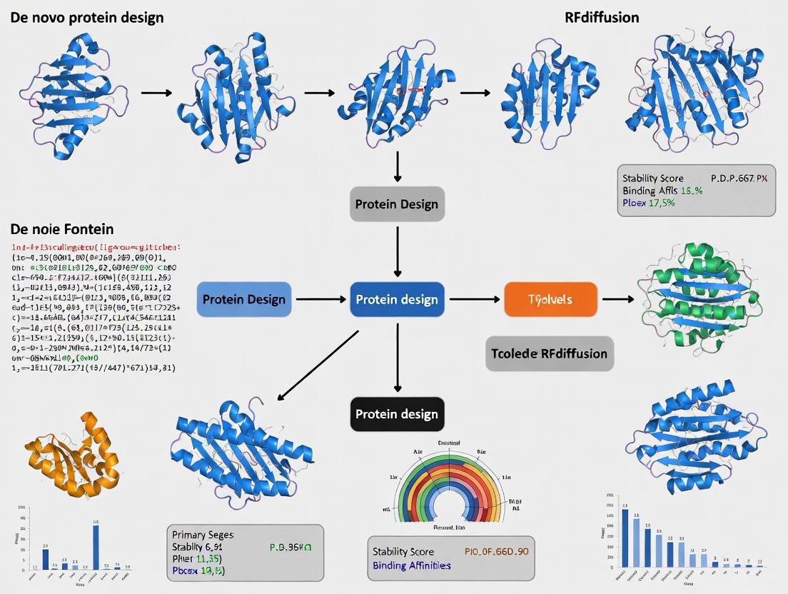
This article provides a comprehensive overview of RFdiffusion, a groundbreaking generative AI model that is transforming the field of de novo protein design.
RFdiffusion: Revolutionizing De Novo Protein Design with Generative AI
Abstract
This article provides a comprehensive overview of RFdiffusion, a groundbreaking generative AI model that is transforming the field of de novo protein design. By fine-tuning the RoseTTAFold structure prediction network for denoising tasks, RFdiffusion enables the computational creation of novel protein structures and functions from simple molecular specifications. We explore the foundational principles of this diffusion-based approach, detail its methodology and diverse applications—from creating protein binders and symmetric assemblies to scaffolding enzyme active sites. The article also addresses practical challenges and optimization strategies for researchers, presents rigorous experimental validation of designed proteins, and compares performance with alternative methods. Aimed at researchers, scientists, and drug development professionals, this review synthesizes current capabilities, acknowledges limitations, and outlines future directions for this rapidly advancing technology in biomedical research and therapeutic development.
The Foundations of RFdiffusion: From Structure Prediction to Generative Design
The field of de novo protein design has been transformed by deep-learning methods, yet a general framework capable of addressing a wide range of design challenges remained elusive. RoseTTAFold, a sophisticated structure prediction network, provided a powerful foundation for understanding protein sequence-structure relationships but was not originally conceived as a generative model [1]. This application note details the conceptual and methodological genesis of adapting RoseTTAFold into RFdiffusion, a generative model for protein design that leverages denoising diffusion probabilistic models (DDPMs) to create novel protein structures and functions from simple molecular specifications [1] [2].
The core innovation lies in repurposing a network that excelled at predicting structure from sequence into one that generates novel, designable protein backbones from noise. By fine-tuning RoseTTAFold on protein structure denoising tasks, researchers obtained a generative model that achieves outstanding performance across diverse challenges, including unconditional protein monomer design, protein binder design, and symmetric oligomer design [1]. The following sections provide a detailed breakdown of the core adaptation framework, its quantitative performance, and practical experimental protocols for implementation and validation.
Core Adaptation Framework
From Structure Prediction to Generative Denoising
The adaptation strategy capitalized on key architectural properties of RoseTTAFold that made it uniquely suited for a diffusion model. Table 1 summarizes the primary modifications required to transition from a structure prediction network to a generative backbone model.
Table 1: Core Adaptations of RoseTTAFold for Generative Modeling
| Component | Function in RoseTTAFold | Adaptation for RFdiffusion | Impact on Design |
|---|---|---|---|
| Primary Input | Protein sequence & optional structural templates [1] | Noised protein backbone coordinates from previous diffusion step [1] | Enables iterative generation from random noise |
| Training Task | Minimize FAPE loss for structure prediction [1] | Minimize mean-squared error (MSE) loss between frame predictions and true structure [1] | Promotes global coordinate frame continuity between denoising steps |
| Network Output | Predicted protein structure from sequence [1] | Denoised structure prediction used to update input for next step [1] | Drives the generative denoising trajectory toward designable backbones |
| Conditioning | Limited to structural templates [1] | Flexible inputs (partial sequence, fixed motifs, fold info) [1] | Enables solution of diverse design challenges from molecular specifications |
The RFdiffusion model represents protein backbones using a frame representation comprising a Cα coordinate and an N-Cα-C rigid orientation for each residue [1]. Training involves corrupting structures from the Protein Data Bank (PDB) with increasing levels of Gaussian noise for up to 200 steps and tasking the network with reversing this process. The use of MSE loss, as opposed to the Frame Aligned Point Error (FAPE) loss used in structure prediction, was found to be crucial for unconditional generation, as it is not invariant to the global reference frame and thus promotes continuity between timesteps [1].
The Denoising Diffusion Workflow
The process of generating a novel protein backbone is a progressive denoising operation. Diagram 1 illustrates the logical flow and data transformation through a single denoising step within the RFdiffusion model.
Diagram 1: A single denoising step in RFdiffusion. The network takes noised residue frames as input, predicts a denoised structure, and updates the frames for the next iteration by moving in the direction of the prediction with controlled noise addition.
The generation process begins with protein backbones initialized as random residue frames. As shown in Diagram 2, the model iteratively refines these random frames over many steps. Early steps prioritize broad structural compatibility, while later steps focus on achieving highly realistic, protein-like geometries [1].
Diagram 2: The high-level generative workflow of RFdiffusion, from random initialization to a final, novel protein backbone.
A critical enhancement to the training and inference process was the implementation of self-conditioning, a strategy inspired by the "recycling" mechanism in AlphaFold2 [1]. This allows the model to condition its predictions on its own outputs from previous denoising steps, which significantly improved performance on both conditional and unconditional protein design tasks by increasing the coherence of predictions within a trajectory [1].
Performance and Validation Data
The performance of RFdiffusion was rigorously benchmarked both computationally and experimentally. A design was considered an in silico success if the AlphaFold2-predicted structure from a single sequence met three criteria: high confidence (mean pAE < 5), global backbone RMSD < 2 Ã…, and motif backbone RMSD < 1 Ã… for any scaffolded functional site [1]. This is a more stringent metric than TM-score-based assessments used in earlier studies.
Table 2: Quantitative Performance Benchmarks of RFdiffusion
| Design Challenge | In Silico Success Rate | Key Experimental Validation | Scale of Experimental Testing |
|---|---|---|---|
| Unconditional Monomer Generation | High structural diversity & confidence (AF2 pLDDT > 90) [1] | High thermostability; CD spectra match designs [1] | 9 designs (200-300 aa) characterized [1] |
| Protein Binder Design | High success rate for complex targets [1] | Cryo-EM structure nearly identical to design model (Influenza hemagglutinin binder) [1] | Hundreds of designed binders tested [1] |
| Symmetric Oligomer Design | High computational accuracy [1] | Structures confirmed by electron microscopy [1] [2] | Hundreds of symmetric assemblies tested [1] |
| Motif Scaffolding | High success on functional site scaffolding [1] | Design of metal-binding proteins & enzyme active sites [1] | Wide range of therapeutic & metal-binding proteins [1] |
The model demonstrated a remarkable ability to generalize beyond the PDB, generating elaborate structures with little overall similarity to known proteins [1]. Its performance was found to significantly outperform existing protein design methods across a broad range of problems, reducing the number of molecules that needed to be tested experimentally to as little as one per design challenge in some cases [2].
Experimental Protocols
Protocol 1: Unconditional Protein Monomer Generation
This protocol describes the procedure for generating novel protein monomers without initial structural constraints.
- Initialization: Initialize a protein chain with the desired length (L) using random residue frames (Cα coordinates and N-Cα-C orientations).
- Denoising Loop: For T timesteps (e.g., 200 steps), perform the following: a. Model Inference: Pass the current noised frames and timestep index to the RFdiffusion network. b. Prediction: The network outputs a prediction of the denoised protein structure. c. Frame Update: Update each residue frame by taking a step towards the network's prediction. Add a controlled amount of noise to generate the input for the next timestep, following the reverse of the diffusion noise schedule.
- Sequence Design: Upon completion of the denoising loop, a protein backbone is generated. Use ProteinMPNN to design sequences that encode this structure. Typically, sample 8 sequences per design [1].
- In Silico Validation: Filter designed sequences using structure prediction networks. a. Prediction: Input each designed sequence into AlphaFold2 or ESMFold to generate a predicted structure. b. Assessment: Calculate the backbone RMSD between the AF2/ESMFold prediction and the original RFdiffusion model. Also check the predicted confidence metrics (pLDDT for AF2; pAE for interface accuracy). A successful design should have a global backbone RMSD < 2 Ã… and high confidence (e.g., mean pAE < 5) [1].
Protocol 2: Functional Motif Scaffolding
This protocol details the process of scaffolding a fixed functional motif (e.g., an enzyme active site or a protein-binding peptide) within a novel protein structure.
- Motif Specification: Define the functional motif by specifying the sequence and 3D coordinates of the motif residues. These residues will be held fixed throughout the diffusion process.
- Conditional Generation: Initialize a full-length protein chain with random frames. During the denoising process, provide the fixed motif coordinates as conditioning information to the RFdiffusion network at each step.
- Progressive Scaffolding: The network learns to build a structured scaffold around the fixed motif, ensuring the overall fold is compatible and the motif is presented in its native, functional conformation.
- Sequence Design and Validation: Use ProteinMPNN for sequence design, with the motif sequence fixed. During in silico validation, ensure that the functional motif in the AF2-predicted structure is nearly identical to the original design (motif backbone RMSD < 1 Ã…), in addition to the global RMSD and confidence filters [1]. This protocol has been successfully applied to scaffold therapeutic and metal-binding motifs [1] and short peptides targeting specific proteins like Keap1 [3].
The Scientist's Toolkit: Essential Research Reagents
The following table lists key computational and experimental tools essential for conducting research with RFdiffusion.
Table 3: Key Research Reagent Solutions for RFdiffusion-Based Design
| Reagent / Tool | Function | Application in RFdiffusion Workflow |
|---|---|---|
| RFdiffusion Software | Generative backbone model | Core engine for generating novel protein structures from noise or molecular specifications [1]. |
| RoseTTAFold Network | Structure prediction network | Provides the underlying architecture and pre-trained understanding of protein physics for the diffusion model [1]. |
| ProteinMPNN | Protein sequence design network | Designs sequences that fold into the protein backbones generated by RFdiffusion [1]. |
| AlphaFold2 / ESMFold | Protein structure prediction | In silico validation of designs; assesses whether designed sequences fold into the intended structures [1]. |
| Cryo-Electron Microscopy | High-resolution structure determination | Experimental validation of complex designs, such as symmetric assemblies and protein binders, at near-atomic resolution [1] [2]. |
| Circular Dichroism (CD) Spectroscopy | Experimental analysis of secondary structure and stability | Confirms that expressed proteins have the designed secondary structure and assesses thermostability [1]. |
| Size-Exclusion Chromatography (SEC) | Biophysical characterization | Assesses the solubility and monomeric state of expressed protein designs [4]. |
| Methyl 3-hydroxyheptadecanoate | Methyl 3-hydroxyheptadecanoate, MF:C18H36O3, MW:300.5 g/mol | Chemical Reagent |
| Heparin disaccharide I-A sodium | Heparin disaccharide I-A sodium, CAS:136098-00-5, MF:C14H18NNa3O17S2, MW:605.4 g/mol | Chemical Reagent |
The adaptation of RoseTTAFold into RFdiffusion represents a paradigm shift in computational protein design. By fine-tuning a state-of-the-art structure prediction network for generative modeling within a diffusion framework, researchers have created a versatile and powerful tool that solves a wide array of design challenges. The detailed protocols and performance data outlined in this application note provide a foundation for researchers to apply and further develop these methods. The experimental success across hundreds of designs—from high-affinity binders to complex symmetric assemblies—confirms that RFdiffusion enables the design of diverse functional proteins from simple molecular specifications, opening new frontiers in therapeutic, vaccine, and nanotechnology development [1] [2].
Diffusion models have emerged as a powerful class of generative artificial intelligence that are revolutionizing de novo protein design. These models draw inspiration from statistical physics, simulating a process where noise is progressively added to data (forward diffusion) and then learning to reverse this process to generate new, structured data from noise (reverse diffusion). In computational biology, this framework has been successfully adapted to create novel protein backbone structures, enabling researchers to design proteins with specific functions and binding capabilities from scratch. The core architectural principle involves training neural networks to denoise protein representations, gradually transforming random initial states into biologically plausible and stable protein folds.
The integration of diffusion models, particularly RFdiffusion, into the de novo protein design pipeline represents a paradigm shift in computational biology. RFdiffusion uses the AlphaFold2 and RoseTTAFold2 frame representation of protein backbones comprising the Cα coordinate and N-Cα-C rigid orientation for each residue [5]. During training, a noising schedule corrupts the protein frames over multiple timesteps toward random prior distributions, with Cα coordinates corrupted with three-dimensional Gaussian noise and residue orientations with Brownian motion. The model then learns to predict the de-noised structure at each timestep, minimizing the mean squared error between the true structure and the prediction [5]. This fundamental architecture has enabled unprecedented capabilities in generating functional protein binders and antibodies with atomic-level accuracy.
Core Architectural Frameworks for Protein Backbone Generation
Coordinate-Based Diffusion (RFdiffusion)
RFdiffusion implements a coordinate-based diffusion approach that operates directly on the three-dimensional structural representation of proteins. The model represents protein backbones using frames consisting of Cα atomic coordinates and local orientational frames defined by the N-Cα-C vectors for each residue [5]. The diffusion process adds noise to both the positional (coordinates) and orientational (frames) components of the structure, with the model learning to reverse this noising process to generate novel protein structures from random noise.
The architectural implementation involves several key components. At inference time, sampling begins from a random residue distribution (XT), and RFdiffusion iteratively de-noises this initial state through a series of steps to generate novel protein structures [5]. For conditional generation tasks such as designing antibodies against specific epitopes, the framework can be fine-tuned with specialized conditioning mechanisms. The framework structure is provided as conditioning input using the template track of RFdiffusion, which represents the framework as a two-dimensional matrix of pairwise distances and dihedral angles between residue pairs [5]. This representation allows three-dimensional structures to be accurately recapitulated while maintaining global-frame invariance.
Angle-Based Diffusion (FoldingDiff)
In contrast to coordinate-based approaches, FoldingDiff employs an angle-based representation that captures protein geometry through internal coordinates rather than Cartesian coordinates. This framework represents protein backbones as a sequence of angle sets comprising three bond angles and three dihedral angles for each residue, effectively describing the relative orientation of all backbone atoms from one residue to the next [6]. This representation inherently embeds translation and rotation invariance, eliminating the need for complex equivariant neural networks.
The FoldingDiff architecture implements a denoising diffusion probabilistic model (DDPM) with a bidirectional transformer backbone that operates directly on the angular representation [6]. During generation, the model starts from random angles corresponding to an unfolded state and iteratively denoises these angles to arrive at a final folded backbone structure. A critical implementation detail involves handling the periodicity of angular values, requiring specialized noising and denoising procedures that wrap values about the domain [-π, π) [6]. This approach mirrors natural protein folding principles, where proteins twist into energetically favorable conformations through angular rotations.
Distilled and Fractional Diffusion Variants
Recent advancements have focused on optimizing the diffusion process for improved computational efficiency and modeling capability. Distilled Protein Backbone Generation explores score distillation techniques to dramatically reduce the number of sampling steps required during inference. By adapting Score identity Distillation (SiD), researchers have demonstrated that few-step generators can achieve more than a 20-fold improvement in sampling speed while maintaining comparable performance to original teacher models [7]. The key to success in this approach lies in combining multistep generation with inference-time noise modulation.
ProT-GFDM introduces fractional stochastic dynamics to protein generation, replacing standard Brownian motion with fractional processes exhibiting superdiffusive properties [8]. This architectural innovation enhances the model's ability to capture long-range dependencies in protein structures, resulting in measurable improvements across multiple quality metrics including a 7.19% increase in density, 5.66% improvement in coverage, and 1.01% reduction in Fréchet inception distance compared to conventional score-based models [8].
Table 1: Comparison of Protein Backbone Diffusion Architectures
| Architecture | Representation | Key Innovation | Performance Advantages |
|---|---|---|---|
| RFdiffusion [5] | Cartesian coordinates (Cα frames) | Fine-tuning for specific applications (e.g., antibodies) | Atomic-level accuracy in designed structures; experimentally validated binders |
| FoldingDiff [6] | Internal angles (6 per residue) | Rotation and translation invariance by design | Realistic angle distributions; rich secondary structure motifs |
| Distilled Diffusion [7] | Cartesian coordinates | Score identity Distillation (SiD) | 20x faster sampling; maintained designability and diversity |
| ProT-GFDM [8] | Cartesian coordinates | Fractional stochastic processes | Better long-range dependency capture; improved density and coverage |
Quantitative Analysis of Model Performance
The evaluation of diffusion models for protein backbone generation employs multiple quantitative metrics to assess the quality, diversity, and biological relevance of generated structures. Designability—the probability that a generated backbone can be realized with a stable amino acid sequence—serves as a crucial benchmark for practical utility. State-of-the-art models now achieve designability rates comparable to natural proteins, with RFdiffusion-based pipelines successfully generating functional antibodies that bind to specific epitopes with nanomolar affinity [5].
Diversity and novelty metrics ensure that models generate structurally varied proteins rather than simply memorizing training examples. Analyses confirm that designed antibodies make diverse interactions with target epitopes and differ significantly from sequences in the training dataset, with no correlation between training dataset similarity and binding success [5]. Self-consistency metrics, which measure the similarity between a designed structure and the AlphaFold2-predicted structure for its designed sequence, provide another important validation signal, though specialized versions of RoseTTAFold2 fine-tuned on antibody structures are often required for accurate assessment of antibody-antigen complexes [5].
Table 2: Key Performance Metrics for Protein Backbone Diffusion Models
| Metric Category | Specific Metrics | Typical Values for State-of-the-Art | Measurement Method |
|---|---|---|---|
| Structural Quality | RMSD to native-like structures, secondary structure element composition | Similar to natural protein distributions [6] | Structural alignment, DSSP |
| Designability | Successful sequence design rate, experimental validation rate | Generation of binders with nanomolar affinity [5] | ProteinMPNN sequence design, experimental binding assays |
| Efficiency | Sampling steps, inference time | 20x speedup with distillation [7] | Computational benchmarking |
| Diversity | Structural diversity, novelty compared to training set | Significant differences from training sequences [5] | Structural clustering, sequence similarity analysis |
Experimental Protocols for Diffusion-Based Protein Design
Protocol 1: De Novo Antibody Design Using RFdiffusion
The design of de novo antibodies against specific epitopes represents one of the most significant applications of diffusion models in protein design. The following protocol outlines the key steps for generating epitope-specific antibodies using fine-tuned RFdiffusion:
Step 1: Framework Selection and Preparation
- Select an appropriate antibody framework based on the desired properties (e.g., VHH for single-domain antibodies, scFv for single-chain variable fragments)
- For VHH designs, commonly use humanized frameworks such as h-NbBcII10FGLA [5]
- Prepare the framework structure and sequence to be provided as conditioning input to RFdiffusion
Step 2: Epitope Specification and Hotspot Residue Definition
- Define the target epitope on the antigen surface
- Identify key hotspot residues that should participate in binding interactions
- Encode these hotspot residues as a one-hot feature to guide the diffusion process toward the specified epitope [5]
Step 3: Conditional Generation with RFdiffusion
- Run the fine-tuned RFdiffusion model with the framework and epitope conditioning
- The model simultaneously designs CDR loop conformations and the rigid-body placement of the antibody relative to the target [5]
- Generate thousands of candidate structures to ensure adequate sampling of the structural space
Step 4: Sequence Design with ProteinMPNN
- For each generated backbone structure, use ProteinMPNN to design complementary amino acid sequences for the CDR loops [5]
- Keep the framework sequence fixed to maintain structural stability
Step 5: In Silico Filtering with Fine-Tuned RoseTTAFold2
- Use a specialized version of RoseTTAFold2 fine-tuned on antibody structures to repredict the structure of designed antibody-antigen complexes [5]
- Filter designs based on self-consistency between the RFdiffusion design and the RoseTTAFold2 prediction
- Assess interface quality using computational metrics such as Rosetta ddG
Step 6: Experimental Validation
- Express filtered designs using high-throughput methods (yeast surface display or E. coli expression)
- Screen for binding using surface plasmon resonance (SPR) or similar techniques
- For promising binders, determine high-resolution structures using cryo-electron microscopy to verify atomic-level accuracy [5]
Protocol 2: Unconditional Backbone Generation with FoldingDiff
For unconditional generation of novel protein backbones without specific binding targets, FoldingDiff provides an angle-based approach:
Step 1: Data Preparation and Preprocessing
- Curate a dataset of protein domains (e.g., from CATH database) with lengths between 40-128 residues [6]
- Convert protein structures from Cartesian coordinates to internal angle representation
- Compute the six angles (three bond angles, three dihedral angles) for each residue
Step 2: Model Training and Configuration
- Implement a denoising diffusion probabilistic model with bidirectional transformer architecture
- Configure periodic noising and denoising procedures appropriate for angular values
- Train the model to predict the noise added at each diffusion step
Step 3: Generation and Reconstruction
- Start from random angles corresponding to an unfolded state
- Apply iterative denoising for T steps (typically 1000) to generate novel angle sets
- Convert the generated angles back to 3D Cartesian coordinates using iterative reconstruction
- Apply structural relaxation to resolve potential atomic clashes
Step 4: Quality Assessment and Filtering
- Evaluate generated structures for realistic angle distributions compared to natural proteins
- Assess structural plausibility using metrics such as Ramachandran plot preferences
- Filter designs based on structural novelty and foldability metrics
Visualization of Key Workflows
Diagram 1: FoldingDiff Angle-Based Generation
Diagram 2: RFdiffusion Antibody Design Pipeline
Research Reagent Solutions
Table 3: Essential Research Reagents and Computational Tools
| Reagent/Tool | Type | Function in Protocol | Implementation Notes |
|---|---|---|---|
| RFdiffusion [5] | Software | Conditional generation of protein structures | Fine-tuned on antibody complexes for epitope-specific design |
| FoldingDiff [6] | Software | Angle-based backbone generation | Uses transformer architecture; rotation-invariant by design |
| ProteinMPNN [5] | Software | Sequence design for generated backbones | Designs amino acid sequences compatible with backbone structures |
| RoseTTAFold2 [5] | Software | Structure prediction and design validation | Fine-tuned version for antibody complex prediction |
| Yeast Surface Display [5] | Experimental platform | High-throughput screening of designed binders | Enables screening of thousands of designs in parallel |
| OrthoRep [5] | Experimental system | In vivo affinity maturation | Enables evolution of binders to single-digit nanomolar affinity |
The integration of self-conditioning and specialized fine-tuning strategies has been pivotal in transforming RFdiffusion from a general protein structure prediction network into a powerful generative model for de novo protein design. These training innovations enable the solution of diverse design challenges—from unconditional protein monomer generation to the atomically accurate design of antibodies—by providing greater control over the generative process and enhancing the quality and reliability of designed proteins [1]. By building upon the deep understanding of protein structure embedded in pre-trained RoseTTAFold (RF) weights, these methods allow RFdiffusion to generate functional proteins that explore regions of the protein universe beyond natural evolutionary constraints [1] [9].
Core Technical Principles of RFdiffusion Training
Foundation in RoseTTAFold Architecture
RFdiffusion is constructed using the RoseTTAFold frame representation, which comprises a Cα coordinate and N-Cα-C rigid orientation for each residue [1]. This representation provides a mathematically robust framework for applying noise and learning denoising operations. The model operates through a denoising diffusion probabilistic model (DDPM) framework, where training involves corrupting protein structures from the Protein Data Bank with increasing levels of noise and training the network to reverse this process [1]. During this training, RFdiffusion learns to generate realistic protein backbones by minimizing a mean-squared error (m.s.e.) loss between frame predictions and the true protein structure without alignment, which promotes continuity of the global coordinate frame between timesteps [1].
Training Infrastructure and Noise Scheduling
The training process utilizes a carefully designed noising schedule that corrupts structures over up to 200 steps [1]. For translations, Cα coordinates are perturbed with 3D Gaussian noise, while residue orientations are corrupted using Brownian motion on the manifold of rotation matrices [1]. This mathematical formulation ensures proper diffusion of both positional and orientational components of the protein structure representation. The model is trained to predict the denoised structure (pX0) at each timestep, with the loss function driving denoising trajectories to match the data distribution at each step, ultimately converging on designable protein backbones [1].
Table 1: Key Components of RFdiffusion Training Infrastructure
| Component | Implementation in RFdiffusion | Purpose |
|---|---|---|
| Base Architecture | Fine-tuned RoseTTAFold structure prediction network | Leverages pre-existing understanding of protein structure |
| Representation | Cα coordinates + N-Cα-C rigid orientations | Mathematically robust framework for noise operations |
| Noise Type (Position) | 3D Gaussian noise on Cα coordinates | Corrupts positional information |
| Noise Type (Orientation) | Brownian motion on SO(3) manifold | Corrupts orientational information |
| Training Loss | Mean-squared error (m.s.e.) without alignment | Maintains global coordinate frame continuity |
| Noising Steps | Up to 200 steps | Progressive corruption of structure |
Self-Conditioning: Enhancing Generative Coherence
Implementation of Self-Conditioning
Self-conditioning represents a significant innovation in the training of RFdiffusion, drawing inspiration from the "recycling" mechanism in AlphaFold2 [1]. In this approach, the model conditions its predictions on outputs from previous timesteps during the denoising trajectory, rather than treating each prediction as independent. This creates a more coherent generative process where structural decisions are informed by prior steps, leading to improved overall quality and designability [1]. The self-conditioning mechanism is implemented by providing the model with its previous predictions as additional inputs during training, establishing a memory mechanism across denoising steps.
Performance Impact of Self-Conditioning
The adoption of self-conditioning has demonstrated substantial improvements in RFdiffusion's performance across multiple design challenges. When evaluated on in silico benchmarks encompassing both conditional and unconditional protein design tasks, the self-conditioning strategy consistently outperformed the canonical approach of making independent predictions at each timestep [1]. This performance improvement is attributed to increased coherence of predictions within self-conditioned trajectories, where structural elements develop more consistently throughout the denoising process [1]. The enhanced coherence translates to higher success rates as measured by computational validation metrics, including AlphaFold2 self-consistency with design models.
Diagram 1: Self-conditioning mechanism in RFdiffusion (Title: Self-Conditioning in RFdiffusion)
Fine-Tuning Strategies for Specialized Applications
Foundational Fine-Tuning from RoseTTAFold Weights
The initial development of RFdiffusion demonstrated that fine-tuning from pre-trained RoseTTAFold weights was dramatically more successful than training from untrained weights for an equivalent duration [1]. This approach leverages the substantial knowledge of protein structure relationships already embedded in RF, repurposing this understanding for generative rather than predictive tasks. The performance advantage of fine-tuning from pre-trained weights was evident across multiple design challenges, establishing this as a foundational principle for developing specialized protein design networks [1].
Task-Specific Fine-Tuning for Antibody Design
A particularly advanced application of fine-tuning is demonstrated in the development of RFdiffusion for de novo antibody design [5] [10]. This specialized variant involves fine-tuning predominantly on antibody complex structures, with modifications to the conditioning approach to maintain antibody framework integrity while designing novel complementarity-determining regions (CDRs). During training, the antibody structure is corrupted while the framework sequence and structure are provided as conditioning input in a global-frame-invariant manner using the template track of RFdiffusion [5]. This approach enables the model to sample alternative rigid-body placements of the antibody relative to the target epitope while preserving the essential immunoglobulin fold.
Epitope-Targeted Fine-Tuning with Hotspot Conditioning
The antibody-design variant of RFdiffusion incorporates an adapted "hotspot" feature that specifies target residues with which CDR loops should interact [5] [10]. This enables precise targeting of specific epitopes while generating diverse CDR-mediated interactions. The fine-tuning process maintains the underlying thermodynamics of interface formation but specializes the network for the distinct structural constraints of antibody-antigen recognition. This approach has enabled the first fully de novo computational design of antibodies targeting user-specified epitopes with atomic-level precision [5].
Table 2: Comparison of Fine-Tuning Strategies for RFdiffusion
| Fine-Tuning Type | Training Data | Conditioning Information | Key Applications | Performance Outcomes |
|---|---|---|---|---|
| Foundation Model | General PDB structures | Secondary structure, functional motifs | Unconditional monomer generation, symmetric oligomers | Far superior to training from scratch [1] |
| Antibody Design | Antibody complex structures | Framework structure, epitope hotspots | VHHs, scFvs, full antibodies targeting specific epitopes | Atomic-level accuracy in CDR loops [5] |
| Binder Design | Protein-protein interfaces | Target structure, interface residues | Protein binders to therapeutic targets | High success rate in experimental validation [1] |
Experimental Protocols for Training RFdiffusion Variants
Protocol: Implementing Self-Conditioning in RFdiffusion Training
Objective: Integrate self-conditioning into RFdiffusion training to improve generative coherence and design success rates.
Materials:
- Pre-trained RoseTTAFold network weights
- Curated Protein Data Bank structures for training
- High-performance computing infrastructure with multiple GPUs
Procedure:
- Initialize Network: Load pre-trained RoseTTAFold weights as starting point for RFdiffusion
- Modify Architecture: Adapt network to incorporate connections for self-conditioning inputs
- Training Loop:
- Sample protein structure (X0) from PDB and random timestep (t)
- Apply t noising steps to generate corrupted structure (Xt)
- For self-conditioning: store previous prediction (pX0{t+1}) from earlier training step
- Provide Xt and optional conditioning information (including previous prediction for self-conditioning) to network
- Compute MSE loss between network prediction (pX0t) and true structure (X0)
- Update network parameters via backpropagation
- Validation: Periodically evaluate on held-out validation set using in silico metrics (AF2 self-consistency, pLDDT, pAE)
Troubleshooting:
- If training instability occurs, adjust learning rate or gradient clipping
- If overfitting observed, increase diversity of training set or implement additional regularization
Protocol: Fine-Tuning RFdiffusion for Antibody Design
Objective: Create specialized RFdiffusion variant for de novo antibody design targeting specific epitopes.
Materials:
- Pre-trained RFdiffusion weights
- Curated antibody-antigen complex structures from PDB
- Target framework sequences for therapeutic antibodies
Procedure:
- Data Curation:
- Collect high-resolution antibody-antigen complex structures
- Annotate framework regions and CDR loops
- Identify epitope residues for hotspot conditioning
- Training Setup:
- Initialize with pre-trained RFdiffusion weights
- Configure template track to provide framework structure as pairwise distances and dihedral angles
- Implement hotspot conditioning for epitope specification
- Specialized Training:
- At each step: sample antibody complex, corrupt antibody structure while preserving target
- Provide framework structure via template track (global-frame-invariant)
- Specify epitope residues via hotspot conditioning
- Train network to recover original antibody structure
- Validation: Use fine-tuned RF2 for antibody structure prediction to assess design quality
Quality Control:
- Verify framework preservation in generated structures
- Assess epitope targeting accuracy through in silico docking
- Experimental validation via yeast display and cryo-EM structure determination
Diagram 2: Fine-tuning workflow for antibody design (Title: Fine-tuning for Antibody Design)
Performance Validation and Metrics
Quantitative Assessment of Training Innovations
The impact of self-conditioning and fine-tuning strategies has been quantitatively evaluated through rigorous in silico benchmarking and experimental validation. For self-conditioning, performance improvements were measured across multiple design challenges, with enhanced performance particularly evident in complex conditional design tasks [1]. Success is typically defined using a stringent computational validation pipeline where designs are considered successful only if the AlphaFold2-predicted structure from the designed sequence shows high confidence (mean pAE < 5), global backbone RMSD < 2 Ã… to the design model, and <1 Ã… RMSD on any scaffolded functional sites [1].
For the antibody-design variant, validation includes both computational and experimental methods. The fine-tuned RFdiffusion successfully generated antibody structures that closely matched input framework structures while targeting specified epitopes with novel CDR loops [5]. Experimental characterization through cryo-EM confirmed atomic-level accuracy in designed CDR conformations, with high-resolution structures nearly identical to design models [5]. Although initial computational designs typically exhibited modest affinity (tens to hundreds of nanomolar Kd), affinity maturation enabled production of single-digit nanomolar binders that maintained intended epitope selectivity [5].
Research Reagent Solutions
Table 3: Essential Research Reagents and Computational Tools
| Reagent/Tool | Function/Purpose | Implementation in RFdiffusion |
|---|---|---|
| RoseTTAFold Pre-trained Weights | Foundation for fine-tuning RFdiffusion | Provides initial parameters; knowledge transfer from structure prediction [1] |
| Protein Data Bank (PDB) Structures | Training data for foundational and specialized fine-tuning | Source of native protein structures for training [1] [11] |
| Antibody Complex Structures | Specialized training data for antibody design | Enables fine-tuning for CDR loop and epitope targeting [5] |
| Template Track (RFdiffusion) | Provides structural constraints during generation | Encodes framework structure as pairwise distances/angles [5] |
| Hotspot Conditioning | Guides generation toward specific interactions | Specifies epitope residues for antibody-target interactions [5] |
| ProteinMPNN | Sequence design for generated backbones | Designs sequences encoding the diffused structures [1] |
| Fine-tuned RoseTTAFold2 | Antibody structure prediction for validation | Filters designs by predicting antibody-antigen complexes [5] |
For decades, the field of protein modeling and design has been constrained by fundamental limitations that restricted our ability to explore the vast functional protein universe. Conventional protein engineering methods, such as directed evolution, remained tethered to existing biological templates, performing local searches within the immense landscape of possible protein sequences and structures [9]. This approach confined discovery to incremental improvements and failed to access genuinely novel functional regions beyond natural evolutionary pathways. Physics-based computational design methods, exemplified by tools like Rosetta, demonstrated early success by operating on Anfinsen's hypothesis that proteins fold into their lowest-energy state [9]. These methods employed fragment assembly and force-field energy minimization to design novel proteins like Top7, a 93-residue protein with a fold not observed in nature [9]. However, these approaches faced two critical constraints: their underlying force fields remained approximate, often resulting in designs that misfolded or failed to function in vitro, and the computational expense was prohibitive for exhaustive sampling of sequence-structure space [9].
The core challenge stems from the astronomical scale of the protein functional universe. For a mere 100-residue protein, there are approximately 10^130 possible amino acid arrangements—exceeding the number of atoms in the observable universe [9]. Within this space, naturally occurring proteins represent an infinitesimally small subset, biased by evolutionary history and assayability [9]. This article examines how the integration of artificial intelligence, specifically RFdiffusion and related technologies, has overcome these historical barriers, enabling the precise de novo design of functional proteins with transformative applications across biotechnology and medicine.
The AI-Driven Paradigm Shift: From Prediction to Generation
The breakthrough began with advancements in protein structure prediction. Deep learning networks like AlphaFold2 and RoseTTAFold solved the long-standing problem of predicting a protein's three-dimensional structure from its amino acid sequence with near-experimental accuracy [12]. These models demonstrated a deep understanding of protein structure, but were inherently analytical rather than generative.
The critical transition from predictive analysis to generative design came with the development of RFdiffusion by the Baker Lab [1] [2]. This approach adapted denoising diffusion probabilistic models (DDPMs)—previously successful in image generation—to protein design. RFdiffusion works by fine-tuning a RoseTTAFold structure prediction network on protein structure denoising tasks [1]. The model learns to iteratively "denoise" a random cloud of atom coordinates into a coherent protein backbone through many steps of refinement [1]. Starting from pure noise, RFdiffusion generates elaborate protein structures with little overall similarity to training data, indicating substantial generalization beyond known Protein Data Bank structures [1]. Following backbone generation, the ProteinMPNN network designs sequences encoding these structures [1].
Table 1: Evolution of RFdiffusion Capabilities
| Model Version | Key Innovation | Design Scope | Experimental Validation |
|---|---|---|---|
| RFdiffusion (2023) | Protein backbone generation via diffusion | Protein monomers, binders, symmetric assemblies | Hundreds of tested designs: binders, assemblies, enzymes [1] |
| RFdiffusion All-Atom | Inclusion of small molecule context | Protein-ligand complexes | Design of proteins binding specific ligands [12] |
| Fine-tuned RFdiffusion (2025) | Specialized for antibody loop design | Antibody variable chains (VHHs, scFvs) | Designed binders to influenza HA, C. difficile TcdB [5] [13] |
| RFdiffusion2 (2025) | Enzyme design from chemical transformations | Enzymes with custom active sites | Active enzymes for 5 distinct reactions [14] |
| RFdiffusion3 (2025) | All-atom co-diffusion of biomolecular complexes | Protein complexes with ligands, DNA, RNA | DNA-binding proteins, cysteine hydrolases [12] |
This foundational technology established a new paradigm for protein design, overcoming previous limitations through several key innovations:
- Global exploration: Unlike previous methods confined to local optimization, RFdiffusion can generate entirely novel folds and topologies not observed in nature [1]
- Conditional generation: The model accepts conditioning information including partial sequences, fold specifications, or fixed functional motifs to guide design toward specific objectives [1]
- Computational efficiency: The approach significantly reduces experimental screening burden, with successful designs often identified with as few as one test per design challenge [2]
Breakthrough Application: De Novo Design of Antibodies
A landmark demonstration of RFdiffusion's capabilities came with the de novo design of antibodies targeting specific epitopes—a problem previously considered intractable for computational methods [5]. Despite antibodies being the dominant class of protein therapeutics, with over 160 licensed globally and a market value expected to reach $445 billion, no method previously existed to design epitope-specific antibodies entirely in silico [5].
Methodology: Fine-Tuning RFdiffusion for Antibody Design
The research team fine-tuned RFdiffusion specifically on antibody complex structures to enable design of novel complementarity-determining regions (CDRs) while maintaining the framework region of therapeutic antibodies [5]. The key methodological innovations included:
- Framework conditioning: The antibody framework structure and sequence were provided as conditioning input using the template track of RFdiffusion, represented as a 2D matrix of pairwise distances and dihedral angles [5]
- Epitope specification: A one-hot encoded "hotspot" feature directed antibody CDRs toward user-specified epitopes with atomic-level precision [5]
- Rigid-body sampling: The model designed both CDR loop conformations and the overall orientation of the antibody relative to the target [5]
Following RFdiffusion generation, ProteinMPNN designed sequences for the CDR loops, and a fine-tuned RoseTTAFold2 network filtered designs by re-predicting antibody-antigen complex structures [5]. This filtering step enriched for experimentally successful binders by assessing structural self-consistency—a metric previously unavailable for antibodies due to AlphaFold2's poor performance on antibody-antigen complexes [5].
Diagram 1: RFdiffusion antibody design workflow. The process begins with target epitope specification and proceeds through conditional generation, sequence design, computational filtering, and experimental validation with affinity maturation.
Experimental Protocol and Validation
The experimental validation followed a rigorous protocol across multiple disease-relevant targets, including C. difficile toxin B (TcdB), influenza haemagglutinin, respiratory syncytial virus (RSV), SARS-CoV-2 receptor-binding domain, and IL-7Rα [5]. The specific methodology included:
Design Generation: RFdiffusion generated antibody variable heavy chains (VHHs) and single-chain variable fragments (scFvs) using a humanized VHH framework (h-NbBcII10FGLA) as the structural basis [5]
High-Throughput Screening: Computationally filtered designs were screened using yeast surface display (assessing ~9,000 designs per target for RSV sites I/III, RBD, and influenza haemagglutinin) [5]
Binding Affinity Assessment: Lower-throughput screening employed E. coli expression and single-concentration surface plasmon resonance (SPR) for 95 designs per target (TcdB, IL-7Rα, and influenza haemagglutinin) [5]
Structural Validation: Cryo-electron microscopy determined binding poses for designed VHHs targeting influenza haemagglutinin and TcdB [5]
Affinity Maturation: The OrthoRep system for in vivo continuous evolution further improved binding affinities of initial designs [5]
Table 2: Experimental Results of De Novo Designed Antibodies
| Target | Design Format | Initial Affinity | After Maturation | Structural Validation |
|---|---|---|---|---|
| Influenza Haemagglutinin | VHH | Tens to hundreds of nM Kd | Single-digit nM Kd | Cryo-EM: nearly identical to design [5] |
| C. difficile TcdB | VHH, scFv | Tens to hundreds of nM Kd | Single-digit nM Kd | Cryo-EM: atomic accuracy of CDRs [5] |
| RSV Sites I/III | VHH | Tens to hundreds of nM Kd | Single-digit nM Kd | High-resolution structure confirmation [5] |
| SARS-CoV-2 RBD | VHH | Tens to hundreds of nM Kd | Single-digit nM Kd | Epitope selectivity maintained [5] |
| PHOX2B peptide–MHC | scFv | Tens to hundreds of nM Kd | Single-digit nM Kd | Combination of heavy/light chains [5] |
The experimental results demonstrated that initial computational designs exhibited modest affinity (tens to hundreds of nanomolar Kd), but affinity maturation enabled production of single-digit nanomolar binders that maintained the intended epitope selectivity [5]. Critically, high-resolution structures confirmed atomic accuracy of the designed complementarity-determining regions, with cryo-EM data for one design verifying the atomically precise conformation of all six CDR loops in a single-chain variable fragment [5].
The Scientist's Toolkit: Essential Research Reagents and Workflows
Implementing RFdiffusion-based protein design requires specific computational and experimental resources. The following table details key research reagent solutions essential for successful design campaigns.
Table 3: Essential Research Reagents and Tools for RFdiffusion Design
| Reagent/Tool | Type | Function | Application Example |
|---|---|---|---|
| RFdiffusion (fine-tuned) | Computational Model | Generates antibody structures with novel CDRs | Designing VHHs and scFvs to target epitopes [5] [13] |
| ProteinMPNN | Computational Tool | Designs sequences for generated structures | Sequence design for RFdiffusion-generated backbones [1] |
| RoseTTAFold2 (fine-tuned) | Computational Model | Filters designs by structure prediction | Assessing design self-consistency and binding confidence [5] |
| Yeast Surface Display | Experimental Platform | High-throughput screening of design libraries | Screening ~9,000 designs per target [5] |
| OrthoRep | Experimental System | In vivo continuous evolution for affinity maturation | Improving initial designs to single-digit nM binders [5] |
| Cryo-EM | Structural Biology | High-resolution structure determination | Validating binding poses and CDR conformations [5] |
| ZH8651 | ZH8651, CAS:73918-56-6, MF:C8H10BrN, MW:200.08 g/mol | Chemical Reagent | Bench Chemicals |
| 2,3-Dihydroxyisovaleric acid | 2,3-Dihydroxy-3-methylbutanoic Acid|Research Chemical | 2,3-Dihydroxy-3-methylbutanoic acid is a key intermediate in branched-chain amino acid biosynthesis. This product is for research use only (RUO). Not for human or veterinary use. | Bench Chemicals |
Advanced Architectures: From RFdiffusion to RFdiffusion3
The RFdiffusion ecosystem has rapidly evolved to address increasingly complex design challenges. RFdiffusion2 introduced specialized capabilities for enzyme design, generating protein backbones with custom active sites from simple descriptions of chemical transformations [14]. This model demonstrated remarkable experimental success, producing active enzymes for five distinct chemical reactions with fewer than 100 designs tested per case—a significant departure from traditional workflows requiring thousands of molecules [14].
The most advanced iteration, RFdiffusion3, represents a quantum leap through its unified all-atom framework [12]. This model operates directly at the atomic level, simultaneously generating protein backbones, sidechains, and complex interactions with ligands, DNA, and other non-protein molecules [12]. Key innovations include:
- All-atom co-diffusion: Concurrently generates proteins and their binding partners from random noise, enabling dynamic mutual adaptation [12]
- Atomic-level conditioning: Imposes precise constraints including hydrogen bonds, solvent accessibility, and symmetry operations [12]
- Computational efficiency: Lightweight transformer-U-Net architecture with sparse attention enables 10-fold speed increase [12]
Experimental validation of RFdiffusion3 included designing a DNA-binding protein for a randomly generated DNA sequence, with one of five tested designs binding with low-micromolar affinity, and engineering a cysteine hydrolase with the best performer achieving kcat/Km of 3557 Mâ»Â¹sâ»Â¹ [12].
Diagram 2: Evolution of RFdiffusion capabilities from protein backbones to all-atom biomolecular design, showing expanding application scope with each iteration.
The development of RFdiffusion and its subsequent specialized versions represents a paradigm shift in protein modeling, effectively overcoming the historical limitations that constrained previous approaches. By transitioning from residue-level approximation to atomic-level precision, these models have closed the resolution gap between computational design and biological function [12]. The experimental success in designing antibodies, enzymes, and DNA-binding proteins purely through computation demonstrates that the field has entered an era where the primary limitation is no longer the design tools themselves, but the creativity and biological insight of researchers applying them [12] [9].
Future advancements will likely focus on integrating these design capabilities with high-throughput experimental validation platforms, creating a continuous feedback loop that further refines computational models. Additional challenges remain, including incorporating post-translational modifications and glycosylation, and improving the prediction and design of conformational dynamics [12]. Nevertheless, RFdiffusion has unequivocally transformed protein engineering from a template-dependent process to a rational design discipline, fundamentally expanding our ability to explore the vast untapped potential of the protein functional universe for therapeutic, catalytic, and synthetic biology applications.
RFdiffusion in Practice: Methodology and Diverse Applications
The de novo design of proteins represents a paradigm shift in biotechnology, moving from predicting natural proteins to creating entirely new proteins with customized structures and functions. RFdiffusion, a deep-learning framework based on a diffusion model, has emerged as a powerful tool for this purpose, enabling researchers to generate diverse protein structures that can be experimentally validated for specific applications, including therapeutic development, enzyme design, and antibody engineering [1] [15]. This application note provides a detailed overview of the RFdiffusion workflow, from its fundamental noise-to-structure generation process to the experimental protocols required for functional validation. The methodology is framed within the broader context of expanding the explorable protein functional universe, moving beyond the constraints of natural evolution to access novel folds and functions [9].
The core innovation of RFdiffusion lies in its adaptation of the RoseTTAFold structure prediction network, which is fine-tuned on protein structure denoising tasks. This transforms it into a generative model capable of creating protein backbones through an iterative denoising process [1]. By starting from random noise and progressively applying denoising steps, RFdiffusion can generate novel, designable protein backbones that fulfill specific design challenges, such as binding to a target protein, forming symmetric assemblies, or scaffolding functional active sites [1].
Core Mechanism: From Noise to Structure
The Denoising Diffusion Process
The RFdiffusion workflow is grounded in Denoising Diffusion Probabilistic Models (DDPMs). The process involves two main phases: a forward noising process and a reverse denoising process. During training, protein structures from the Protein Data Bank (PDB) are progressively corrupted with Gaussian noise over a series of timesteps (T), disrupting their coordinates and orientations [1] [5]. The network learns to predict the original, uncorrupted structure from any given noised state.
At inference time, the process is reversed to generate new proteins:
- Initialization: The process begins with a set of random residue frames, where each frame consists of a Cα coordinate and an N-Cα-C orientation [1].
- Iterative Denoising: RFdiffusion takes these random frames and makes a prediction of the denoised structure. Each residue frame is then updated by moving in the direction of this prediction, with a controlled amount of noise added back to generate the input for the next step [1].
- Structure Formation: Initially, the predictions from the random frames do not resemble proteins. However, over many denoising steps (up to 200), the possible structures from which the input could have arisen become more defined, and the output converges on a coherent, protein-like backbone structure [1].
A key technical aspect is the use of a mean-squared error (m.s.e.) loss during training, which promotes continuity in the global coordinate frame between timesteps, unlike the Frame Aligned Point Error (FAPE) loss used in standard RoseTTAFold training [1]. Furthermore, the incorporation of self-conditioning—allowing the model to condition its predictions on its own outputs from previous timesteps—significantly improves the coherence and quality of the generated structures compared to canonical diffusion approaches [1].
Conditioning for Functional Design
The true power of RFdiffusion for applied research lies in its ability to accept conditioning information, which guides the generation process to meet specific design objectives. This is analogous to text-guided image generation models [1]. The network can be conditioned on various inputs provided at the individual residue, inter-residue, or 3D coordinate levels, enabling precise control over the final output.
Table: Common Conditioning Strategies in RFdiffusion
| Conditioning Type | Input Provided | Design Application |
|---|---|---|
| Fixed Functional Motifs | 3D coordinates of a specific motif (e.g., an enzyme active site) [1] | Scaffolding active sites into new protein folds [14] |
| Partial Structure | A portion of the protein structure is held fixed [1] [5] | Designing binders or antibodies by keeping the framework fixed and designing flexible loops [5] |
| Target Epitope | Structure of a target protein with specified "hotspot" residues [5] | De novo design of antibodies or protein binders to a specific epitope [5] |
| Symmetry Operators | Mathematical definition of rotational or translational symmetry [1] | Design of symmetric oligomers and higher-order protein assemblies [1] |
| Fold Information | Secondary structure and block-adjacency information [1] | Topology-constrained protein monomer design |
The following diagram illustrates the complete workflow, integrating the core denoising process with key conditioning strategies and downstream experimental steps.
Detailed Methodologies and Experimental Protocols
Protocol 1: De Novo Protein Monomer Design
This protocol details the generation of a novel protein fold without a specific functional site, testing the model's ability to explore uncharted regions of the protein structural universe [1] [9].
Procedure:
- Unconditional Generation: Run RFdiffusion without any conditioning input, starting from completely random residue frames [1].
- Structure Generation: Allow the model to perform iterative denoising for the full number of timesteps (e.g., 200) to produce a final backbone structure.
- Sequence Design: Input the generated backbone into ProteinMPNN to design a protein sequence that stabilizes the fold. Typically, 8 sequences are sampled per design for diversity [1].
- In Silico Validation:
- Use AlphaFold2 or ESMFold to predict the structure of the designed sequence.
- Define a successful design by three criteria [1]:
- High confidence prediction (mean pAE < 5).
- Global backbone RMSD < 2 Ã… compared to the RFdiffusion model.
- For any scaffolded functional site, local backbone RMSD < 1 Ã….
Expected Outcomes: Successful designs will be diverse, spanning alpha, beta, and mixed alpha-beta topologies, and will often show little overall structural similarity to proteins in the PDB, demonstrating generalization beyond the training data [1]. Experimental characterization of such designs via circular dichroism should reveal spectra consistent with the designed secondary structure and high thermal stability [1].
Protocol 2: De Novo Antibody Design
This protocol leverages a specialized version of RFdiffusion fine-tuned on antibody complex structures to design antibodies targeting specific epitopes [5].
Procedure:
- Framework Specification: Select a well-characterized antibody framework (e.g., a humanized VHH framework for single-domain antibodies). Provide its sequence and structure as a fixed conditioning input to the model via the template track, which encodes pairwise distances and orientations [5].
- Epitope Conditioning: Provide the structure of the target antigen, with specific "hotspot" residues on the epitope marked to direct the designed CDR loops [5].
- Structure Generation: Run the fine-tuned RFdiffusion. The model will sample different rigid-body docking positions and generate novel conformations for the Complementarity-Determining Regions (CDRs) while keeping the framework stable.
- Sequence Design: Use ProteinMPNN to design the sequences for the generated CDR loops.
- In Silico Filtering:
- Use a specialized, fine-tuned version of RoseTTAFold2 that is provided with the target structure and epitope information to re-predict the structure of the designed antibody-antigen complex [5].
- Filter designs where the predicted structure is highly confident and nearly identical to the designed model.
- Perform in silico cross-reactivity analysis to check for off-target binding [5].
Expected Outcomes: Initial computational designs may exhibit modest binding affinity (nanomolar to hundreds of nanomolar Kd). Affinity maturation (e.g., using OrthoRep) can subsequently produce single-digit nanomolar binders while maintaining epitope specificity [5]. Cryo-electron microscopy structures of successful designs confirm atomic-level accuracy in CDR loop conformations and binding poses [5].
Protocol 3: Enzyme Active Site Scaffolding
This protocol uses RFdiffusion2, an advanced version of the model, to scaffold a functional enzyme active site (a "theozyme") into a stable, novel protein backbone [14].
Procedure:
- Theozyme Input: Define the desired catalytic site by specifying the key residues and their geometric arrangements necessary for the chemical reaction. RFdiffusion2 can work with minimally defined catalytic sites without requiring pre-set rotamers or indexed atomic positions, offering greater flexibility [14].
- Conditioned Generation: Condition RFdiffusion2 on this theozymal motif. The model will generate a complete protein backbone that encapsulates the active site.
- Sequence Design: Use ProteinMPNN to design the full protein sequence, including both the active site and the stabilizing scaffold.
- In Silico Validation: Validate designs using the Atomic Motif Enzyme (AME) benchmark. Confirm that the designed structure is designable and that the active site geometry is preserved [14].
Expected Outcomes: RFdiffusion2 has demonstrated a high success rate in lab tests, producing active enzymes for distinct reactions (e.g., retroaldolase, hydrolases) while testing fewer than 100 designs per case—a significant reduction compared to traditional methods that require screening thousands of variants [14]. Designed metallohydrolases have shown orders-of-magnitude higher activity than previous engineered versions [14].
Table: Quantitative Performance of RFdiffusion Across Design Challenges
| Design Challenge | In Silico Success Metric | Experimental Success / Activity | Key Citation |
|---|---|---|---|
| Protein Monomers | AF2 confidence (pAE <5) & global RMSD <2Ã… [1] | High stability; CD spectra match design [1] | [1] |
| Protein Binders | Similar to monomer, plus interface RMSD <1Ã… [1] | Cryo-EM confirms near-identical complex [1] | [1] |
| De Novo Antibodies (VHHs) | Fine-tuned RF2 confidence & low interface RMSD [5] | Initial Kd in nM range; affinity maturation to sub-10 nM [5] | [5] |
| Enzymes (RFdiffusion2) | Solved 41/41 cases in AME benchmark [14] | Active enzymes with <<100 designs tested per reaction [14] | [14] |
The Scientist's Toolkit: Essential Research Reagents and Materials
The following table catalogues key computational and experimental reagents essential for implementing the RFdiffusion workflow.
Table: Essential Research Reagents and Computational Tools
| Reagent / Tool Name | Type | Function in Workflow | Key Features |
|---|---|---|---|
| RFdiffusion | Deep Learning Model | Core generative engine for protein backbone structures. | Fine-tuned from RoseTTAFold; uses diffusion model; accepts diverse conditioning inputs [1]. |
| RFdiffusion2 | Deep Learning Model | Advanced version for enzyme design and other applications. | Employs flow matching; handles unindexed atomic motifs for flexible active site scaffolding [14]. |
| ProteinMPNN | Deep Learning Model | Designs amino acid sequences for a given protein backbone. | Fast, robust sequence design; high experimental success rate for stabilizing designed folds [1]. |
| AlphaFold2 (AF2) | Validation Tool | In silico validation of designed structures via self-consistency. | Predicts structure from sequence; used to check if designed sequence folds into intended structure [1] [16]. |
| Fine-tuned RoseTTAFold2 | Validation Tool | Specialized structure prediction for antibody-antigen complexes. | Critical for filtering antibody designs; requires holo target and epitope info for accuracy [5]. |
| Rosetta | Software Suite | Physics-based energy calculations (ddG) for interface quality. | Evaluates the energetic favorability of designed protein-protein interfaces [5]. |
| Yeast Surface Display | Experimental Platform | High-throughput screening of designed binders (e.g., antibodies). | Allows screening of thousands of designs to identify binders for a given target [5]. |
| Surface Plasmon Resonance (SPR) | Analytical Instrument | Quantifies binding affinity (Kd) and kinetics of designed proteins. | Provides quantitative data on the strength and character of target binding [5]. |
| Cryo-Electron Microscopy | Structural Biology Tool | Experimental high-resolution structure determination of complexes. | Gold-standard verification that the designed protein-target complex matches the computational model [1] [5]. |
| 2-Pyridinecarbothioamide | Pyridine-2-carbothioamide|Research Chemical|CAS 5346-38-3 | Bench Chemicals | |
| 11-Deoxy-16,16-dimethyl-PGE2 | 11-Deoxy-16,16-dimethyl-PGE2, MF:C22H36O4, MW:364.5 g/mol | Chemical Reagent | Bench Chemicals |
The RFdiffusion workflow represents a mature and powerful framework for the de novo design of functional proteins. By leveraging a noise-to-structure generative process guided by precise conditioning, it enables the creation of proteins that meet exact research and therapeutic specifications. The detailed protocols for monomer, antibody, and enzyme design provide a roadmap for researchers to apply this technology. As these tools continue to evolve, they are poised to dramatically accelerate the exploration of the protein functional universe, paving the way for bespoke biomolecules with tailored functionalities for medicine and biotechnology [15] [9].
RFdiffusion represents a transformative advancement in de novo protein design, enabling researchers to generate novel protein structures through conditional guidance rather than unconditional generation. This guided diffusion approach allows precise control over generated structures by incorporating molecular specifications as conditioning information during the denoising process. By fine-tuning the RoseTTAFold structure prediction network on protein structure denoising tasks, RFdiffusion obtains a generative model of protein backbones that achieves outstanding performance across diverse design challenges when provided with appropriate conditioning inputs [1]. The conditioning mechanism operates by providing auxiliary information to the network during the iterative denoising process, steering the generation toward structures that fulfill specific functional or structural requirements.
The power of RFdiffusion's conditioning strategies lies in their ability to solve a wide range of design challenges, including de novo binder design, symmetric oligomer generation, functional motif scaffolding, and enzyme active site design [1] [2]. This capability has profound implications for biomedical research and therapeutic development, as it enables the computational generation of proteins with atom-level precision for specific applications. By leveraging different conditioning strategies, researchers can now design proteins that target specific epitopes with atomic accuracy, scaffold functional motifs within novel protein structures, and create symmetric assemblies with precisely positioned functional elements [5] [17].
Core Conditioning Mechanisms in RFdiffusion
Architectural Foundation
RFdiffusion builds upon the RoseTTAFold2 (RF2) architecture, which provides a robust foundation for processing three-dimensional structural information. The network employs a three-track architecture that jointly reasons about sequence, distance, and coordinate information, enabling it to handle complex structural relationships essential for conditional protein design [18]. During training, RFdiffusion learns to reverse a corruption process where protein structures are progressively noisy through the addition of Gaussian noise to Cα coordinates and Brownian motion perturbations to residue orientations [1]. This training regimen enables the network to generate novel, designable protein backbones when conditioned on specific molecular specifications.
A critical technical aspect of RFdiffusion is its use of mean-squared error (MSE) loss rather than the frame-aligned point error (FAPE) loss typically used in structure prediction networks like AlphaFold2. The MSE loss promotes continuity of the global coordinate frame between timesteps, which is essential for maintaining consistency throughout the diffusion process [18]. Additionally, the implementation of self-conditioning, where the model conditions on previous predictions between timesteps, significantly improves performance compared to canonical diffusion approaches where predictions at each timestep are independent [1]. This self-conditioning strategy increases coherence within denoising trajectories and contributes to the method's exceptional performance.
Conditioning Input Representation
RFdiffusion accepts various types of conditioning information that are integrated through different network pathways. The primary conditioning mechanism utilizes the template track of RF2 to provide structural information as a two-dimensional matrix of pairwise distances and dihedral angles between residues [5]. This representation encodes three-dimensional structural relationships while remaining invariant to global rotations and translations, which is essential for flexible docking applications. For epitope-specific antibody design, researchers can provide "hotspot" residues on the target protein using a one-hot encoded feature that directs the generated CDR loops to interact with specified regions [5].
Table: Core Conditioning Input Types in RFdiffusion
| Conditioning Type | Representation Format | Network Pathway | Primary Applications |
|---|---|---|---|
| Structural Templates | Pairwise distances and dihedral angles | Template track | Framework preservation, motif scaffolding |
| Sequence Masks | One-hot encoded residue specifications | 1D sequence track | Active site design, sequence constraints |
| Hotspot Residues | One-hot encoded interface residues | 2D/3D tracks | Binder design, epitope targeting |
| Symmetry Operators | Symmetry-specific transformations | 3D coordinate track | Symmetric oligomer generation |
| Fold Information | Secondary structure & block adjacency | 2D track | Topology-constrained design |
The contig string system provides a flexible language for specifying complex design requirements, enabling researchers to define which portions of a structure should be fixed versus designed, specify connectivity between segments, and control structural sampling [19]. For example, a contig string of [5-15/A10-25/30-40] would direct RFdiffusion to build 5-15 residues N-terminally of motif A10-25 from an input PDB, followed by 30-40 residues C-terminally, with the length ranges randomly sampled during each inference cycle to explore diverse solutions [19].
Key Conditioning Strategies and Applications
Epitope-Specific Antibody Design
The conditioning strategy for de novo antibody design represents one of RFdiffusion's most sophisticated applications. By fine-tuning the network predominantly on antibody complex structures and providing the antibody framework as conditioning input, researchers can generate novel complementarity-determining regions (CDRs) that target user-specified epitopes with atomic-level precision [5]. The framework structure is provided in a global-frame-invariant manner using the template track, allowing RFdiffusion to design both the CDR loop conformations and the overall rigid-body placement of the antibody relative to the target [5].
This approach has demonstrated remarkable success in designing single-domain antibodies (VHHs) targeting disease-relevant proteins including influenza haemagglutinin, Clostridium difficile toxin B (TcdB), respiratory syncytial virus sites, SARS-CoV-2 receptor-binding domain, and IL-7Rα [5]. Experimental validation through cryo-electron microscopy confirmed that designed VHHs bind in the intended pose with atomic accuracy in their CDR regions. Although initial computational designs typically exhibit modest affinity (tens to hundreds of nanomolar Kd), subsequent affinity maturation can produce single-digit nanomolar binders that maintain the intended epitope specificity [5].
Functional Motif Scaffolding
Motif scaffolding represents a fundamental conditioning strategy where RFdiffusion generates novel protein structures that encapsulate and display functional motifs while preserving their structural integrity and function. This approach conditions the diffusion process on fixed motif coordinates, requiring the network to generate complementary structural elements that stabilize the motif without altering its functional conformation [1] [18]. The contig mapping system enables precise specification of which motif residues should remain fixed and which regions should be designed, with control over the length ranges of connecting segments [19].
In benchmark tests across 25 motif scaffolding challenges derived from recent literature, RFdiffusion successfully solved 23 problems, significantly outperforming previous methods [18]. The method has demonstrated particular utility in enzyme active site scaffolding, where it can generate novel scaffolds around specified catalytic residues, creating functional enzymes with novel topologies not found in nature [1]. This capability was further extended to symmetric functional motif scaffolding, where RFdiffusion designs symmetric oligomers that precisely position functional motifs for enhanced binding or catalysis [18].
Table: Performance Metrics for RFdiffusion Conditioning Strategies
| Application Domain | Conditioning Input | Success Rate | Experimental Validation |
|---|---|---|---|
| Antibody Design | Framework + hotspot residues | 19% experimental binders [18] | Cryo-EM structures at atomic resolution [5] |
| Motif Scaffolding | Fixed motif coordinates | 23/25 benchmark problems [18] | High-resolution design confirmation [1] |
| Symmetric Oligomers | Symmetry operators + motif | 87/608 designs [18] | SEC, nsEM validation [18] |
| Enzyme Design | Active site residues | 15% active designs [17] | Catalytic efficiency up to 2.2×10âµ Mâ»Â¹sâ»Â¹ [17] |
| Protein Binders | Target surface + hotspots | Picomolar affinity achieved [2] | Crystal structures with <1.5Ã… RMSD [17] |
Symmetric Assembly Design
Symmetric oligomer design represents another powerful conditioning strategy where RFdiffusion generates protein assemblies with cyclic, dihedral, or cubic symmetries. This approach conditions the diffusion process on symmetry operators that enforce equivalent relationships between subunits throughout the generation process [18]. The network employs explicit resymmetrization during each denoising step and leverages the equivariant properties of the underlying architecture to maintain symmetry [1].
This conditioning strategy has produced diverse symmetric assemblies with applications as vaccine platforms, delivery vehicles, and catalysts [18]. Experimental characterization of 608 designed symmetric assemblies revealed that at least 87 matched the intended oligomeric states based on size-exclusion chromatography, with further validation through negative-stain electron microscopy [18]. The method has been particularly successful in designing symmetric scaffolds that position functional motifs with precise geometry, such as C3-symmetric trimers that match ACE2 binding sites on the SARS-CoV-2 spike protein or C4-symmetric assemblies with histidine residues arranged for metal ion coordination [18].
Experimental Protocols
Protocol 1: De Novo Binder Design with Hotspot Conditioning
This protocol outlines the standard workflow for designing de novo binders targeting specific epitopes on protein targets using hotspot conditioning in RFdiffusion.
Materials:
- Target protein structure in PDB format
- RFdiffusion software (available via GitHub [19] or web server [20])
- ProteinMPNN for sequence design
- RoseTTAFold2 or AlphaFold2 for structure validation
Procedure:
Target Preparation and Epitope Selection:
- Obtain a high-quality structure of your target protein. If necessary, crop the target around the desired binding site to reduce computational complexity, being cautious to avoid exposing hydrophobic core residues [20].
- Identify specific hotspot residues on the target that should participate in binding interactions. These will be provided as conditioning information to guide the diffusion process.
Conditioning Configuration:
- Configure the contig string to specify the binder length range and target interaction. For example:
'contigmap.contigs=[<LENGTH_MIN>-<LENGTH_MAX>]'where the length range is determined based on the target epitope size [19]. - Specify hotspot residues using the format
ChainResidueNumber(e.g.,A100for residue 100 on chain A) [20]. - Set appropriate symmetry conditions if designing symmetric binders.
- Configure the contig string to specify the binder length range and target interaction. For example:
RFdiffusion Execution:
- Run RFdiffusion with the configured conditioning parameters. The standard implementation uses 50 diffusion steps, providing a balance between quality and computational efficiency [20].
- Generate multiple designs (typically 100-1000) to sample diverse solutions to the design problem.
Sequence Design and Filtering:
- Process generated backbones with ProteinMPNN to design optimal sequences for each structure.
- Filter designs using fine-tuned RoseTTAFold2 or AlphaFold2 to assess self-consistency and binding pose accuracy [5].
- Select top candidates based on predicted interface quality (e.g., Rosetta ddG) and structural accuracy.
Experimental Characterization:
- Express selected designs using appropriate expression systems (E. coli, yeast, or mammalian systems).
- Screen for binding using yeast surface display or surface plasmon resonance (SPR).
- Validate binding affinity and specificity for top candidates.
- Determine high-resolution structures of complexes using cryo-EM or X-ray crystallography to confirm design accuracy [5].
Protocol 2: Motif Scaffolding with Structural Conditioning
This protocol describes the process of scaffolding functional motifs within novel protein structures using RFdiffusion's structural conditioning capabilities.
Materials:
- Motif structure in PDB format (can be a small fragment or functional site)
- RFdiffusion with motif scaffolding capabilities
- Structure visualization software for analyzing results
Procedure:
Motif Preparation:
- Prepare a structure file containing the functional motif to be scaffolded. This can range from a few critical residues (e.g., an enzyme active site) to larger structural elements.
- Identify which residues should remain fixed during the design process and which can be flexible.
Contig Configuration:
- Define a contig string that specifies the N-terminal and C-terminal extensions around the motif. For example:
'contigmap.contigs=[<N_TERM_MIN>-<N_TERM_MAX>/<MOTIF_REGION>/<C_TERM_MIN>-<C_TERM_MAX>]'[19]. - Use length ranges (e.g., 5-15 residues) to allow sampling of different scaffold sizes.
- For complex motifs, specify chain breaks using
/0in the contig string.
- Define a contig string that specifies the N-terminal and C-terminal extensions around the motif. For example:
Conditioned Diffusion:
- Run RFdiffusion with the motif structure as input and the configured contig string.
- Utilize the "inpainting" capability to fix the motif coordinates while generating novel structural elements around them.
- Generate multiple designs to sample different topological solutions for scaffolding the motif.
Design Validation:
- Assess design quality using structure prediction tools (AlphaFold2 or RoseTTAFold2) to verify that the designed sequences fold into the intended structures.
- Check for proper burial and preservation of the functional motif geometry.
- Evaluate structural stability using molecular dynamics or Rosetta relaxation if available.
Experimental Validation:
- Express and purify designed proteins.
- Verify correct folding using circular dichroism or NMR.
- Assess function retention through activity assays specific to the scaffolded motif.
- Determine high-resolution structures to confirm design accuracy when possible.
Research Reagent Solutions
Table: Essential Research Reagents and Resources for RFdiffusion Experiments
| Resource | Type | Function | Availability |
|---|---|---|---|
| RFdiffusion Software | Computational tool | Protein backbone generation with conditioning | GitHub: RosettaCommons/RFdiffusion [19] |
| ProteinMPNN | Computational tool | Sequence design for generated backbones | Publicly available |
| RoseTTAFold2 | Computational tool | Structure prediction and design validation | Publicly available |
| AlphaFold2/3 | Computational tool | Structure prediction and validation | Publicly available |
| Model Weights | Pre-trained models | Specialized RFdiffusion models for different tasks | UW Protein Design Bank [19] |
| SE3-Transformer | Software library | Equivariant neural network backend | Conda installation [19] |
| Neurosnap Server | Web service | RFdiffusion without local installation | neurosnap.ai [20] |
| Example Scaffolds | Data resource | Pre-curated scaffolds for binder design | Included in RFdiffusion repository [19] |
Discussion and Future Perspectives
The conditioning strategies implemented in RFdiffusion represent a paradigm shift in computational protein design, moving from unconstrained generation to precise molecular specification. The ability to direct protein design through epitope targeting, motif scaffolding, and symmetric assembly has dramatically expanded the scope of problems accessible to computational methods. Experimental validations consistently demonstrate that RFdiffusion-generated proteins achieve atomic-level accuracy, with cryo-EM structures confirming design accuracy and functional assays verifying intended activities [5] [17].
Future developments in conditioning strategies will likely focus on increasingly sophisticated specification mechanisms, including multi-state conditioning for designing conformational switches, temporal conditioning for dynamic systems, and integration with experimental data from cryo-EM or mass spectrometry. The recent extension to antibody design demonstrates how specialized fine-tuning can expand RFdiffusion's capabilities to complex molecular recognition problems that were previously intractable to computational methods [5]. As these methods continue to mature, conditioned protein design will play an increasingly central role in therapeutic development, synthetic biology, and basic biological research.
The integration of RFdiffusion with closed-loop experimental validation systems represents a particularly promising direction, where experimental measurements of stability, function, and expression are continuously fed back to improve design models [17]. This iterative refinement process will further enhance the success rates of conditioned protein design and enable the tackling of increasingly ambitious design challenges, from artificial cellular systems to smart therapeutics with precisely programmed functions.
The advent of RFdiffusion represents a transformative development in the field of de novo protein design, providing researchers with a powerful and versatile deep-learning framework for generating novel protein structures and functions. By fine-tuning the RoseTTAFold (RF) structure prediction network on protein structure denoising tasks, RFdiffusion functions as a generative model that can create protein backbones from simple molecular specifications [1] [21]. This technology outperforms previous protein design methods across a remarkably broad spectrum of challenges, enabling the creation of proteins with potential applications in medicine, vaccines, and advanced materials [2] [22].
RFdiffusion adapts the principles of denoising diffusion probabilistic models (DDPMs)—highly successful in image and language generation—to the complex geometry of protein structures [1] [18]. The model is trained to reverse a corruption process, gradually denoising random initial residue frames into coherent, designable protein backbones through many iterative steps [1]. Its conditional generation capabilities allow researchers to guide the design process toward specific objectives, such as creating proteins that bind to therapeutic targets or assemble into symmetric nanomaterials [1]. Following backbone generation, sequences for these structures are typically designed using ProteinMPNN, which encodes the structures into amino acid sequences that fold into the intended conformations [1] [19].
The experimental validation of RFdiffusion has been extensive, with hundreds of designed symmetric assemblies, metal-binding proteins, and protein binders characterized in the laboratory [1]. The accuracy of the method is confirmed by structural techniques such as cryogenic electron microscopy, which has shown near-identical matches between design models and experimental structures [1] [21]. With the code freely available to the research community, RFdiffusion has become an accessible tool for scientists worldwide to explore innovative solutions to challenges in biomedicine and biotechnology [19] [22].
Application Note 1: Protein Binders
The design of high-affinity protein binders that specifically interact with therapeutic targets represents one of the most impactful applications of RFdiffusion. This capability addresses a fundamental challenge in molecular biology and drug development: creating proteins from scratch that can recognize and bind to biologically relevant targets such as hormones, cytokines, and viral proteins [1] [22]. RFdiffusion has demonstrated remarkable success in this domain, achieving an experimental success rate of 19% across five therapeutically relevant targets—a two-order-of-magnitude improvement over previous Rosetta-based methods [18]. In one notable achievement, researchers created a picomolar binder generated through pure computation, highlighting the precision and power of this approach [2].
Technical Mechanism
RFdiffusion generates protein binders through a conditional denoising process guided by specific information about the target. The model is fine-tuned to accept "interface hotspots" as conditioning information—residues on the target protein that define the desired binding interface [18]. These hotspots guide the diffusion process to generate binder backbones that form complementary surfaces at the specified location. For additional control, researchers can further condition the generation on secondary structure and block-adjacency information, directing the model to produce binders with particular structural features or folds [18].
The process begins with the target structure and specified interface residues. During the diffusion trajectory, RFdiffusion generates a novel protein chain that evolves to form stereochemically complementary interactions with the target at the specified interface [1] [18]. The denoising process ensures that the generated backbone is both designable (able to be encoded by a real amino acid sequence) and capable of forming favorable interactions with the target protein. The subsequent use of ProteinMPNN to design sequences for these backbones results in proteins that not only adopt the intended structure but also frequently exhibit high-affinity binding to their targets [1].
Key Experimental Results
RFdiffusion has been experimentally validated against multiple therapeutically relevant targets. The table below summarizes key results from binder design campaigns:
Table 1: Experimental Validation of RFdiffusion-Designed Protein Binders
| Target Protein | Biological Significance | Experimental Validation | Key Findings |
|---|---|---|---|
| Influenza A H1 Hemagglutinin (HA) [1] [18] | Viral surface protein | Cryo-EM structure of complex | Near-identical match between design model and experimental structure |
| Interleukin-7 Receptor-α (IL-7Rα) [18] | Immunoregulatory signaling | Binding affinity measurements | Successful generation of binders to two different sites on the same target |
| Programmed Death-Ligand 1 (PD-L1) [18] | Immune checkpoint protein | Binding affinity measurements | High-affinity binders for potential cancer immunotherapy |
| Insulin Receptor (InsR) [2] [18] | Metabolic regulation | Binding affinity measurements | Created proteins binding more tightly than prior designed molecules |
| Tropomyosin Receptor Kinase A (TrkA) [18] | Neurotrophin receptor | Binding affinity measurements | High-affinity binders for potential neurological applications |
Detailed Protocol
Step 1: Target Preparation and Hotspot Selection
- Obtain target structure: Acquire a high-resolution structure of the target protein from the Protein Data Bank (PDB) or through homology modeling. Ensure the structure includes all relevant residues in the binding region of interest.
- Select interface hotspots: Identify specific residues on the target protein that will form the binding interface. These residues should be chosen based on their solvent accessibility, functional importance, and ability to participate in complementary interactions. Selection can be guided by computational tools that analyze surface properties or known ligand interactions.
- Format the target PDB: Prepare the target structure file by removing irrelevant ligands, solvents, and ensuring consistent chain identifiers. The file should contain only the protein atoms that will be presented to RFdiffusion.
Step 2: RFdiffusion Execution for Binder Generation
- Activate the environment:
- Run the inference script with target and hotspot specifications: Example: To generate a binder of 100-120 residues targeting residues 15-25 and 30-40 on chain A:
Step 3: Sequence Design with ProteinMPNN
- Input the generated backbones from RFdiffusion into ProteinMPNN for sequence design.
- Sample multiple sequences per backbone (typically 8 sequences per design as in previous work [1]) to increase the probability of identifying stable, well-folded variants.
- Select sequences based on ProteinMPNN scores and structural metrics for experimental testing.
Step 4: In Silico Validation
- Predict structures of designed sequences using AlphaFold2 or ESMFold.
- Validate designs by comparing predictions to design models using the success criteria:
Diagram 1: Protein Binder Design Workflow
Application Note 2: Symmetric Assemblies
RFdiffusion enables the de novo design of complex symmetric protein assemblies with unprecedented sophistication and success rates. These assemblies include structures with cyclic (Cn), dihedral (Dn), tetrahedral, octahedral, and even icosahedral symmetries, which have potential applications as vaccine platforms, delivery vehicles, and catalytic nanomaterials [1] [18] [23]. The technology overcomes limitations of previous methods that were largely restricted to cyclic symmetries, opening new possibilities for creating protein nanomaterials with custom geometries and functions [1] [23]. In one impressive demonstration, researchers designed and experimentally characterized 608 symmetric assemblies, with at least 87 matching the intended oligomeric states based on size-exclusion chromatography [18].
Technical Mechanism
The ability of RFdiffusion to generate symmetric assemblies stems from both the equivariant nature of the underlying RoseTTAFold architecture and explicit symmetry enforcement during the generation process [1] [18]. The model's inherent rotational equivariance means that transformations applied to the input result in predictable transformations to the output, naturally accommodating symmetric arrangements. During symmetric assembly generation, RFdiffusion is provided with explicit n-copies of symmetrical starting points, and resymmetrization is performed at each denoising step to maintain the target symmetry throughout the trajectory [18].
For higher-order symmetries that are poorly represented in the Protein Data Bank, researchers augmented RFdiffusion with an additional inter- and intrachain contact potential to guide the formation of proper interfaces between subunits [23]. This approach allows the generation of complex architectures that expand beyond natural protein folds, creating proteins with minimal structural similarity to known structures while maintaining high designability confirmed by structure prediction tools [1] [23].
Key Experimental Results
The experimental characterization of RFdiffusion-generated symmetric assemblies has demonstrated the remarkable accuracy and diversity of this approach:
Table 2: Experimentally Validated Symmetric Assemblies Designed with RFdiffusion
| Symmetry Type | Structural Features | Experimental Validation | Success Rate |
|---|---|---|---|
| Dihedral (D2) [23] | Multi-axis symmetry | SEC, nsEM | 38 designs with expected molecular weights |
| Dihedral (D3) [23] | Three-fold with perpendicular two-folds | SEC, nsEM 3D reconstruction | 7 designs with expected molecular weights |
| Dihedral (D4) [23] | Four-fold with perpendicular two-folds | SEC, nsEM 2D class averages | 3 designs with expected molecular weights |
| Cyclic (C3-C6) [18] | Single rotation axis | SEC, nsEM | Multiple designs across symmetry groups |
| Tetrahedral [18] [23] | Four three-fold axes | SEC characterization | Successful assembly confirmed |
| Icosahedral [18] [23] | Twelve five-fold axes | SEC characterization | Successful assembly confirmed |
Detailed Protocol
Step 1: Symmetry Definition and Initialization
- Select symmetry type: Choose the desired point group symmetry (e.g., C3, D2, tetrahedral) based on the intended application. Consider the size constraints and functional requirements of the assembly.
- Define symmetry parameters: Specify the symmetry operations that will generate the complete assembly from the asymmetric unit. This includes rotation axes, translation vectors, and any screw axes if applicable.
- Prepare symmetry configuration: Create the necessary configuration files that define the symmetry operations for RFdiffusion. The model contains built-in support for common symmetry groups.
Step 2: Symmetric Generation with RFdiffusion
- Activate the environment (if not already active):
- Run symmetric inference using the appropriate symmetry mode: Example: To generate a C3-symmetric trimer with 100-residue protomers:
- For higher-order symmetries with limited natural examples, additional conditioning on an inter- and intrachain contact potential may be applied to guide interface formation [23].
Step 3: Sequence Design and Symmetry Enforcement
- Apply ProteinMPNN with symmetric constraints to ensure identical sequences for all chains in symmetric positions.
- Maintain symmetry during sequence design by treating equivalent positions across chains as coupled, guaranteeing that the same amino acid is assigned to symmetric positions.
- Generate multiple sequence variants (typically 5-8 per backbone) to increase the probability of obtaining well-behaved assemblies.
Step 4: Structural Validation and Experimental Characterization
- Predict assembly structures using AlphaFold2 with multimer mode or specialized symmetric structure prediction tools.
- Validate symmetry maintenance by checking RMSD between symmetric subunits and comparing interface geometries to design models.
- Perform initial experimental characterization using size-exclusion chromatography (SEC) to assess oligomeric state and negative stain electron microscopy (nsEM) to visualize overall architecture.
Diagram 2: Symmetric Assembly Design Workflow
Application Note 3: Enzymes
The application of RFdiffusion to enzyme design represents a groundbreaking advancement in computational catalysis, enabling the creation of custom protein catalysts for specific chemical transformations. The recent development of RFdiffusion2 has dramatically improved capabilities in this domain, removing long-standing barriers to creating catalysts for applications such as plastic degradation and pharmaceutical manufacturing [14]. Unlike previous methods that required experts to hand-pick full sets of atomic details for active sites, RFdiffusion2 can scaffold minimally defined catalytic sites (known as theozymes) into completely novel protein structures [14]. This flexibility was demonstrated through successful design campaigns for multiple distinct catalytic sites, including retroaldolase, cysteine hydrolase, and zinc hydrolase activities [14].
Technical Mechanism
RFdiffusion2 introduces several technical innovations that enhance its capabilities for enzyme design. The model uses flow matching training and can infer rotamers and residue indices, allowing it to handle unindexed atomic motifs with greater flexibility [14]. It operates from a simple input—a description of the desired chemical transformation—and generates complete protein backbones with active sites precisely arranged to catalyze the reaction. Rather than requiring predefined atomic positions and rotamer states, the model can scaffold theozymes (theoretical enzyme active sites) directly into novel protein folds, enabling greater diversity in scaffold architecture and active site geometry [14].
The key advancement in RFdiffusion2 is its ability to work from minimal active site definitions while maintaining the precise atomic geometries necessary for catalytic function. This approach guides the backbone generation process to create pockets that position key catalytic residues and substrate-interacting atoms in optimal orientations for transition state stabilization [14]. The subsequent sequence design with tools like ProteinMPNN then encodes these functional geometries into amino acid sequences that fold into stable, catalytically active enzymes.
Key Experimental Results
RFdiffusion2 has demonstrated exceptional performance in both computational benchmarks and experimental validation:
Table 3: RFdiffusion2 Performance in Enzyme Design
| Design Challenge | Benchmark Performance | Experimental Success | Catalytic Efficiency |
|---|---|---|---|
| Atomic Motif Enzyme (AME) Benchmark (41 cases) [14] | 100% success (41/41) | N/A | N/A |
| Retroaldolase Design [14] | Previous best: 16/41 | Active enzymes confirmed | Quantified rate enhancement |
| Cysteine Hydrolase Design [14] | Not specified | Active enzymes confirmed | Quantified rate enhancement |
| Zinc Hydrolase Design [14] | Not specified | Active enzymes confirmed | Orders-of-magnitude higher activity than previous designs |
| Metallohydrolase Design [14] | Not specified | Characterized in preprint | Significantly improved activity |
Detailed Protocol
Step 1: Active Site Definition (Theozyme Construction)
- Define reaction mechanism: Outline the complete chemical transformation, including bond breaking/formation, proton transfers, and any intermediate states.
- Identify catalytic residues: Determine which amino acid side chains (e.g., His, Asp, Ser, Lys) or cofactors are required for catalysis and their precise geometric arrangement.
- Construct theozyme: Build a minimal model of the active site with transition state analog geometry, including:
- Catalytic residues with proper atom positioning
- Substrate or transition state model
- Essential cofactors or metal ions
- Key hydrogen bonds and electrostatic interactions
Step 2: Enzyme Scaffolding with RFdiffusion2
- Prepare theozyme input: Format the active site definition as a set of 3D coordinates with specified residue types and key atomic positions.
- Run RFdiffusion2 for active site scaffolding: Example: To scaffold a theozyme consisting of residues A10-15 with a 200-250 residue protein:
Step 3: Sequence Design and Active Site Optimization
- Apply ProteinMPNN to generate sequences for the scaffolded structures.
- Use constrained sequence design to maintain the exact identities of key catalytic residues while optimizing the surrounding scaffold for stability and function.
- Generate multiple sequence variants (8-10 per backbone) to explore different chemical environments around the active site.
Step 4: Validation and Kinetic Characterization
- Predict structures of designed sequences using AlphaFold2 to verify maintenance of active site geometry.
- Express and purify designed enzymes for experimental characterization.
- Measure catalytic activity using appropriate substrate assays under steady-state conditions.
- Determine kinetic parameters (kcat, KM) and compare to known catalysts or uncatalyzed reaction rates.
Diagram 3: Enzyme Design Workflow
The Scientist's Toolkit
Research Reagent Solutions
Successful implementation of RFdiffusion-based protein design requires several key computational tools and resources. The table below details essential components of the RFdiffusion workflow:
Table 4: Essential Research Reagents and Computational Tools for RFdiffusion
| Tool/Resource | Function | Application Notes |
|---|---|---|
| RFdiffusion Code [19] | Core generative model for protein backbone design | Available on GitHub; requires specific environment setup with SE3-Transformer |
| RoseTTAFold2 Weights [19] [18] | Pretrained weights providing base knowledge of protein structure | Essential for initializing RFdiffusion; multiple specialized checkpoints available |
| ProteinMPNN [1] [19] | Sequence design for generated backbones | Typically samples 8 sequences per design; crucial for encoding structures into stable sequences |
| AlphaFold2 [1] [18] | Structure prediction for in silico validation | Primary tool for assessing design success; high pAE confidence and low RMSD indicate successful designs |
| Google Colab Notebook [19] [22] | Cloud-based implementation for accessibility | Lower barrier to entry; suitable for initial exploration without local setup |
| Docker Image [19] | Containerized environment for local deployment | Maintained by Rosetta Commons; ensures reproducibility and simplifies dependency management |
| (R)-Benzyl mandelate | (R)-Benzyl mandelate, CAS:97415-09-3, MF:C15H14O3, MW:242.27 g/mol | Chemical Reagent |
| Mag-Fura-2 AM | Mag-Fura-2 AM, CAS:130100-20-8, MF:C30H30N2O19, MW:722.6 g/mol | Chemical Reagent |
Implementation Considerations
When establishing an RFdiffusion workflow, researchers should consider several practical aspects. The computational requirements are significant, with GPU acceleration essential for practical runtime—generating a 100-residue protein takes approximately 11 seconds on an NVIDIA RTX A4000 GPU [23]. For local installation, careful attention must be paid to matching CUDA versions and PyTorch compatibility [19]. The model weights are available for different design tasks (base, complex, inpainting), so selecting the appropriate checkpoint for the specific application is crucial [19].
For researchers new to RFdiffusion, beginning with the Google Colab implementation provides a gentler introduction to the technology without the complexities of local environment management [19] [22]. The Rosetta Commons-maintained documentation and examples are invaluable resources for understanding the contig string system that controls conditional generation [19]. As with any generative model, successful application of RFdiffusion requires iterative experimentation and refinement of conditioning strategies, particularly for novel design challenges not extensively covered in the existing literature.
The integration of RFdiffusion for protein structure generation and ProteinMPNN for subsequent sequence design represents a transformative pipeline in de novo protein design. This synergistic combination addresses the fundamental challenge in computational biology: creating novel proteins with predetermined structures and functions. RFdiffusion functions as a generative backbone architect, producing protein skeletons through a process inspired by diffusion models in image generation [1] [2]. Starting from random noise, it progressively refines structures through denoising steps until coherent protein backbones emerge. This capability enables researchers to generate protein structures for diverse applications, including monomer design, protein binders, symmetric oligomers, and enzyme active sites [1].
However, these AI-generated backbones lack amino acid sequences, creating what is essentially a "scaffold puzzle" that ProteinMPNN solves. As a sequence design specialist, ProteinMPNN operates inversely to structure prediction tools like AlphaFold [24]. While AlphaFold predicts structure from sequence, ProteinMPNN designs sequences that will fold into a given backbone structure [25] [26]. This complementary relationship forms a robust workflow where RFdiffusion creates structural blueprints and ProteinMPNN populates them with physiologically plausible amino acid sequences, enabling the rapid computational design of proteins with tailored properties for therapeutic, industrial, and research applications [27] [28].
Detailed Workflow and Protocols
RFdiffusion Structure Generation Protocol
The protein design process begins with structure generation using RFdiffusion. The following protocol outlines the key steps for generating novel protein backbones:
- System Setup: Clone the RFdiffusion repository from GitHub and download the required weight files. Configure the computational environment using Conda, ensuring proper installation of dependencies like SE(3)-Transformers [29].
- Model Selection: Choose the appropriate RFdiffusion model checkpoint based on the design objective:
- Structure Generation Execution: Run inference with parameters tailored to the specific design goal. For unconditional monomer generation (creating a novel protein without specific constraints), use a simple contig string to specify length (e.g.,
'contigmap.contigs=[150-150]'). For complex tasks like binder design, provide additional conditioning information [29]:inference.input_pdb: Target protein structure file.'contigmap.contigs': Specifies fixed target regions and binder length ranges.'ppi.hotspot_res': Defines key interaction residues on the target surface.
- Output Processing: Successful execution generates predicted backbone structures in PDB format, accompanied by trajectory files documenting the denoising process for analysis [29].
Table 1: Key RFdiffusion Parameters for Different Design Applications
| Application | Critical Parameters | Example Values | Expected Output |
|---|---|---|---|
| Unconditional Monomer | contigmap.contigs |
[150-150] |
Novel protein backbone of specified length |
| Protein Binder Design | inference.input_pdb, contigmap.contigs, ppi.hotspot_res |
[A1-150/0 50-80], [A59,A83,A91] |
Binder backbone interacting with target hotspots |
| Symmetric Oligomer | inference.symmetry |
C4 (for 4-fold cyclic symmetry) |
Symmetric protein assembly backbone |
ProteinMPNN Sequence Design Protocol
Once backbone generation is complete, the following protocol guides the sequence design phase with ProteinMPNN:
- Input Preparation: Prepare the RFdiffusion-generated backbone PDB files as primary input. For symmetric oligomers or complexes, ensure the structure file reflects the complete assembly [25].
- Symmetry Handling: For symmetric designs, utilize the
tied_positions_dictparameter. This "ties" symmetric positions together, ensuring they receive identical amino acids during sequence design, which is crucial for maintaining symmetry in the final protein [25]. - Sequence Constraints: Apply biological constraints through optional parameters:
fixed_position_dict: Specify positions that must maintain specific amino acids (e.g., to preserve catalytic residues or known binding motifs).omit_AA_dict: Exclude specific amino acid types from particular positions (e.g., excluding cysteine to prevent unwanted disulfide bond formation) [25].
- Sequence Generation Execution: Run ProteinMPNN, typically generating multiple sequence candidates (e.g., 8-16) per backbone to maximize the chances of identifying stable, foldable variants [1] [29].
- Output Analysis: The tool generates FASTA files containing amino acid sequences designed to fold into the input backbone structure. These sequences are ready for downstream validation steps [25].
Diagram 1: RFdiffusion-ProteinMPNN integration workflow. The process begins with structure generation and progresses through sequence design to experimental validation.
Validation and Experimental Characterization Protocol
The final phase involves computational and experimental validation to confirm design success:
- In Silico Validation: Use structure prediction networks (AlphaFold2, ESMFold) to verify that designed sequences fold into the intended structures. A successful design typically shows high confidence (pLDDT > 80, pAE < 5) and low RMSD (<2 Ã…) when compared to the design model [1] [29]. Perform molecular dynamics (MD) simulations to assess stability under simulated physiological conditions [27].
- Experimental Characterization: Express and purify designed proteins for biochemical analysis. Employ techniques including:
- Circular Dichroism: Verify secondary structure content and thermal stability [1].
- Single-Molecule Force Spectroscopy: Directly measure mechanical strength, as demonstrated with the SuperMyo proteins which exhibited unprecedented mechanical stability [27].
- X-ray Crystallography/Cryo-EM: Validate high-resolution structures of designed proteins and complexes [1].
- Functional Assays: Test designed proteins for intended functions (e.g., binding affinity, enzymatic activity) [1] [28].
Performance Data and Applications
Quantitative Performance Metrics
The RFdiffusion-ProteinMPNN pipeline demonstrates remarkable performance across diverse design challenges:
Table 2: Experimental Success Rates of RFdiffusion Designs
| Design Category | Number Tested | Experimental Success Rate | Key Performance Metrics |
|---|---|---|---|
| Unconditional Monomers | 9 (6x300aa, 3x200aa) | 100% (folded, stable) | Extreme thermostability; CD spectra matching designs [1] |
| Symmetric Assemblies | Hundreds | High (structures confirmed) | Diverse architectures; accurate symmetry [1] |
| Protein Binders | Multiple targets | High (binding confirmed) | Picomolar affinity for influenza hemagglutinin [1] [2] |
| SuperMyo Series | Multiple variants | 100% (enhanced stability) | 4x mechanical strength (1050 pN); withstands 121°C [27] |
The pipeline's robustness is further evidenced by the SuperMyo project, where systematic increase of β-strand hydrogen bonds from 4 to 33 produced a linear relationship with mechanical strength, culminating in SuperMyo-F553 with an unfolding force of 1050 pN—four times stronger than its natural template [27]. These designs demonstrated exceptional thermal resilience, maintaining structure and function after exposure to 121°C sterilization temperatures and even 150°C for one hour [27].
Application-Specific Case Studies
- Enzyme Design and Optimization: RFdiffusion enables precise scaffolding of functional motifs. The next-generation RFdiffusion2 model dramatically improves this capability, achieving 100% success rate (41/41 cases) in generating backbones with specified active sites, compared to 39% with the original RFdiffusion [30]. This advance opens new possibilities for designing custom catalysts.
- Therapeutic Protein Design: The pipeline excels at creating protein binders against therapeutic targets. Researchers have successfully designed binders targeting the insulin receptor and influenza hemagglutinin, with the latter confirmed via cryo-EM to be nearly identical to the computational model [1] [2].
- Stable Protein Materials: The integration enables creation of hyperstable protein materials. By employing multi-round iterative design, researchers developed the SuperMyo series that forms hydrogels stable at 121°C, unlike natural proteins that denature at 80°C [27]. These materials enable applications requiring extreme conditions, such as sterilizable biomedical implants.
The Scientist's Toolkit
Table 3: Essential Research Reagents and Computational Tools
| Tool/Reagent | Function/Purpose | Application Notes |
|---|---|---|
| RFdiffusion Software | Generative backbone structure design | Requires NVIDIA GPU; multiple model checkpoints for different tasks [29] |
| ProteinMPNN | Amino acid sequence design for given backbones | Handles symmetry via tied_positions; allows sequence constraints [25] [26] |
| AlphaFold2/ESMFold | Structure prediction for validation | Critical for in silico validation of designed sequences [27] [29] |
| Molecular Dynamics Software | Simulation of protein dynamics and stability | Assesses stability under physiological conditions [27] |
| pET Expression Vectors | Recombinant protein expression in E. coli | Standard workflow for experimental characterization [1] |
| Size Exclusion Chromatography | Protein purification and complex verification | Confirms proper folding and oligomeric state [1] |
| Circular Dichroism Spectrometer | Secondary structure and thermal stability analysis | Verifies structural content and measures stability [1] |
| Cryo-Electron Microscope | High-resolution structure determination | Gold standard for experimental validation [1] |
| N-Methylindan-2-amine hydrochloride | N-Methylindan-2-amine hydrochloride, CAS:10408-85-2, MF:C10H14ClN, MW:183.68 g/mol | Chemical Reagent |
| Acetalin-1 | Ac-Arg-Phe-Met-Trp-Met-Thr-NH2 Research Peptide | Research-grade Ac-Arg-Phe-Met-Trp-Met-Thr-NH2 for melanocortin receptor studies. For Research Use Only. Not for human, veterinary, or household use. |
Implementation Guidelines and Troubleshooting
Successful implementation of this pipeline requires attention to several practical considerations:
- Computational Requirements: RFdiffusion requires significant GPU resources (NVIDIA GPUs with ≥8GB memory recommended). The environment configuration is specific, requiring CUDA 11.1 and specialized PyTorch versions in some cases [29].
- Binder Design Optimization: For challenging targets, consider:
- Target Truncation: Artfully truncate large target proteins to reduce computational burden while retaining key interaction regions [29].
- Hotspot Selection: Choose 3-6 key hydrophobic residues on target surfaces as interaction hotspots; the model expects more contacts than specified [29].
- Diversity Generation: Generate thousands of backbone designs (≥10,000 recommended) followed by extensive AF2 screening to identify optimal candidates [29].
- Validation Strategy: Always employ a multi-tier validation approach:
- In silico folding with AlphaFold2/ESMFold
- Molecular dynamics simulations for stability assessment
- Experimental characterization of selected designs
- Troubleshooting Common Issues: Address poor folding predictions by generating more design variants. Improve binding interfaces by adjusting hotspot residues and using the Complex_beta model for diverse topologies beyond alpha-helices [29].
The integration of RFdiffusion and ProteinMPNN represents a paradigm shift in de novo protein design, moving the field from selective modification of natural proteins to truly rational design of novel functional proteins. As these tools continue to evolve—exemplified by RFdiffusion2's improved active site design capabilities—they promise to unlock new therapeutic, industrial, and research applications across the biomedical sciences.
Application Note: De Novo Design of Antibodies with RFdiffusion
Background and Significance
The computational de novo design of antibodies that target specific epitopes with atomic-level precision represents a paradigm shift in therapeutic discovery. Traditional methods rely on animal immunization or screening of random libraries, processes that are laborious, time-consuming, and can fail to identify antibodies interacting with therapeutically relevant epitopes [5]. Prior to this advancement, computational methods were primarily focused on the optimization of existing antibodies (affinity maturation) rather than the initial discovery of epitope-specific binders [5] [31]. The fine-tuning of the RFdiffusion network for antibody design now enables the generation of novel antibody variable heavy chains (VHHs), single-chain variable fragments (scFvs), and full antibodies that bind to user-specified epitopes entirely through computational means [5].
Key Performance Data
The following table summarizes experimental results from design campaigns against four disease-relevant epitopes, demonstrating the method's broad applicability.
Table 1: Experimental Characterization of Designed VHH Binders
| Target Antigen | Experimental Validation Method | Initial Affinity (Kd) | Affinity after Maturation | Epitope Specificity Confirmed |
|---|---|---|---|---|
| Influenza Haemagglutinin | Cryo-electron microscopy, Yeast display | Tens to hundreds of nanomolar | Single-digit nanomolar | Yes [5] |
| C. difficile Toxin B (TcdB) | Cryo-electron microscopy, SPR | Tens to hundreds of nanomolar | Single-digit nanomolar | Yes [5] |
| Respiratory Syncytial Virus (Sites I & III) | Yeast surface display | Not specified (Binders identified) | Not reported | Not specified [5] |
| SARS-CoV-2 RBD, IL-7Rα | Yeast display, SPR | Not specified (Binders identified) | Not reported | Not specified [5] |
Experimental Protocol: De Novo VHH Design and Validation
Part A: Computational Design of VHHs using Fine-Tuned RFdiffusion
- Input Specification: Define the target protein structure and select specific epitope residues. Choose a stable, humanized VHH framework (e.g., h-NbBcII10FGLA) to provide the structural scaffold for the designed complementarity-determining regions (CDRs) [5].
- Conditioned Generation: Run the fine-tuned RFdiffusion network, providing the following conditioning inputs [5]:
- Target & Framework Structure: Provide the target structure and the VHH framework structure using the template track, which encodes information as a global-frame-invariant 2D matrix of pairwise distances and dihedral angles.
- Epitope "Hotspots": Provide a one-hot encoded feature specifying the residues the designed CDRs should interact with, directing the generation towards the desired epitope.
- Sequence Design: Feed the generated antibody backbone structures (variable regions) into ProteinMPNN to design the amino acid sequences for the CDR loops [5].
- In Silico Filtering: Use a fine-tuned version of RoseTTAFold2 (RF2) to re-predict the structure of the designed VHH-antigen complexes. Filter for designs where the predicted structure is highly confident and closely matches the original RFdiffusion model (high self-consistency) [5].
Part B: Experimental Screening and Affinity Maturation
- Library Construction and Screening: Clone the filtered sequences into a yeast surface display vector. Induce expression and screen for binding to the target antigen using fluorescence-activated cell sorting (FACS) [5].
- Characterization: Express soluble VHHs from selected clones. Quantify binding affinity and kinetics using surface plasmon resonance (SPR) [5].
- Affinity Maturation: For initial binders with modest affinity (nanomolar Kd), use a continuous evolution system like OrthoRep to generate and select for higher-affinity variants while maintaining epitope specificity [5].
- Structural Validation: Determine the high-resolution structure of the designed VHH in complex with its antigen using cryo-electron microscopy (cryo-EM) or X-ray crystallography to confirm atomic-level accuracy of the designed CDR loops and binding pose [5].
Application Note: De Novo Design of Metal-Binding Proteins
Background and Significance
Designing proteins that bind metal ions with high specificity and functionality is critical for applications in catalysis, sensing, and renewable energy. A key challenge is accurately predicting and constructing the three-dimensional coordination geometry of metal-binding sites. RFdiffusion has demonstrated success in this domain, enabling the design of novel metal-binding proteins and symmetric assemblies [1] [22]. Complementary to these structure-based approaches, bioinformatic tools like the MetalSite-Analyzer (MeSA) have been developed to design minimal biomimetic metal-binding peptides by analyzing the conserved sequence motifs of enzymatic active sites [32].
Key Performance Data
The table below summarizes results from different computational strategies for designing metal-binding proteins and peptides.
Table 2: Performance of Metal-Binding Protein and Peptide Design Strategies
| Design Method / System | Key Outcome | Validation Method | Reported Accuracy / Performance |
|---|---|---|---|
| RFdiffusion (General metal-binding proteins) | Successful design of novel metal-binding proteins and symmetric architectures [1] [22] | Experimental characterization of structures and functions for hundreds of designs [1] | High computational and experimental success rates [1] |
| ESMBind (Prediction pipeline) | Predicts binding sites and 3D coordinates for 7 metal ions (Zn²âº, Ca²âº, Mg²âº, etc.) [33] | Comparison with ground truth data from BioLip database [33] | 10-fold cross-validation: MCC=0.89, Precision/Recall/F1 >95% for top 6 ions [33] |
| MeSA Tool (H4pep peptide for Cu²âº) | An 8-residue peptide (HTVHYHGH) forms a Cu²âº(H4pep)â‚‚ complex with β-sheet structure, capable of Oâ‚‚ reduction [32] | UV-visible, CD and NMR spectroscopy, catalytic activity measurements [32] | Proof-of-concept catalytic activity for Oâ‚‚ reduction; stable β-sheet complex in solution [32] |
Experimental Protocol: Designing a Biomimetic Catalytic Peptide
Part A: Bioinformatics-Driven Peptide Design using MeSA
- Input and Fragment Extraction: Provide the PDB code of the target metalloenzyme (e.g., laccase, 3tbc) to the MeSA web server (https://metalsite-analyzer.cerm.unifi.it/). Select the target metal site (e.g., the trinuclear Cu cluster) [32].
- Conservation Analysis: MeSA will extract the sequence fragments contributing to metal coordination and run a PSI-BLAST search to generate multiple sequence alignments and a conservation profile for each fragment [32].
- Rational Peptide Design: Combine the consensus sequences from the shortest identified fragments. Modify the sequence based on structural analysis (e.g., ensuring the peptide can form an antiparallel β-sheet that supports the geometry of the original metal site) to arrive at a minimal peptide ligand (e.g., H4pep: HTVHYHGH) [32].
Part B: Experimental Validation of Metallopeptide
- Peptide Synthesis: Synthesize the designed peptide using solid-phase peptide synthesis (SPPS) and purify via reverse-phase high-pressure liquid chromatography (RP-HPLC) [32].
- Metal Binding Assay:
- UV-Visible Spectroscopy: Titrate a Cu²⺠solution into the peptide solution at pH 5.6. Monitor the appearance of d-d transition bands between 500-800 nm, characteristic of histidine-copper coordination [32].
- Circular Dichroism (CD) Spectroscopy: Record CD spectra of the peptide with and without added Cu²âº. A shift towards a β-sheet conformation upon metal addition confirms successful folding and complex formation [32].
- Catalytic Activity Test: Perform electrochemical measurements or a colorimetric assay to test the designed metallopeptide's ability to catalyze the target reaction, such as the reduction of Oâ‚‚, comparing its activity to the native enzyme or a negative control [32].
The Scientist's Toolkit: Research Reagent Solutions
Table 3: Essential Research Reagents and Software for De Novo Design
| Item Name | Type | Function in Research |
|---|---|---|
| RFdiffusion | Software | A generative diffusion model fine-tuned for de novo protein design, enabling the creation of antibodies and metal-binding proteins from scratch [5] [1] [22]. |
| ProteinMPNN | Software | A message-passing neural network that designs amino acid sequences for a given protein backbone structure, crucial for generating functional sequences for RFdiffusion outputs [31]. |
| RoseTTAFold2 (Fine-tuned) | Software | A structure prediction network fine-tuned on antibody complexes, used for in silico validation and filtering of designed antibody-antigen complexes [5]. |
| Yeast Surface Display | Experimental Platform | A high-throughput screening system to identify designed antibody variants that bind to a target antigen from a large library of candidates [5]. |
| OrthoRep | Experimental Platform | A continuous in vivo evolution system used for affinity maturation of initial designed binders to achieve single-digit nanomolar affinity [5]. |
| MetalSite-Analyzer (MeSA) | Web Tool / Software | A bioinformatics tool that identifies conserved metal-binding motifs from protein databases to inform the design of minimal biomimetic peptides [32]. |
| ESMBind | Software | A deep learning framework that predicts metal-ion binding sites and their 3D coordinates in protein structures, integrating ESM-2 and ESM-IF models [33]. |
Optimizing RFdiffusion: Addressing Challenges and Improving Success Rates
The advent of RFdiffusion has marked a transformative period in de novo protein design, enabling the computational generation of novel protein structures and complexes with high experimental success rates [2]. This powerful generative model, built upon structure prediction networks, has demonstrated remarkable capabilities across a broad range of design challenges including protein binder design, symmetric oligomer design, and enzyme active site scaffolding [2]. However, as the technology moves from proof-of-concept demonstrations to real-world biochemical applications, researchers have identified significant challenges with low-affinity designs and inconsistent recombinant expression that can limit practical utility [34]. This application note examines these challenges within the broader context of RFdiffusion research, providing quantitative analysis, detailed protocols, and strategic frameworks to enhance experimental success rates for researchers and drug development professionals.
Quantitative Analysis of Design Challenges
Documented Performance Limitations
Recent independent evaluations of RFdiffusion reveal specific limitations in generating functional protein binders. A systematic study designing binders for six different targets—Strep-TagII and five eukaryotic proteins (STAT3, FGF4, EGF, PDGF-BB, and CD4)—demonstrated modest success rates [34].
Table 1: Experimental Success Rates for RFdiffusion-Generated Binders
| Target Category | Targets Tested | Designs per Target | Successful Binders | Success Rate | Primary Limitation |
|---|---|---|---|---|---|
| Peptide Tag (Strep-TagII) | 1 | 5 | 2 | 40% | Lower sensitivity than antibodies [34] |
| Eukaryotic Proteins | 5 | 5 | 0 | 0% | Low expression or undetectable affinity [34] |
| Overall | 6 | 30 | 2 | 6.7% | Affinity and expression issues [34] |
While two Strep-TagII binders functioned in Western blot assays and even outperformed streptavidin, none matched the sensitivity of anti-Strep-TagII antibodies. For the five eukaryotic protein targets, all designs failed due to low expression, nonspecific binding, or undetectable affinity, despite structural diversity in the generated candidates [34].
Advanced Methods and Improved Success Rates
The field is rapidly evolving with new methodologies specifically designed to overcome these limitations. Research on antibody design with fine-tuned RFdiffusion networks demonstrates that initial computational designs often exhibit modest affinity (tens to hundreds of nanomolar Kd) [5]. Subsequent affinity maturation can improve these to single-digit nanomolar binders while maintaining epitope selectivity [5].
Table 2: Performance Comparison of Protein Design Methods
| Method | Primary Application | Reported Success Rate | Key Innovation | Experimental Validation |
|---|---|---|---|---|
| Standard RFdiffusion + ProteinMPNN | General protein design | ~3% (enzyme design) [35] | Diffusion model for backbone generation | High computational success [2] |
| RFdiffusion (fine-tuned for antibodies) | VHH/antibody design | High fraction of binders with modest affinity [5] | Epitope-specific conditioning | Cryo-EM confirmation of binding pose [5] |
| EnhancedMPNN (ResiDPO) | Enzyme & binder design | 17.57% (from 6.56% baseline) [35] | Direct designability optimization | Nearly 3-fold increase in success rate [35] |
| RFdiffusion3 (All-Atom) | Complex biomolecular interactions | 90% (enzyme active site scaffolding) [12] | All-atom co-diffusion | Functional cysteine hydrolase designed [12] |
Notably, the introduction of Residue-level Designability Preference Optimization (ResiDPO) to create EnhancedMPNN has demonstrated a nearly 3-fold increase in the in silico design success rate on a challenging enzyme design benchmark, rising from 6.56% to 17.57% [35]. This approach directly addresses the designability gap by optimizing for structural foldability rather than native sequence recovery.
Experimental Protocols for Validation and Optimization
Protocol 1: Initial Validation of Novel Binders
This protocol outlines the experimental workflow for validating RFdiffusion-generated protein binders, based on methodologies successfully employed in recent studies [5] [34].
Materials and Reagents
- RFdiffusion-designed protein sequences
- Cloning vector (e.g., pET series for E. coli expression)
- Competent E. coli cells (e.g., BL21(DE3) for expression)
- Target antigen(s) for binding assays
- Surface Plasmon Resonance (SPR) equipment or ELISA reagents
- Yeast surface display system (optional for screening)
Procedure
Sequence Optimization and Cloning
- Codon-optimize designed sequences for the expression system of choice
- Synthesize genes or generate via PCR
- Clone into appropriate expression vectors with relevant tags (e.g., His-tag, AviTag)
Small-Scale Expression Screening
- Transform plasmids into expression cells
- Inoculate 5 mL cultures and grow to mid-log phase
- Induce protein expression with appropriate inducer (e.g., 0.5 mM IPTG for E. coli)
- Incubate for 16-24 hours at appropriate temperature (e.g., 18-25°C)
- Harvest cells and lyse via sonication or chemical methods
- Analyze soluble fraction by SDS-PAGE
Protein Purification
- Scale up expression of promising constructs
- Purify proteins using affinity chromatography (e.g., Ni-NTA for His-tagged proteins)
- Further purify by size-exclusion chromatography if needed
- Determine concentration and assess purity
Binding Characterization
- For SPR: Immobilize target antigen on chip surface
- Inject purified binder at varying concentrations (e.g., 0.1-1000 nM)
- Measure association and dissociation rates
- Calculate equilibrium dissociation constant (Kd)
- For ELISA: Coat plates with target antigen, add serial dilutions of binder, detect with tag-specific antibody
Specificity Assessment
- Test binding against unrelated proteins to assess nonspecific interactions
- Evaluate cross-reactivity with homologous targets if relevant
Protocol 2: Affinity Maturation of Initial Hits
When initial designs show binding but modest affinity (typically in the nanomolar range), this affinity maturation protocol can be employed to achieve therapeutic-grade binders [5].
Materials and Reagents
- Primary binder sequence
- Error-prone PCR kit or oligonucleotide library
- Yeast display or phage display system
- Fluorescently labeled antigen for sorting
- Flow cytometer with cell sorting capability
Procedure
Library Generation
- Design focused mutagenesis strategy targeting CDR loops
- Generate library using error-prone PCR or synthesized oligos
- Clone into display vector (yeast or phage)
Selection Process
- Transform library into appropriate host cells
- Induce surface expression of variants
- Label with antigen at decreasing concentrations over selection rounds
- Sort highest-binding population using FACS (for yeast display)
- Collect 0.1-1% of population with highest binding signal
Characterization of Improved Variants
- Sequence clones from sorted populations
- Express and purify lead variants
- Characterize affinity using SPR as in Protocol 1
- Test for maintained epitope specificity
Validation
- Validate top binders with secondary method (e.g., BLI, ITC)
- Assess structural integrity via circular dichroism or thermal shift
- Confirm epitope specificity through competition assays
The Scientist's Toolkit: Research Reagent Solutions
Table 3: Essential Research Reagents for RFdiffusion-Based Protein Design
| Reagent / System | Function | Application Notes |
|---|---|---|
| RFdiffusion & Fine-Tuned Variants | Generative protein structure design | Fine-tuned versions available for antibodies, all-atom design [5] [12] |
| ProteinMPNN/EnhancedMPNN | Sequence design for given backbones | EnhancedMPNN improves designability nearly 3-fold [35] |
| Yeast Surface Display | High-throughput binder screening | Can screen ~9,000 designs per target [5] |
| OrthoRep | In vivo continuous evolution | Enables affinity maturation without retransformation [5] |
| AlphaFold2/3 & RoseTTAFold | Structure prediction for validation | Fine-tuned RF2 improves antibody complex prediction [5] |
| Surface Plasmon Resonance | Quantitative binding affinity measurement | Essential for determining Kd values of designs [5] |
| Cryo-Electron Microscopy | High-resolution structural validation | Confirms binding pose at atomic accuracy [5] |
Strategic Framework for Mitigating Design Challenges
Addressing Low-Affinity Designs
The recurring issue of low-affinity designs stems from several factors in the standard RFdiffusion pipeline. Strategic approaches to mitigate this challenge include:
Incorporating All-Atom Design: RFdiffusion3's all-atom co-diffusion approach enables more precise modeling of molecular interactions, leading to higher-quality interfaces and improved success rates in designing functional proteins [12].
Leveraging Fine-Tuned Networks: Specialized RFdiffusion networks trained on antibody complexes demonstrate markedly improved performance for specific applications, enabling epitope-specific targeting while maintaining framework stability [5].
Implementing Advanced Filtering: Fine-tuned RoseTTAFold networks for antibody validation can distinguish true binders from decoys and accurately predict complex structures, enabling better enrichment of successful designs before experimental testing [5].
Integrating Affinity Maturation: Planning for subsequent affinity maturation using systems like OrthoRep provides a pathway to improve initial modest-affinity binders (tens to hundreds of nanomolar) to single-digit nanomolar affinities suitable for therapeutic applications [5].
Overcoming Expression Issues
Poor recombinant expression remains a significant barrier to practical application of RFdiffusion designs. Evidence suggests several strategies can improve expression outcomes:
Framework Stabilization: Providing stable framework structures as conditioning input during the design process helps maintain structural integrity while allowing diversity in complementary-determining regions [5].
Designability-Focused Sequence Design: Moving beyond sequence recovery optimization to explicit designability optimization, as implemented in ResiDPO, significantly improves the likelihood that designed sequences will fold correctly into their target structures [35].
Multi-Scale Expression Screening: Implementing tiered expression screening—from high-throughput yeast display to E. coli expression and purification—allows efficient identification of designs with favorable biophysical properties [5] [34].
While RFdiffusion represents a breakthrough in computational protein design, challenges with low-affinity designs and expression issues remain significant considerations for researchers. The quantitative data presented herein reveals success rates between 6.7% for independent binder validation to over 90% for specific tasks like enzyme active site scaffolding with advanced methods. Critically, the field is rapidly evolving with solutions such as fine-tuned networks for specific applications, all-atom co-diffusion in RFdiffusion3, and designability-focused sequence optimization with ResiDPO. By implementing the detailed protocols and strategic frameworks outlined in this application note, researchers can systematically address these challenges and advance the development of novel protein therapeutics and reagents with enhanced success rates.
RFdiffusion represents a transformative advance in de novo protein design, leveraging a deep-learning framework based on diffusion models to generate novel protein structures and functions. By fine-tuning the RoseTTAFold structure prediction network on protein structure denoising tasks, researchers have obtained a generative model capable of creating protein backbones for a wide array of design challenges [1]. This methodology has demonstrated remarkable success in unconditional protein monomer design, protein binder design, symmetric oligomer design, and enzyme active site scaffolding [1] [21].
The computational process of RFdiffusion involves a sophisticated workflow where the model progressively denoises random residue frames through multiple iterations. Starting from completely random configurations, the network makes denoised predictions at each step, updating residue frames by moving in the direction of this prediction with controlled noise addition. This iterative refinement process gradually converges on realistic, designable protein backbones [1]. The entire workflow encompasses structure generation, sequence design, and computational validation, each with distinct hardware demands and optimization considerations crucial for research efficiency.
Computational Hardware Infrastructure
Core Hardware Requirements
The computational burden of RFdiffusion necessitates specialized hardware configurations to achieve practical research timelines. Based on the model architecture and inference patterns, the following hardware specifications are recommended:
Graphics Processing Units (GPUs): RFdiffusion heavily utilizes GPU acceleration for both training and inference. Large-scale memory GPUs (≥24GB VRAM) are essential for handling the complex neural network computations and extensive parameter sets. Multi-GPU setups significantly reduce experiment turnaround times, particularly for complex design tasks such as enzyme active site scaffolding or binder generation [1] [14].
Central Processing Units (CPUs): High-core-count CPUs (≥32 cores) with strong single-thread performance support data preprocessing, model I/O operations, and parallel execution of validation workflows. The CPU-GPU communication bandwidth becomes a critical factor in overall system performance.
System Memory and Storage: Large system RAM (≥128GB) accommodates the substantial memory footprint during data preprocessing and model loading. High-speed NVMe storage arrays (≥10TB) enable efficient handling of extensive protein structure databases and rapid access to model checkpoints, which can exceed several gigabytes each [1].
Network Infrastructure: For distributed training scenarios, high-throughput interconnects (InfiniBand or 100GbE) minimize communication latency between nodes, ensuring efficient scaling across multiple servers.
Table 1: Recommended Hardware Configurations for RFdiffusion Workloads
| Component | Minimum Configuration | Recommended Configuration | Large-Scale Research |
|---|---|---|---|
| GPU | Single GPU (24GB VRAM) | 4-8 GPUs (40GB+ VRAM each) | Multi-node, 8+ GPUs per node |
| GPU Memory | 24 GB | 40-80 GB total | 160+ GB total |
| System Memory | 64 GB | 128 GB | 512 GB - 1 TB |
| Storage | 2 TB NVMe SSD | 10 TB NVMe RAID | High-performance parallel file system |
| CPU Cores | 16 cores | 32 cores | 64+ cores |
Performance Benchmarking and Scaling
Experimental data from RFdiffusion implementations demonstrate clear performance scaling with computational resources. Protein backbone generation times show near-linear improvement with additional GPUs for models of moderate complexity (200-300 residues). However, performance gains diminish for smaller proteins due to fixed overheads in data transfer and synchronization [1].
The memory footprint during inference scales approximately quadratically with protein length due to the self-attention mechanisms in the underlying RoseTTAFold architecture. This relationship makes memory capacity a crucial constraint for designing large protein complexes or symmetric assemblies [1].
Table 2: Performance Characteristics for Different Protein Design Tasks
| Design Task | Typical Runtime (Single GPU) | Memory Footprint | Parallelization Efficiency |
|---|---|---|---|
| Protein Monomers (≤300 aa) | 2-10 minutes | 12-18 GB | ~85% (4 GPUs) |
| Protein Binders | 10-30 minutes | 18-24 GB | ~80% (4 GPUs) |
| Symmetric Oligomers | 15-45 minutes | 22-32 GB | ~75% (4 GPUs) |
| Enzyme Active Sites | 20-60 minutes | 24-36 GB | ~70% (4 GPUs) |
Performance Tuning and Optimization Strategies
Software and Algorithmic Optimizations
Several software-level optimizations can significantly enhance RFdiffusion performance without requiring hardware upgrades:
Mixed Precision Training: Utilizing AMP (Automatic Mixed Precision) with Float16 operations reduces memory consumption by approximately 40% while maintaining model accuracy. This enables larger batch sizes or more complex designs within the same GPU memory constraints.
Model Pruning and Quantization: For inference workloads, applying pruning techniques to remove redundant parameters and quantizing weights to 8-bit integers can reduce model size by 60-70% with minimal accuracy impact, dramatically decreasing load times and memory requirements.
Caching and Data Pipeline Optimization: Preprocessing and caching of input structural templates and multiple sequence alignments eliminates redundant computation across design iterations. Optimized data loaders with prefetching ensure continuous GPU utilization without stalling.
Gradient Checkpointing: Selectively recomputing intermediate activations during backward passes rather than storing them all trades computation for memory, effectively reducing memory usage by 20-30% at the cost of a 15-25% increase in computation time.
Workflow-Specific Optimization
Different stages of the RFdiffusion pipeline benefit from targeted optimizations:
Structure Generation Phase: The diffusion process itself, involving iterative denoising steps, benefits from optimized kernel implementations for the geometric transformations used in protein frame representations. Specialized CUDA kernels for rotation operations can provide 2-3x speedups for this computationally intensive component [1].
Sequence Design with ProteinMPNN: Following structure generation, ProteinMPNN-based sequence design [1] constitutes a significant portion of the workflow. Efficient batching of multiple sequence design tasks improves GPU utilization, while CPU-parallelization for single-sequence designs increases throughput.
Validation with AlphaFold2: The computational validation of designs using structure prediction networks like AlphaFold2 represents the most resource-intensive phase [1] [36]. Strategic approaches include running validation in parallel on multiple GPUs, using reduced precision models where appropriate, and employing early stopping for low-confidence predictions.
Research Reagent Solutions
Table 3: Essential Research Reagents and Computational Tools for RFdiffusion Experiments
| Reagent/Tool | Function | Application in Workflow |
|---|---|---|
| RFdiffusion Model | Generative backbone design | Core structure generation from specifications |
| ProteinMPNN | Sequence design | Optimizes sequences for generated backbones [1] |
| AlphaFold2 | Structure validation | Predicts folded structure of designed proteins [1] [36] |
| ESMFold | Alternative validation | Rapid structure prediction for design verification [1] |
| PyRosetta | Energy calculation | Computes physicochemical properties and stability |
| PDB Datasets | Training data | Provides structural templates and motifs |
Experimental Protocols for Performance Benchmarking
Protocol 1: GPU Memory and Runtime Profiling
Objective: Quantify memory utilization and execution time for standard design tasks across hardware configurations.
- Setup: Implement monitoring using
nvidia-smitracking and custom timing wrappers around RFdiffusion calls. - Execution: Run standardized design tasks (monomer generation, binder design) with batch sizes from 1-8 designs.
- Data Collection: Record peak GPU memory usage, total execution time, and CPU utilization at 1-second intervals.
- Analysis: Correlate resource utilization with design complexity (protein length, symmetry) and identify performance bottlenecks.
Protocol 2: Multi-Node Scaling Efficiency
Objective: Measure parallelization efficiency across multiple GPUs and nodes.
- Setup: Configure distributed training environment with MPI or NCCL communications.
- Execution: Execute fixed design tasks while varying the number of GPUs (1, 2, 4, 8).
- Metrics Calculation: Compute strong scaling efficiency as Tâ‚/(N×Tâ‚™), where Tâ‚™ is runtime with N GPUs.
- Optimization: Tune communication parameters (batch sizes, gradient accumulation) to maximize throughput.
Protocol 3: Inference Optimization Validation
Objective: Validate accuracy-efficiency tradeoffs for optimization techniques.
- Baseline Establishment: Generate reference designs using full-precision model without optimizations.
- Optimized Execution: Generate identical designs using mixed precision, pruning, and quantization.
- Quality Assessment: Compare design quality metrics (pAE, RMSD, confidence scores) between baseline and optimized runs.
- Performance Comparison: Calculate speedup factors and memory reduction while quantifying any quality impact.
Workflow Visualization
Strategies for Improving Functional Success Rates in Binder Design
The de novo design of protein binders represents a transformative frontier in therapeutic development. This application note details proven strategies, with a focus on the RFdiffusion platform, to enhance the experimental success rates of computationally designed binders. We summarize quantitative performance data across diverse targets, provide step-by-step protocols for key methodologies, and outline a robust experimental pipeline from in silico design to functional validation, providing researchers with a structured framework for developing target-specific binders.
Despite the central role of antibodies and protein binders in modern medicine, traditional discovery methods rely on immunization, random library screening, or isolation from patients, processes that are laborious, time-consuming, and often fail to identify binders to therapeutically relevant epitopes [5]. Computational design, particularly using fine-tuned RFdiffusion networks, has emerged as a powerful alternative, enabling the de novo generation of binders with atomic-level precision [5] [37]. However, achieving high functional success rates requires more than just generating designs; it necessitates an integrated strategy combining specialized network fine-tuning, rigorous computational filtering, and strategic experimental screening and optimization. This note details these strategies within the context of RFdiffusion research, providing a roadmap for improving the probability of experimental success.
Core Design Strategies and Quantitative Performance
Fine-Tuning RFdiffusion for Antibody Design
The standard RFdiffusion network, while successful for designing binders with regular secondary structures, is unable to design antibodies de novo [5]. Specialized fine-tuning is required to address the unique challenges of antibody design:
- Objective: Enable RFdiffusion to design novel complementarity-determining regions (CDRs) that target user-specified epitopes while maintaining the structure of a therapeutic antibody framework.
- Method: RFdiffusion is fine-tuned predominantly on antibody complex structures. The framework sequence and structure are provided as conditioning input via the template track, which encodes this information in a global-frame-invariant manner. This allows the network to design CDR loops and the overall rigid-body placement of the antibody relative to the target simultaneously [5].
- Epitope Specification: A one-hot encoded 'hotspot' feature is used to specify the target epitope, directing the designed CDRs towards the desired binding site [5].
In Silico Filtering with Fine-Tuned RoseTTAFold2
AlphaFold2 is known to fail in accurately predicting antibody-antigen structures, preventing its use as a reliable filter [5]. To address this, RoseTTAFold2 (RF2) can be fine-tuned specifically on antibody structures.
- Rationale: Providing the target structure and epitope location during training enables the fine-tuned RF2 to more accurately model the antibody-antigen complex and distinguish true binders from decoys.
- Application: Using this fine-tuned RF2 to repredict the structure of RFdiffusion-designed binders enriches for designs that are confidently predicted to bind in a manner nearly identical to their designed structure, which correlates with a higher likelihood of experimental success [5].
Success Rates Across Design Platforms
The table below summarizes the functional success rates for de novo binder design as reported for RFdiffusion and other contemporary platforms.
Table 1: Experimental Success Rates of De Novo Binder Design Platforms
| Design Platform | Binder Type | Targets | Experimental Success Rate | Key Affinity (Kd) Achieved | Citation |
|---|---|---|---|---|---|
| RFdiffusion (Fine-tuned) | VHHs, scFvs, Full Antibodies | Influenza HA, C. difficile TcdB, RSV, SARS-CoV-2 RBD, IL-7Rα | Binding confirmed for multiple designs per target (4 disease-relevant epitopes) | Initial designs: tens-hundreds of nM. After maturation: single-digit nM | [5] |
| BindCraft | General Protein Binders | PD-1, PD-L1, IFNAR2, Allergens, Cas9 | 10-100% (e.g., 13/53 for PD-1, 7/9 for PD-L1, 3/9 for IFNAR2) | Sub-nM (PD-1), 615 nM (PD-L1) | [38] |
| PepMLM | Linear Peptide Binders | NCAM1, AMHR2, Huntington's disease targets | 38% hit rate (in silico, outperforming RFdiffusion for peptides) | N/A (Demonstrated specific binding & degradation) | [39] |
Detailed Experimental Protocols
Protocol: De Novo VHH Design and Screening with RFdiffusion
This protocol details the workflow for designing and validating single-domain antibodies (VHHs) [5].
Workflow Diagram
Materials and Reagents
Table 2: Key Research Reagent Solutions for RFdiffusion Binder Design
| Item | Function/Description | Example/Specification |
|---|---|---|
| Therapeutic VHH Framework | Provides the constant structural scaffold for designs. | Humanized VHH framework (e.g., h-NbBcII10FGLA) [5]. |
| Yeast Surface Display System | High-throughput screening of designed binder libraries. | Used to screen ~9,000 designs per target [5]. |
| OrthoRep System | A platform for in vivo continuous evolution and affinity maturation. | Enables development of single-digit nanomolar binders from initial modest-affinity designs [5]. |
| Surface Plasmon Resonance (SPR) | Label-free analysis of binding kinetics and affinity (Kd). | Used for lower-throughput screening and characterization [5]. |
Procedure
- Input Specification: Select a stable, well-characterized VHH framework (e.g., h-NbBcII10FGLA). Define the target protein structure and the specific epitope residues using the 'hotspot' conditioning feature in RFdiffusion [5].
- Backbone Generation: Use the fine-tuned RFdiffusion network to generate thousands of de novo VHH backbones. The network is conditioned on the provided framework and epitope to sample novel CDR loop conformations and rigid-body placements [5].
- Sequence Design: Design sequences for the generated backbones using ProteinMPNN, focusing computational effort on the variable CDR loop regions [5].
- Computational Filtering: Filter the designed VHHs using a fine-tuned version of RoseTTAFold2. Select designs that are confidently re-predicted to bind the target in a pose nearly identical to the original design [5].
- Experimental Screening:
- Option A (High-Throughput): Clone filtered designs into a yeast surface display vector. Screen a large library (e.g., 9,000 designs) for target binding using flow cytometry [5].
- Option B (Lower-Throughput): Express a smaller subset of designs (e.g., 95 designs) in E. coli. Purify the binders and test for binding using single-concentration Surface Plasmon Resonance (SPR) [5].
- Affinity Maturation: For confirmed binders with modest affinity (tens to hundreds of nM), employ an orthogonal replication system (OrthoRep) for rapid in vivo affinity maturation. This can enhance affinity to single-digit nanomolar levels while maintaining epitope selectivity [5].
- Validation: Characterize top binders using SPR for precise kinetic analysis. Validate the binding pose and atomic-level accuracy of the designed CDRs using cryo-electron microscopy (cryo-EM) [5].
Protocol: Specificity and Cross-reactivity Analysis
Ensuring binder specificity is critical for therapeutic applications.
Procedure
- In Silico Specificity Screening: Use the fine-tuned RoseTTAFold2 to perform cross-reactivity analysis. Predict the binding of designed VHHs against a panel of unrelated proteins. Designs that are rarely predicted to bind off-targets are prioritized [5].
- Experimental Competition Assay: To confirm the intended binding site and epitope specificity, perform a competition assay with a known functional antibody or binder targeting the same site.
- Immobilize the target antigen on an SPR sensor chip or similar platform.
- Saturate the target with the designed binder.
- Inject the well-characterized competing antibody. A significant reduction in the signal from the second antibody indicates overlapping binding sites, confirming epitope specificity [38].
The integration of fine-tuned deep learning networks for both design (RFdiffusion) and validation (RoseTTAFold2) establishes a robust framework for achieving high functional success rates in de novo binder design [5]. The quantitative data demonstrates that while initial computational designs can yield binders with modest affinity, they provide an excellent starting point for further optimization. The subsequent application of high-throughput screening and affinity maturation is a critical step in obtaining high-affinity, therapeutically relevant binders [5].
The strategies outlined here—specialized network fine-tuning, sophisticated computational filtering, and tiered experimental screening—collectively address the major bottlenecks in de novo binder generation. By adopting this integrated pipeline, researchers can systematically improve the odds of transitioning from in silico designs to functionally validated, high-affinity binders against a wide array of challenging therapeutic targets.
Within the field of de novo protein design, the rise of deep learning generative models like RFdiffusion has enabled the creation of novel proteins with unprecedented structural and functional diversity [1] [21]. However, a significant challenge persists: the transition from in silico designs to experimentally viable constructs. A substantial proportion of newly designed molecules, despite impeccable computational metrics, exhibit poor expression, low solubility, or inadequate stability in biological assays, hindering their characterization and application, particularly in drug development [40] [41].
This application note details protocols for integrating essential stability and solubility filters into a standard RFdiffusion-based design pipeline. By implementing these filters, researchers can prioritize designs with a higher probability of experimental success, streamlining the development of functional proteins for therapeutic and biotechnological applications. The guidance herein is framed within the context of a broader research thesis on advancing RFdiffusion methodologies, aiming to bridge the gap between computational prediction and empirical validation.
Background
The Solubility and Stability Challenge in Drug Development
In modern drug development, a staggering 70-90% of new drug candidates face significant solubility challenges, which directly threaten their bioavailability and efficacy [40]. A drug with insufficient solubility cannot adequately dissolve in the gastrointestinal tract, failing to reach systemic circulation and its intended target. While this statistic pertains to small-molecule drugs, the underlying principle is equally critical for biologic therapeutics, including designed proteins. Insufficient solubility and stability can cause promising candidates to be dropped from the development pipeline, representing a major bottleneck [41].
The RFdiffusion Design Revolution
RFdiffusion represents a transformative advance in protein design. It is a deep-learning generative model that fine-tunes the RoseTTAFold structure prediction network on protein structure denoising tasks [1] [2]. Operating in a manner analogous to image-generative AI, RFdiffusion begins with random noise and iteratively denoises it to produce novel, designable protein backbones conditioned on user specifications [21]. Its demonstrated capabilities include de novo design of protein monomers, symmetric oligomers, and—with specialized fine-tuning—antigen-binding proteins and enzymes [5] [14].
The power of this method is evidenced by its experimental success; for some design challenges, only a single design needed testing to find a functional protein, a significant leap from traditional methods that often required screening tens of thousands of candidates [2]. Subsequent iterations, like RFdiffusion2, have further expanded its capabilities to include designing enzymes from simple descriptions of chemical reactions [14].
Integrated Pipeline: Workflow and Filtering Points
The following workflow diagram illustrates the enhanced protein design pipeline, highlighting the critical points for integrating stability and solubility filters.
Diagram 1: Enhanced protein design pipeline with integrated filters. The workflow shows key stages from design to experimental validation, with critical filtration points (red) that enable iterative refinement of designs.
The Scientist's Toolkit: Key Research Reagents and Solutions
The following table catalogues essential reagents and materials required for implementing the stability and solubility filters described in this protocol.
Table 1: Research Reagent Solutions for Stability and Solubility Screening
| Item | Function/Application in Protocol |
|---|---|
| RFdiffusion Software | Core generative model for de novo protein backbone design [1] [2]. |
| RoseTTAFold2 (RF2) / AlphaFold2 (AF2) | Structure prediction networks for computational validation and self-consistency metrics [1] [5]. |
| ProteinMPNN | Deep learning-based sequence design tool for generating sequences that fold into a given backbone structure [1]. |
| 96-/384-Well Microplates | Miniaturized platforms for high-throughput solubility and stability screening assays with minimal protein consumption [41]. |
| Surface Plasmon Resonance (SPR) | Label-free technique for quantifying binding affinity (KD) and kinetics of designed binders [5]. |
| Yeast Display System | High-throughput platform for screening libraries of designed protein binders [5]. |
| Dynamic Light Scattering (DLS) | Assesses particle size distribution and aggregation state of protein solutions, indicating solubility. |
| Circular Dichroism (CD) Spectrophotometer | Determines secondary structure and measures thermal stability (Tm) of designed proteins [1]. |
| Lipid-Based Excipients | Used in formulation screening to enhance the solubility of poorly soluble compounds [40]. |
| Size-Exclusion Chromatography (SEC) | Purifies proteins based on size and assesses monomeric state and homogeneity. |
Quantitative Data from Experimental Characterization
Data from recent experimental characterizations of RFdiffusion-designed proteins provide benchmarks for expected success rates and biophysical properties.
Table 2: Experimental Success Rates for RFdiffusion-Generated Proteins
| Design Campaign | Design Type | Designs Tested | Experimental Success Rate | Key Metric | Reference |
|---|---|---|---|---|---|
| General Monomers | Unconditional | 9 (reported subset) | 100% (9/9) | High Thermostability, Correct CD Spectrum | [1] |
| Ig-like Folds (IGF) | Complex Fold | 19 | 37% (7/19) | Soluble & Monodisperse | [42] |
| β-Barrel Folds (BBF) | Complex Fold | 25 | 24% (6/25) | Folded & Monomeric | [42] |
| TIM-Barrel Folds (TBF) | Complex Fold | 25 | 20% (5/25) | Folded & Monomeric | [42] |
| VHH Binders | Antigen-Binding | Various Epitopes | Affinities from tens to hundreds of nM Kd | Identified via Yeast Display/SPR | [5] |
Table 3: Biophysical Properties of Successfully Designed Proteins
| Property | Measurement Technique | Typical Result for Successful Designs | Significance |
|---|---|---|---|
| Thermal Stability (Tm) | Circular Dichroism (CD) | Often >80°C, sometimes >95°C [1] [42] | Indicates a well-packed, folded core and high robustness. |
| Secondary Structure | Circular Dichroism (CD) | Spectrum matches design model (e.g., mixed alpha–beta) [1] | Confirms the designed fold is adopted in solution. |
| Solution State | Size-Exclusion Chromatography (SEC) | Monodisperse, single peak [42] | Indicates homogeneity and lack of significant aggregation. |
| Binding Affinity (Kd) | Surface Plasmon Resonance (SPR) | nM to pM range after affinity maturation [5] [2] | Validates functional efficacy of designed binders. |
| Atomic Accuracy | Cryo-Electron Microscopy (cryo-EM) | Near-identical to design model (<1 Ã… backbone RMSD) [5] | Ultimate validation of computational design accuracy. |
Detailed Experimental Protocols
Protocol 1: In Silico Stability and Self-Consistency Filter
Principle: This primary filter assesses whether a ProteinMPNN-designed sequence is predicted to fold back into the intended RFdiffusion-generated structure, a strong indicator of a stable, designable protein.
Procedure:
- Input: Generate protein backbones using RFdiffusion, conditioned on the design objective (e.g., binder design, symmetric assembly) [1].
- Sequence Design: Use ProteinMPNN to generate multiple candidate sequences for each designed backbone. It is standard to sample at least eight sequences per design [1].
- Structure Re-prediction: Submit each designed sequence to a structure prediction network. For general proteins, AlphaFold2 (AF2) or RoseTTAFold (RF) are used. For antibody designs, use a fine-tuned version of RoseTTAFold2 trained on antibody-antigen complexes [5].
- Analysis and Filtering:
- Calculate the backbone root-mean-square deviation (RMSD) between the RFdiffusion design model and the AF2/RF2 prediction.
- Examine confidence metrics: mean predicted aligned error (pAE) and per-residue pLDDT.
- Success Criteria: Define a successful design as one where the AF2-predicted structure has a high confidence (mean pAE < 5), a global backbone RMSD < 2 Ã… to the design model, and any scaffolded functional site (e.g., an active site or binding interface) within 1 Ã… RMSD [1]. For stringent filtering, select only top-ranking designs based on these metrics.
Protocol 2: Solubility and Developability Filter
Principle: This filter uses sequence-based analysis and physicochemical property calculations to flag designs with a high propensity for aggregation or poor solubility.
Procedure:
- Input: Sequences that have passed Protocol 1.
- Sequence Analysis:
- Calculate the isoelectric point (pI). Prefer designs with a pI significantly away from the experimental buffer pH (e.g., pI ~5-9 for neutral buffers) to minimize insolubility.
- Compute the grand average of hydropathy (GRAVY) index. Avoid designs with strongly positive GRAVY scores, which indicate high hydrophobicity.
- Use algorithms like TANGO or AGGRESCAN to predict aggregation-prone regions (APRs). Designs with significant APRs, especially in solvent-exposed regions, should be deprioritized.
- Surface Property Analysis:
- From the predicted structure, analyze the electrostatic surface potential. Highly charged, uneven patches can indicate poor solubility.
- Check for an abundance of exposed hydrophobic residues, a key driver of aggregation.
- Filtering: There are no absolute thresholds; instead, rank designs by their combined profile. Prefer designs with a near-neutral pI, negative GRAVY score, and minimal predicted APRs.
Protocol 3: High-Throughput Experimental Solubility Screening
Principle: This miniaturized, automated assay rapidly evaluates the empirical solubility of multiple protein designs using milligram quantities, providing an early experimental filter [41].
Procedure:
- Cloning and Expression:
- Clone gene sequences for passed designs into an appropriate expression vector (e.g., pET series for E. coli).
- Perform small-scale expression in a 96-deep-well block. Induce protein expression and grow cultures for a standardized period.
- Lysis and Clarification:
- Lyse cells using chemical or enzymatic methods (e.g., lysozyme) compatible with automation.
- Centrifuge to separate soluble protein (supernatant) from insoluble inclusion bodies and cell debris (pellet).
- Parallel Purification:
- Use an automated liquid handler to transfer the soluble supernatant to a plate containing affinity resin (e.g., Ni-NTA for His-tagged proteins).
- Perform wash and elution steps in the 96-well format.
- Solubility and Yield Assessment:
- Measure the protein concentration in the elution fraction using a UV-vis plate reader (e.g., via A280).
- Success Criteria: A defined yield threshold (e.g., >5 mg/L of culture) is a practical indicator of acceptable solubility and expression.
- Optional: Analyze the elution fraction by dynamic light scattering (DLS) in a plate-based reader to check for monodispersity and absence of large aggregates.
The integration of robust stability and solubility filters into the RFdiffusion design pipeline is a critical step towards translating computational breakthroughs into real-world applications. The protocols outlined here—spanning computational self-consistency checks, sequence-based developability assessments, and miniaturized experimental screening—provide a concrete framework for researchers to efficiently triage designs. By adopting this stratified filtration strategy, scientists can significantly enrich their experimental pipelines with designs that are not only structurally accurate but also exhibit the high solubility and stability required for advanced therapeutic and biotechnological development, thereby accelerating the entire cycle of de novo protein design.
The field of de novo protein design, particularly with tools like RFdiffusion, has demonstrated remarkable success in generating novel protein structures and functions. However, the experimental success rate for computationally designed proteins often remains low, sometimes falling below 1% [43]. This discrepancy between computational abundance and experimental validation creates a significant bottleneck in the design pipeline. Rather than representing mere setbacks, these experimental failures constitute a rich source of information that can drive algorithmic improvements. Systematic analysis of design failures provides crucial insights that are fundamentally expanding the capabilities of RFdiffusion and related protein design platforms, enabling researchers to address persistent challenges such as Type I failures (where designed sequences fail to fold into intended monomer structures) and Type II failures (where properly folded monomers fail to bind their intended targets) [44]. This protocol outlines standardized methodologies for collecting, analyzing, and implementing learning from experimental failures to refine protein design algorithms, with particular focus on RFdiffusion-based approaches.
The transition from heuristic-guided design to data-driven engineering represents a paradigm shift in computational protein science. By establishing rigorous protocols for failure analysis, the field can accelerate the creation of more reliable design workflows with significantly higher experimental success rates. This document provides detailed application notes and experimental protocols for researchers seeking to implement these approaches within their RFdiffusion research programs, with specific emphasis on quantitative metrics, experimental validation methodologies, and iterative model refinement strategies that leverage failure data to drive algorithmic improvements.
Quantitative Analysis of Design Failures: Metrics and Interpretation
Established Metrics for Predicting Design Success and Failure
Comprehensive analysis of experimental failures begins with understanding and applying standardized metrics for assessing design quality. Recent large-scale meta-analyses have evaluated over 200 structural and energetic features across thousands of designed proteins to identify the most reliable predictors of experimental success [43]. The table below summarizes the key metrics that have demonstrated value in distinguishing successful designs from failures, based on analysis of 3,766 computationally designed binders tested against 15 different targets.
Table 1: Key Predictive Metrics for Assessing Protein Design Success
| Metric Category | Specific Metric | Interpretation | Optimal Threshold | Predictive Power |
|---|---|---|---|---|
| Interface Quality | AF3 ipSAE_min | Stringent interface evaluation focusing on highest-confidence binding regions | Lower values preferred (<5-10) | 1.4x higher average precision vs. ipAE [43] |
| Structural Integrity | RMSD_binder | Deviation between design model and AF3-predicted structure | <2.0 Ã… | Filters Type I failures [43] |
| Shape Complementarity | Sc | Surface fit between binder and target | >0.6-0.7 | Higher values indicate better interface packing [43] |
| Monomer Confidence | pLDDT | Per-residue confidence score from AlphaFold | >70-80 | Discriminates folded vs. misfolded structures [44] |
| Complex Confidence | pAE | Predicted aligned error for complex structures | Lower values preferred | Moderate predictive power for binding [44] |
| Energetic Favorability | Rosetta ddG | Binding energy difference between bound and unbound states | < -7.5 REU | Traditional filter with moderate predictive value [44] |
Statistical Framework for Failure Classification
Effective failure analysis requires a standardized statistical approach for categorizing and prioritizing design failures. The following protocol establishes a systematic framework for failure classification:
Establish Baseline Performance: For any new target, generate and test an initial set of 50-100 designs using standard RFdiffusion parameters to determine baseline success rates specific to that target class.
Quantitative Failure Categorization: Classify failures according to the following hierarchy:
- Type I Failures (Folding): Designs where pLDDT < 70 and RMSD_binder > 2.0Ã… indicate failure to adopt intended monomer structure [44].
- Type II Failures (Binding): Designs with good monomer metrics (pLDDT > 75, RMSDbinder < 1.5Ã…) but poor interface metrics (ipSAEmin > 10, Sc < 0.6) [43].
- Partial Failures: Designs that show weak activity (e.g., binding affinity > 10μM when sub-nanomolar was targeted) despite reasonable computational metrics.
Target-Specific Threshold Adjustment: Calculate target-specific metric thresholds by analyzing the distribution of values for each target class and identifying the 25th percentile of successful designs for each key metric.
Multivariate Analysis: Apply simple linear models incorporating the most predictive metrics (AF3 ipSAEmin, interface shape complementarity, and RMSDbinder) to generate a composite failure prediction score [43].
Figure 1: Experimental Failure Analysis Workflow. This diagram illustrates the standardized protocol for categorizing and analyzing design failures to inform algorithmic improvements.
Experimental Protocols for Failure Characterization
Comprehensive Structural Validation Protocol
Purpose: To definitively characterize Type I failures (folding inaccuracies) in RFdiffusion-designed proteins through structural determination.
Materials:
- Purified designed proteins (expression and purification following standard protocols)
- Crystallization screens (commercial sparse matrix screens)
- Cryo-EM equipment for structures challenging to crystallize
- SEC-MALS for oligomeric state determination
- CD spectroscopy for secondary structure validation
Procedure:
Expression and Purification:
- Express designed proteins in E. coli BL21(DE3) or mammalian Expi293F system based on complexity
- Purify using immobilized metal affinity chromatography followed by size exclusion chromatography
- Confirm purity >95% by SDS-PAGE
- Determine concentration using UV absorbance at 280nm
Biophysical Characterization:
- Perform circular dichroism spectroscopy from 190-260nm to confirm secondary structure
- Conduct thermal denaturation studies to determine melting temperature (Tm)
- Analyze oligomeric state using analytical SEC with multi-angle light scattering
Structure Determination:
- Set up crystallization trials using commercial sparse matrix screens
- For proteins failing to crystallize after 3 months, proceed to cryo-EM analysis
- Collect X-ray diffraction data at home source or synchrotron
- Solve structures using molecular replacement with design models as search models
- Refine structures to Rwork/Rfree < 0.20/0.25
Structural Analysis:
- Calculate Cα RMSD between design model and experimental structure
- Identify regions of structural deviation >2.0Ã…
- Analyze side-chain rotamer accuracy in binding interfaces
- Document backbone geometry anomalies
Data Interpretation: Compare experimental structures with computational predictions to identify systematic inaccuracies in RFdiffusion's structural sampling or energy function. Focus particularly on loop regions, which often show higher divergence [45].
Functional Binding Assays for Interface Validation
Purpose: To characterize Type II failures where designs adopt correct structures but fail to bind intended targets.
Materials:
- Biosensor instrumentation (Surface Plasmon Resonance or Bio-Layer Interferometry)
- Target protein with high purity (>90%)
- 96-well plates for ELISA setup
- Fluorescence polarization equipment for affinity measurements
Procedure:
Biosensor Binding Assays:
- Immobilize target protein on CMS biosensor chip via amine coupling
- Establish baseline signal in running buffer (HBS-EP)
- Inject serial dilutions of designed binders (1nM-10μM)
- Use single-cycle kinetics method for affinity determination
- Include reference flow cell for background subtraction
- Analyze data using 1:1 binding model with mass transport limitation
Solution-Based Binding Validation:
- Perform fluorescence polarization with labeled target or binder
- Conduct isothermal titration calorimetry for thermodynamic profiling
- Implement AlphaScreen/AlphaLISA for high-sensitivity detection
Competition Assays:
- Test designed binders against known functional antibodies
- Determine if designs bind to intended epitopes via epitope binning
Quality Control:
- Include positive and negative controls in all assays
- Perform technical duplicates for each measurement
- Ensure coefficient of variation <15% for replicate measurements
Data Interpretation: Correlate binding affinity (KD) and kinetics (kon, koff) with computational interface metrics like ipSAE_min and shape complementarity to identify thresholds predictive of functional binding [43].
Implementation Guide: Integrating Failure Analysis into RFdiffusion Workflows
Closed-Loop Learning Pipeline
The most significant algorithmic improvements emerge from systematically incorporating failure analysis into an iterative design-build-test-learn cycle. The following protocol establishes a standardized approach for this integration:
Failure Database Creation:
- Implement a structured database to catalog all designed sequences, computational metrics, and experimental outcomes
- Include fields for target information, design parameters, structural models, and quantitative experimental measurements
- Tag each design with failure classification (Type I, Type II, partial success, full success)
Batch Design with Systematic Variation:
- Generate design sets with controlled variations in RFdiffusion parameters (diffusion steps, noise levels, conditioning strategies)
- For each target, create 3-5 design batches with different architectural constraints
- Include both backbone-focused and all-atom design approaches where appropriate [12]
Prioritized Experimental Testing:
- Select designs for experimental testing using stratified sampling across computational metric ranges
- Specifically include designs with intermediate metric values to refine threshold determination
- Allocate 20-30% of testing capacity to designs predicted to fail based on current models
Regular Model Retraining:
- Update predictive models as additional experimental data accumulates
- Fine-tune RFdiffusion using successful designs as positive examples and failure modes as negative examples
- Adjust loss function weights to penalize observed failure modes
Table 2: Research Reagent Solutions for Failure Analysis in Protein Design
| Reagent Category | Specific Product/Platform | Application in Failure Analysis | Key Features |
|---|---|---|---|
| Structure Prediction | AlphaFold3 [43] | Assessment of monomer folding and complex formation | ipSAE metric for interface quality |
| Protein Design | RFdiffusion All-Atom [12] | Generation of protein-small molecule complexes | Atomic-level co-diffusion for precise interactions |
| Sequence Design | ProteinMPNN [44] | Rapid sequence optimization | Enhanced computational efficiency vs. Rosetta |
| Validation Software | Rosetta [44] | Energy-based assessment of designs | ddG calculations for binding energy |
| Expression System | Expi293F | Production of complex protein designs | Proper folding for eukaryotic proteins |
| Biosensor Platform | Biacore 8K | Kinetic characterization of binding failures | High-throughput binding kinetics |
Algorithm-Specific Improvement Strategies
Addressing Type I Failures (Folding):
- Implement structural relaxation protocols using molecular dynamics before experimental testing
- Incorporate folding stability predictors (e.g., AGRO) alongside RFdiffusion
- Adjust backbone sampling parameters to favor more compact, well-packed architectures
- Apply structural motif libraries from successful designs to guide sampling
Addressing Type II Failures (Binding):
- Enhance interface sampling through targeted diffusion around binding sites [12]
- Incorporate explicit small molecule and nucleic acid partners during diffusion process [12]
- Implement multi-state design strategies to favor binding-competent conformations
- Add negative design constraints to disfavor off-target interactions
Figure 2: Closed-Loop Learning from Experimental Failures. This workflow demonstrates how systematic failure analysis creates a feedback cycle for continuous improvement of RFdiffusion algorithms.
Case Studies: Success Stories from Failure Analysis
Antibody Design Improvements Through Failure Analysis
Recent work on de novo antibody design demonstrates the power of systematic failure analysis. Initial designs generated with RFdiffusion showed correct overall folds but failed to achieve high-affinity binding due to inaccurate complementarity-determining region (CDR) loops [45]. Through detailed structural characterization of these failures, researchers identified specific sampling limitations in loop region generation. By fine-tuning RFdiffusion with explicit attention to CDR loop flexibility and incorporating failure examples as negative constraints, the team achieved significantly improved success rates. The key improvement was implementing atomic-level co-diffusion that simultaneously models antibody and antigen, allowing for more natural adaptation at the binding interface [45].
Enzyme Active Site Design
In enzyme design applications, initial failures often stemmed from inaccurate positioning of catalytic residues despite correct overall scaffolds. Analysis of these failures revealed that RFdiffusion's backbone-centric approach sometimes placed key functional atoms suboptimally for catalysis [12]. The introduction of RFdiffusion3 addressed this through all-atom diffusion that explicitly models side-chain conformations during the generation process, resulting in a dramatic improvement from 5% to 90% success in scaffolding catalytic motifs [12]. This breakthrough emerged directly from systematic analysis of why earlier designs failed to achieve catalytic activity despite adopting stable folds.
Future Directions: Emerging Opportunities in Failure-Driven Optimization
The ongoing development of RFdiffusion and related protein design platforms continues to be heavily influenced by failure analysis. Several promising directions emerge from current research:
High-Throughput Characterization: Development of automated platforms for rapid expression, purification, and functional screening of thousands of designs, generating the comprehensive failure datasets needed for robust machine learning.
Multi-Scale Modeling: Integration of molecular dynamics simulations with RFdiffusion to identify and correct conformational sampling limitations that lead to failures.
Explainable AI: Implementation of interpretable machine learning approaches that not only predict failures but provide structural insights into why certain designs fail, enabling more targeted improvements.
Cross-Target Generalization: Creation of failure prediction models that transfer learning across different target classes, reducing the need for extensive target-specific testing.
As the field progresses, the systematic analysis of experimental failures will continue to drive algorithmic improvements in RFdiffusion, gradually increasing success rates and expanding the scope of designable proteins. By adopting the standardized protocols outlined in this document, research teams can accelerate this progress, transforming protein design from an artisanal process to a predictable engineering discipline.
Validating RFdiffusion: Experimental Proof and Method Comparison
In the field of de novo protein design, the computational generation of novel proteins represents only the initial phase of a comprehensive research pipeline. The critical subsequent step is rigorous experimental validation, which confirms that the designed proteins not only adopt the intended structures but also perform the desired functions. This application note details the key methodologies for the experimental validation of proteins designed with RFdiffusion, focusing specifically on structural determination using cryo-electron microscopy (cryo-EM) and the functional assays that quantify biological activity. RFdiffusion represents a transformative advance in computational protein design, enabling the generation of novel protein structures—including monomers, symmetric oligomers, and protein binders—through a deep-learning-based diffusion model fine-tuned from the RoseTTAFold structure prediction network [1]. The validation protocols outlined herein are essential for transitioning these computational designs into validated tools for therapeutic development, basic research, and biotechnology.
Quantitative Validation Metrics for RFdiffusion-Based Designs
The experimental characterization of hundreds of RFdiffusion-designed symmetric assemblies, metal-binding proteins, and protein binders has established a quantitative framework for assessing design success. The following table summarizes key validation metrics obtained from published studies.
Table 1: Experimental Success Metrics for RFdiffusion-Designed Proteins
| Design Category | Experimental Success Rate | Key Validation Method | Representative Structural Accuracy | Functional Affinity (Kd) |
|---|---|---|---|---|
| Protein Binders | High (hundreds characterized) | Cryo-EM complex structure | Near-identical to design model [1] | Tens to hundreds of nM (initial designs); single-digit nM after maturation [5] |
| Symmetric Assemblies | High (hundreds characterized) | Cryo-EM, X-ray crystallography | Atomic accuracy confirmed [1] | N/A |
| Antibodies (VHHs/scFvs) | Multiple epitopes targeted | Cryo-EM, SPR, yeast display | Atomic-level precision in CDR loops [5] | Modest initial affinity, improvable via OrthoRep [5] |
The accuracy of the RFdiffusion method is powerfully demonstrated by the cryo-EM structure of a designed binder in complex with influenza haemagglutinin, which was found to be nearly identical to the design model [1]. For antibody designs, high-resolution structural data has confirmed atomically accurate design of complementarity-determining regions (CDRs), with all six CDR loops adopting the intended conformations in successful designs [5].
Cryo-EM Structural Validation Protocols
Sample Preparation Strategies for cryo-EM
Successful cryo-EM structural determination requires careful sample optimization. For RFdiffusion-designed proteins, the following preparation strategies have proven effective:
Complex Formation and Purification: Incubate the designed protein with its target partner at a 1.2:1 molar ratio (binder:target) for 30 minutes at 4°C in a buffer containing 20 mM HEPES pH 7.5, 150 mM NaCl. Purify the complex by size exclusion chromatography using a Superose 6 Increase 10/300 GL column. Assess complex formation and monodispersity by analytical SEC and native mass spectrometry [5].
Grid Preparation and Vitrification: Apply 3.5 μL of the purified complex at 0.8-1.2 mg/mL concentration to glow-discharged (30 seconds at 15 mA) Quantifoil R1.2/1.3 or R0.6/1.0 Au 300 mesh grids. Blot for 3-4 seconds at 100% humidity and 4°C before plunge-freezing in liquid ethane using a Vitrobot Mark IV. Cryo-EM grids can be stored in liquid nitrogen until data collection [46] [47].
Strategies for Small Proteins: For proteins under 50 kDa, which present challenges for traditional cryo-EM, employ fusion strategies to increase effective particle size:
- Coiled-Coil Fusion: Fuse the target protein to the APH2 coiled-coil motif, which can be targeted by nanobodies to create a larger complex [46].
- Scaffold Fusion: Utilize fusion partners like the artificial trimeric protein scaffold HR00C3_2 or DARPin-based cages that provide symmetric amplification [46].
- Nanobody Recruitment: Engineer fusion proteins that can be bound by known nanobodies or megabodies, significantly increasing particle size and complexity [46].
Data Collection and Processing
Data Acquisition: Collect cryo-EM data using a 300 keV cryo-electron microscope (such as Titan Krios or similar) equipped with a direct electron detector (e.g., Gatan K3 or Falcon 4). Collect movies in super-resolution mode at a nominal magnification of 105,000x, corresponding to a physical pixel size of 0.825-0.85 Ã…. Use a total exposure of 50-60 eâ»/Ų fractionated into 40-50 frames with defocus ranges of -0.8 to -2.2 μm [47].
Image Processing Workflow: Process the collected data through the following steps:
- Motion Correction & CTF Estimation: Use MotionCor2 for beam-induced motion correction and CTFFIND-4.1 or Gctf for contrast transfer function estimation [47].
- Particle Picking: Employ reference-free picking methods (e.g., CryoLOO or Topaz) followed by several rounds of 2D classification to remove junk particles [47].
- Ab Initio Reconstruction and Heterogeneous Refinement: Generate initial models without templates, followed by multiple rounds of heterogeneous refinement to separate conformational and compositional heterogeneity [47].
- Non-uniform Refinement and Local Motion Correction: Apply these techniques to achieve the highest possible resolution, typically 3.0-3.5 Ã… for well-behaved samples [47].
- Model Building and Refinement: Use the RFdiffusion design model as an initial reference for building the atomic model into the cryo-EM density map. Iteratively refine the model using Coot and ISOLDE, followed by real-space refinement in Phenix [47].
Table 2: Key Reagents and Equipment for Cryo-EM Validation
| Research Reagent/Equipment | Function in Validation Pipeline | Example Specifications |
|---|---|---|
| Titan Krios Microscope | High-resolution data collection | 300 keV, Gatan K3 detector |
| Superose 6 Increase Column | Complex purification | 10/300 GL, 2.4 mL bed volume |
| Quantifoil Grids | Sample support for imaging | R1.2/1.3 Au 300 mesh |
| Vitrobot Mark IV | Sample vitrification | Standardized plunge-freezing |
| RELION/CRYOSPARC Software | Image processing | Versions 3.1+ / 3.3+ |
| Coot Software | Model building and refinement | Version 0.9.4.1+ |
| APH2 Coiled-Coil Module | Scaffold for small protein imaging | Enables nanobody recruitment |
The following workflow diagram illustrates the complete cryo-EM validation pipeline for RFdiffusion-designed proteins:
Figure 1: Cryo-EM validation workflow for de novo designed proteins. The process begins with the computational design model and proceeds through sample preparation, data collection, and processing to produce a validated atomic structure.
Functional Assays for Validated Designs
Binding Affinity and Specificity Assays
Surface Plasmon Resonance (SPR): Immobilize the target antigen on a Series S CM5 sensor chip using standard amine coupling to achieve 50-100 response units. Use HBS-EP+ (10 mM HEPES, 150 mM NaCl, 3 mM EDTA, 0.05% v/v Surfactant P20, pH 7.4) as running buffer. Inject designed antibodies at concentrations ranging from 0.5 nM to 500 nM with a flow rate of 30 μL/min and contact time of 120 seconds. Determine kinetic parameters (ka, kd) using a 1:1 binding model, and calculate equilibrium dissociation constants (KD) from the ratio kd/ka [5].
Yeast Surface Display Screening: Clone designed antibody sequences into the pYDS vector for surface expression. Induce expression in EBY100 yeast strain with SG-CAA medium at 20°C for 24-48 hours. Label yeast cells with 50-100 nM biotinylated antigen, followed by staining with streptavidin-PE and anti-c-Myc-FITC. Use flow cytometry to sort double-positive populations, which indicate binding clones. For affinity maturation, integrate error-prone PCR using OrthoRep system to generate diversified libraries [5].
Thermodynamic Stability assays
Circular Dichroism (CD) Spectroscopy: Dilute designed proteins to 0.1-0.2 mg/mL in 10 mM potassium phosphate buffer (pH 7.0). Record far-UV CD spectra (190-260 nm) at 20°C using a 1 mm path length cuvette. For thermal denaturation experiments, monitor ellipticity at 222 nm while increasing temperature from 20°C to 95°C at a rate of 1°C/min. Calculate melting temperatures (Tm) from the inflection point of the denaturation curve. RFdiffusion-designed proteins frequently exhibit exceptional thermostability, a hallmark of well-designed structures [1].
Differential Scanning Calorimetry (DSC): Perform additional thermal stability measurements using DSC with a scanning rate of 1°C/min from 20°C to 120°C at protein concentrations of 0.5-1.0 mg/mL in PBS buffer. Analyze the thermograms to determine transition midpoints and unfolding enthalpies [1].
Integrated Validation Workflow
The complete validation pathway for RFdiffusion-designed proteins integrates both structural and functional assessments, creating a rigorous framework for confirming design success. The following diagram illustrates this integrated approach:
Figure 2: Integrated validation workflow combining functional screening with high-resolution structural biology. This pathway ensures comprehensive characterization of de novo designed proteins.
This integrated validation approach has been successfully applied to numerous RFdiffusion-designed proteins, including binders targeting influenza haemagglutinin and Clostridium difficile toxin B (TcdB), with high-resolution structural data confirming atomic-level accuracy of the designed binding interfaces [5]. The combination of computational design with rigorous experimental validation represents a powerful pipeline for creating novel protein therapeutics and tools with predefined structures and functions.
The experimental validation protocols detailed in this application note provide a comprehensive framework for confirming the structural accuracy and functional capability of proteins designed with RFdiffusion. The integration of cryo-EM for high-resolution structural validation with binding assays and stability measurements creates a robust pipeline for transitioning computational designs to experimentally verified biomolecules. As RFdiffusion and related protein design methods continue to advance, these validation approaches will remain essential for translating in silico innovations into real-world applications across therapeutics, biotechnology, and basic research.
The emergence of RFdiffusion has substantially advanced the field of de novo protein design, enabling researchers to tackle increasingly complex biomolecular engineering challenges. As these computational methods transition from academic research to practical applications in drug discovery and biotechnology, systematic performance benchmarking across different design categories becomes essential. This application note provides a comprehensive quantitative analysis of RFdiffusion success rates across key protein design challenges, alongside detailed experimental protocols for implementing and validating these designs in research settings. By synthesizing performance data from multiple studies and providing standardized methodologies, we aim to establish a framework for cross-platform comparison and accelerate the adoption of these tools in therapeutic development pipelines.
Quantitative Performance Analysis
Success Rates Across Design Challenges
Table 1: Experimental success rates of RFdiffusion across protein design categories
| Design Challenge | Experimental Success Rate | Key Performance Metrics | Primary Validation Methods |
|---|---|---|---|
| Unconditional Monomer Design | High (in silico) | ~90% designability (scRMSD <2Ã…, pLDDT >80) [1] | AF2 self-consistency, ESMFold validation, CD spectroscopy, thermal stability assays [1] |
| Protein Binders (de novo) | 11.6% overall (varies by target) [43] | Binding affinity (nM range), interface quality metrics | Yeast surface display, SPR/BLI, cryo-EM validation [1] [43] |
| Antibody Design (VHHs) | Modest initial affinity (tens-hundreds nM) [5] | Kd values, epitope specificity, structural accuracy | Cryo-EM complex validation, affinity maturation, SPR [5] |
| Symmetric Assemblies | High structural accuracy [1] | Cryo-EM resolution, interface complementarity | Cryo-EM, structural comparison to design models [1] |
| Motif Scaffolding | Variable (depends on motif complexity) [1] | Functional site preservation, overall fold stability | Functional assays, structural validation [1] |
Predictive Metrics Correlating with Experimental Success
Table 2: Computational metrics predictive of experimental success for RFdiffusion designs
| Metric Category | Specific Metrics | Predictive Power | Optimal Thresholds |
|---|---|---|---|
| Structure Prediction Confidence | AF3 ipSAE_min, pLDDT, pAE [43] | High (1.4x improvement over previous metrics) [43] | ipSAE_min <5, pLDDT >80, pAE <5 [1] [43] |
| Interface Quality | Shape complementarity, Rosetta ddG [5] [43] | Moderate to high | High shape complementarity, favorable ddG [43] |
| Design Consistency | scRMSD, model confidence [48] | High for fold stability | scRMSD <2Ã…, high confidence on functional sites [1] [48] |
| Composite Metrics | Simple linear models (2-3 features) [43] | Highest predictive value | Combination of ipSAEmin, shape complementarity, RMSDbinder [43] |
Experimental Protocols
Core RFdiffusion Workflow for Binder Design
Protocol: De Novo Binder Design with RFdiffusion
Purpose: Generate novel protein binders targeting specific epitopes on protein targets of interest.
Materials:
- RFdiffusion software (fine-tuned for binder design)
- Target protein structure (PDB format or AlphaFold prediction)
- Computational resources (GPU recommended)
- ProteinMPNN for sequence design
- AlphaFold3 or fine-tuned RoseTTAFold2 for validation
Procedure:
- Target Preparation
- Obtain or generate high-quality structure of target protein
- Identify and specify residues comprising the target epitope
- Format input structure for RFdiffusion compatibility
Conditional Sampling
- Configure RFdiffusion with hotspot residues specifying the epitope
- Set sampling parameters (number of designs, symmetry if applicable)
- Generate backbone structures using diffusion process
- Typical generation: 500-5,000 backbone designs per target
Sequence Design
- Process generated backbones with ProteinMPNN
- Sample multiple sequences (typically 8 per backbone) [1]
- Filter sequences based on confidence metrics and properties
Computational Validation
- Predict structures of designed sequences using AlphaFold3 or fine-tuned RF2
- Calculate interface quality metrics (ipSAE_min, shape complementarity)
- Apply composite scoring: 0.6 × ipSAEmin + 0.3 × shape complementarity + 0.1 × (1 - RMSDnorm) [43]
- Select top candidates (typically 50-200) for experimental testing
Antibody Design Protocol
Purpose: Design de novo antibodies (VHHs, scFvs) targeting specific epitopes with atomic-level precision.
Materials:
- RFdiffusion fine-tuned on antibody structures [5]
- Therapeutic antibody framework (e.g., h-NbBcII10FGLA for VHHs) [5]
- Target antigen structure with defined epitope
Procedure:
- Framework Selection
- Select appropriate antibody framework (VHH for single-domain, scFv for full binding domains)
- Provide framework sequence and structure as conditioning input to RFdiffusion
CDR and Dock Design
- Configure RFdiffusion to design CDR loops while maintaining framework structure
- Enable rigid-body placement sampling between antibody and target
- Use template track to provide framework structure in global-frame-invariant manner [5]
Validation with Fine-Tuned RF2
- Use antibody-specialized RF2 for structure prediction
- Provide holo target structure and epitope information during prediction
- Filter designs based on CDR prediction accuracy and interface quality [5]
Affinity Maturation
- For initial binders with modest affinity (tens-hundreds nM Kd)
- Implement OrthoRep or other mutagenesis systems for affinity maturation [5]
- Screen matured libraries for single-digit nM binders while maintaining epitope specificity
The Scientist's Toolkit
Table 3: Essential research reagents and computational tools for RFdiffusion workflows
| Tool/Reagent | Type | Primary Function | Application Notes |
|---|---|---|---|
| RFdiffusion | Software | Protein backbone generation | Base version for general design; fine-tuned versions available for antibodies, symmetric assemblies [1] [5] |
| ProteinMPNN | Software | Protein sequence design | Generates sequences for designed backbones; typically sample 8 sequences per design [1] |
| AlphaFold3 | Software | Structure prediction | Key for validation; use ipSAE_min for interface assessment [43] |
| Fine-tuned RF2 | Software | Antibody-antigen prediction | Specialized for antibody design validation; requires holo target and epitope info [5] |
| Yeast Surface Display | Experimental | Binder screening | High-throughput screening (≈9,000 designs per target) [5] |
| SPR/BLI | Experimental | Affinity measurement | Quantifies binding kinetics for validated hits [5] |
| Cryo-EM | Experimental | Structural validation | Atomic-level validation of complex structures [1] [5] |
| OrthoRep | Experimental | Affinity maturation | In vivo mutagenesis system for antibody optimization [5] |
Advanced Applications and Workflows
Multi-State and Large Protein Design
Protocol: Large Protein and Multi-State Design
Purpose: Design proteins exceeding 400 residues or proteins adopting distinct folds under different conditions.
Materials:
- SALAD (Sparse All-Atom Denoising) models for large proteins [48]
- Structure editing scripts for applying constraints
- Multi-condition validation pipeline
Procedure:
- Large Protein Design
- Utilize SALAD models for proteins up to 1,000 residues [48]
- Generate backbones with sparse attention architecture (O(N·K) complexity)
- Validate with extended structure prediction methods
Structure Editing
- Apply symmetry constraints by editing input noise and output structures [48]
- Embed functional motifs by replacing residue coordinates
- Implement multi-state constraints through iterative editing
Multi-State Validation
- Predict structures under different conditions (e.g., cleaved/uncleaved)
- Verify distinct folds meet design specifications
- Experimental validation under corresponding conditions
Performance Optimization Guidelines
Computational Filtering Strategy
Based on meta-analysis of 3,766 experimentally tested binders, implement the following filtering cascade:
- Primary Filter: AF3 ipSAE_min < 5.0 (highest predictive power) [43]
- Secondary Filter: Interface shape complementarity > 0.6 [43]
- Tertiary Filter: RMSD_binder < 2.0 Ã… (structural integrity check) [43]
- Composite Score: Linear combination of above metrics for final ranking
This approach increases average precision by 1.4-fold compared to single metrics and can boost experimental success rates from baseline <1% to >10% overall [43].
Target-Specific Considerations
- Epitope Accessibility: Success rates vary significantly based on target epitope characteristics
- Framework Selection: For antibodies, use highly stable, humanized frameworks as base
- Symmetry Exploitation: Symmetric assemblies show particularly high success rates [1]
- Motif Complexity: Minimal functional motifs require more extensive sampling than structured domains
This benchmarking analysis demonstrates that while RFdiffusion achieves impressive success rates across diverse design challenges, appropriate computational filtering and experimental validation protocols are essential for translating in silico designs to functional proteins. The provided protocols and metrics establish a standardized framework for evaluating design performance across different applications, enabling more efficient resource allocation and higher experimental success rates in therapeutic protein development.
RFdiffusion vs. Traditional Protein Design Methods
The field of de novo protein design aims to create novel proteins with specified structures and functions, a capability with profound implications for therapeutic development, enzyme engineering, and synthetic biology. For decades, this field was dominated by traditional physics-based and evolutionary methods. However, the recent emergence of generative AI models, particularly RFdiffusion, represents a paradigm shift in capabilities and approach. This document details the key differentiators between RFdiffusion and traditional methods, provides application notes for its use, and outlines experimental protocols for validating computationally designed proteins.
Comparative Analysis: Core Methodologies and Performance
The table below summarizes the fundamental distinctions between RFdiffusion and traditional protein design methodologies.
Table 1: Comparison of RFdiffusion and Traditional Protein Design Methods
| Aspect | Traditional Methods (Rational Design & Directed Evolution) | RFdiffusion (Generative AI) |
|---|---|---|
| Underlying Principle | Relies on physics-based energy functions (Rosetta) or random mutagenesis with screening [49]. | A denoising diffusion model fine-tuned from the RoseTTAFold structure prediction network [1]. |
| Design Process | Often iterative and sequential (e.g., backbone design followed by sequence design) [49]. | End-to-end generation of protein backbones from noise in a single, conditional process [1]. |
| Conditioning & Control | Requires explicit manual specification of constraints and energy functions for new tasks. | Accepts flexible conditioning inputs (motifs, symmetry, targets) via contig strings and template tracks [5] [19]. |
| Diversity of Output | Explores a limited conformational space due to high computational cost [1]. | Generates highly diverse outputs, exploring novel folds beyond the training data [1]. |
| Computational Throughput | Can be slow and resource-intensive, especially for large proteins or complexes. | Faster than hallucination-based approaches; enables high-throughput design [48] [50]. |
| Experimental Success | Proven but can be labor-intensive, especially directed evolution [49]. | High experimental success rates demonstrated for monomers, binders, and symmetric assemblies [1] [5]. |
Quantitative performance comparisons further highlight RFdiffusion's capabilities. It has been successfully used to generate elaborate protein structures with low similarity to known proteins, and designs of up to 600 residues have been validated to fold as intended by structure prediction networks like AlphaFold2 and ESMFold [1]. In binder design, it has produced single-digit nanomolar binders after affinity maturation, with cryo-electron microscopy structures confirming atomic-level accuracy [5].
Table 2: In-silico and Experimental Performance Metrics
| Design Task | In-silico Validation Metric | Experimental Result |
|---|---|---|
| Unconditional Monomer Generation | AF2/ESMFold prediction scRMSD < 2 Å, pLDDT > 80 [48] [1]. | High thermostability; CD spectra matching designed mixed alpha–beta topologies [1]. |
| Protein Binder Design | Self-consistent complex structure with low interface RMSD [5]. | Cryo-EM confirmation of binding pose; affinities in nanomolar range, matured to single-digit nanomolar [5] [50]. |
| Antibody Design | Fine-tuned RF2 re-prediction confidence and low interface ddG [5]. | Validation via yeast display; SPR-confirmed binding to disease-relevant epitopes (e.g., Influenza HA, C. difficile TcdB) [5] [13]. |
Application Notes: Key RFdiffusion Workflows
Workflow Diagram: RFdiffusion for Protein Design
The following diagram illustrates the core process of generating proteins using RFdiffusion, from task specification to experimental characterization.
Protocol 1: Unconditional Protein Monomer Generation
Purpose: To generate a novel, stable protein backbone de novo without specific constraints.
Methodology:
- Setup: Clone the RFdiffusion repository and install dependencies using the provided Conda environment (
SE3nv) [19]. - Execution: Run the inference script, specifying the desired protein length and output.
The argument
[150-150]specifies a fixed length of 150 residues [19]. - Sequence Design: Use ProteinMPNN to generate sequences for the designed backbones. Typically, 8 sequences are sampled per backbone [1].
- In-silico Validation:
Protocol 2: Motif Scaffolding and Binder Design
Purpose: To generate a functional protein that incorporates a specific structural motif (e.g., an enzyme active site) or binds to a target protein at a specified epitope.
Methodology:
- Input Preparation: Prepare a PDB file containing the target motif or protein structure.
- Contig Definition: Formulate a contig string that defines the scaffolding strategy.
- Example: To scaffold residues 10-25 on chain A of an input PDB with 5-15 new residues on the N-terminus and 30-40 on the C-terminus, use:
- For binder design, the contig specifies the target chain (fixed) and the binder chain (to be generated) [19].
- Execution: Run the inference script with the input PDB and contig string.
- Downstream Processing: Follow the same sequence design (ProteinMPNN) and in-silico validation steps as in Protocol 1. For binders, the validation focuses on the accuracy of the designed interface [1].
Advanced Application: De Novo Antibody Design
Purpose: To design antibody variable domains (e.g., VHHs, scFvs) that bind a specific antigen epitope.
Methodology:
- Model: Use a version of RFdiffusion that has been fine-tuned on antibody complex structures [5] [13].
- Conditioning: The model is conditioned on a fixed framework structure (provided via the template track) and one-hot encoded "hotspot" residues defining the target epitope. This allows the model to design novel CDR loops and the rigid-body docking orientation [5].
- Filtering: Use a separately fine-tuned RoseTTAFold2 network for antibody complex prediction to re-predict the structure of designed antibody-antigen complexes. Designs with high prediction confidence and low interface RMSD are selected for experimental testing [5].
- Experimental Screening: Designed sequences are synthesized and screened for binding using methods like yeast surface display. Initial hits can be further optimized via affinity maturation (e.g., using OrthoRep) [5] [13].
The following table lists key computational and experimental tools essential for a protein design pipeline utilizing RFdiffusion.
Table 3: Key Research Reagents and Resources for RFdiffusion-based Design
| Item / Resource | Function / Purpose | Availability / Citation |
|---|---|---|
| RFdiffusion Code | Core generative model for protein backbone design. | GitHub: RosettaCommons/RFdiffusion [19] |
| Pre-trained Weights | Model parameters required for inference (e.g., Base_ckpt.pt, Complex_base_ckpt.pt). |
Downloaded via UW Institute for Protein Design [19] |
| ProteinMPNN | Protein sequence design network for generating sequences for designed backbones. | N/A [48] [1] |
| AlphaFold2 / ESMFold | Protein structure prediction networks for in-silico validation of designs via self-consistency metrics. | N/A [48] [1] |
| Fine-tuned RF2 (Antibodies) | Specialized structure prediction network for filtering and validating designed antibody-antigen complexes. | Described in Bennett et al., 2025 [5] |
| Yeast Surface Display | High-throughput experimental screening method for identifying binding proteins from designed libraries. | Used in antibody validation [5] |
| OrthoRep | A system for in vivo continuous evolution and affinity maturation of designed proteins. | Used to mature initial designs to single-digit nanomolar binders [5] [13] |
RFdiffusion represents a transformative advancement in de novo protein design. Its core strength lies in its generality and the fine-grained control it offers through conditioning, enabling researchers to tackle a wide array of design challenges—from generating novel monomers to scaffolding functional motifs and designing precise protein binders and antibodies—from a single, unified platform. By leveraging the powerful prior of a structure prediction network and a generative diffusion process, it overcomes the sampling limitations and labor-intensive cycles of traditional methods. When coupled with robust in-silico validation and modern experimental screening techniques, RFdiffusion establishes a new, high-throughput paradigm for creating functional proteins with atomic-level accuracy.
Comparison with Alternative Deep Learning Approaches
Within the rapidly evolving field of de novo protein design, deep learning generative models have emerged as powerful tools for creating novel protein structures and functions. While several approaches exist, this application note provides a structured comparison between RFdiffusion and other leading deep learning methodologies, focusing on their architectural principles, design capabilities, and practical applications in therapeutic development. The analysis is framed within a broader research thesis investigating RFdiffusion's unique capacity for atomically accurate antibody design—a capability that remains largely unrealized by other contemporary methods. We provide detailed experimental protocols for implementing RFdiffusion-based antibody design and key resources to facilitate adoption within research and development workflows.
Comparative Analysis of Deep Learning Protein Design Methods
Table 1: Quantitative comparison of key deep learning approaches for protein design.
| Method | Architectural Basis | Structural Representation | Key Design Capabilities | Experimental Success Rates | Notable Limitations |
|---|---|---|---|---|---|
| RFdiffusion | Fine-tuned RoseTTAFold structure prediction network [1] | Cα coordinate + N-Cα-C rigid orientation frame [1] | De novo antibodies, epitope-specific binders, symmetric assemblies, functional sites [5] [1] | Cryo-EM validation of designed antibody-antigen complexes; 4/5 binders matched design model [5] [13] | Initial designs may require affinity maturation (tens to hundreds of nanomolar Kd) [5] |
| Genie 2 | Frenet-Serret frames from triplets of adjacent Cα atoms [51] | Frenet-Serret frames during denoising [51] | Novel protein structure generation, broad coverage of structural space [51] | Lower designability scores compared to RFdiffusion (per FPD analysis) [51] | Optimized for diversity over designability, potentially lower experimental success [51] |
| Chroma | Correlated noise modeling polymer chain scaling [51] | Residue frames (rotation + translation) [51] | Structure generation with physics-inspired noise correlations [51] | Not specifically reported in comparative studies [51] | Less coverage of loop-rich regions compared to RFdiffusion [51] |
| Protpardelle | Atomic coordinate-based (non-rotationally equivariant) [51] | Atomic coordinates per residue [51] | All-atom protein generation without frame representation [51] | Not specifically reported in comparative studies [51] | Non-equivariant architecture may limit structural generalization [51] |
| Multiflow | Discrete flow-matching for joint sequence-structure prediction [51] | Residue frames with joint sequence-structure objective [51] | Simultaneous sequence and structure generation [51] | Not specifically reported in comparative studies [51] | Newer method with less extensive experimental validation [51] |
Specialized RFdiffusion Framework for Antibody Design
Key Methodological Innovations
RFdiffusion distinguishes itself through several key innovations that enable its breakthrough performance in antibody design:
Epitope-Specific Conditioning: The fine-tuned RFdiffusion network incorporates a one-hot encoded 'hotspot' feature that specifies residues the antibody complementarity-determining regions (CDRs) should interact with, enabling precise targeting of user-specified epitopes [5]. This allows researchers to direct computational designs toward therapeutically relevant regions on target antigens.
Framework Preservation: The method conditions on a fixed antibody framework structure and sequence while designing novel CDR loops, ensuring that designs maintain favorable biophysical properties and developability characteristics [5]. This is achieved through the template track of RFdiffusion, which provides the framework structure as a 2D matrix of pairwise distances and dihedral angles in a global-frame-invariant manner [5].
Rigid-Body Docking Sampling: Unlike traditional antibody modeling approaches, RFdiffusion simultaneously designs both the CDR loop conformations and the overall rigid-body placement of the antibody relative to the target epitope [5]. This comprehensive sampling strategy enables the discovery of novel binding geometries not accessible through conventional methods.
Experimental Validation and Performance
RFdiffusion-designed antibodies have been experimentally validated across multiple disease-relevant targets. For influenza hemagglutinin and Clostridium difficile toxin B (TcdB), cryo-electron microscopy confirmed that designed variable heavy chains (VHHs) bound in the intended pose with atomic-level accuracy [5] [13]. High-resolution structures verified the atomic accuracy of designed CDRs, with initial computational designs exhibiting modest affinities (tens to hundreds of nanomolar Kd) that were improved to single-digit nanomolar through affinity maturation using OrthoRep [5]. The method has also been successfully extended to single-chain variable fragments (scFvs) targeting TcdB and a PHOX2B peptide-MHC complex, with high-resolution data verifying accurate conformation of all six CDR loops [5].
Detailed Protocol for De Novo Antibody Design Using RFdiffusion
Stage 1: Preparation of Input Structures and Epitope Specification
Table 2: Research reagent solutions for RFdiffusion antibody design.
| Reagent/Resource | Function in Protocol | Specifications & Alternatives |
|---|---|---|
| Target Antigen Structure | Provides the binding surface for antibody design [5] | PDB file with resolved epitope region; AlphaFold2/3 predictions acceptable for uncharacterized targets |
| Antibody Framework | Scaffold for CDR loop design [5] | Humanized VHH framework (e.g., h-NbBcII10FGLA) for nanobodies; human scFv framework for full antibodies |
| Fine-tuned RFdiffusion Network | Generative model for antibody structure design [5] [13] | Available under MIT license from Baker Lab GitHub repository |
| Epitope Residue Selection | Defines target interaction site [5] | 5-15 contiguous or discontinuous residues specifying the functional epitope |
| ProteinMPNN | Sequence design for generated backbones [1] | Available separately; designs sequences for RFdiffusion-generated structures |
Step 1: Target Preparation
- Obtain a high-resolution structure of the target antigen (from PDB or computational prediction)
- Identify and select surface residues composing the target epitope (5-15 residues recommended)
- Prepare a clean PDB file with the epitope residues marked using B-factor columns or chain segmentation
Step 2: Framework Selection
- Select an appropriate antibody framework based on desired design format (VHH for nanobodies, scFv for full antibodies)
- For VHH designs, the humanized h-NbBcII10FGLA framework has been extensively validated [5]
- Prepare the framework structure file, ensuring CDR loops are removed or truncated at stem regions
Stage 2: RFdiffusion Sampling and Sequence Design
Step 3: Conditional Generation
- Configure RFdiffusion with the target antigen and specified epitope residues as primary inputs
- Provide the antibody framework structure through the template track as pairwise distances and dihedral angles
- Set sampling parameters: 256 diffusion steps typically provides optimal diversity-quality balance
- Generate 500-1000 backbone designs for initial screening
Step 4: Sequence Design with ProteinMPNN
- For each generated backbone, sample 8 sequences using ProteinMPNN with default parameters [1]
- Apply sequence filters to maintain humanization where required for therapeutic applications
- Select top designs based on ProteinMPNN confidence scores and structural consensus
Stage 3: Computational Validation and Filtering
Step 5: Structure Prediction Validation
- Use fine-tuned RoseTTAFold2 (specifically trained on antibody structures) to repredict structures of designed sequences [5]
- Filter designs based on confidence metrics (pAE < 5) and structural similarity to original design (<2Ã… backbone RMSD)
- Perform in silico cross-reactivity analysis against unrelated proteins to assess specificity [5]
Step 6: Interface Quality Assessment
- Calculate binding affinity estimates using Rosetta ddG or similar interface energy metrics
- Select 50-200 top designs for experimental characterization based on composite scores
Discussion and Strategic Implementation
Comparative Advantages for Therapeutic Development
RFdiffusion's specialized architecture provides distinct advantages for drug development applications. The method's capacity to target specific epitopes with atomic accuracy enables precise targeting of functional sites, such as receptor interaction domains or enzyme active sites [5]. This precision is particularly valuable for designing antibodies against conserved epitopes in viral proteins or allosteric sites for modulating protein function. Additionally, the framework-preserving approach ensures designs maintain favorable developability profiles, reducing late-stage attrition due to solubility, stability, or immunogenicity issues.
Integration with Existing Development Workflows
The RFdiffusion pipeline interfaces efficiently with established antibody development processes. Initial computational designs with modest affinity (tens to hundreds of nanomolar Kd) serve as excellent starting points for affinity maturation platforms like OrthoRep, which has demonstrated capability to improve binders to single-digit nanomolar range while maintaining epitope specificity [5]. This integrated approach—combining de novo design with directed evolution—accelerates the development timeline from target identification to therapeutic candidates.
Future Directions and Methodological Evolution
While RFdiffusion represents a significant advance, methodological evolution continues. Current research focuses on improving design success rates for complex functional motifs involving loops and mixed alpha-beta structures, which remain challenging for most generative models [51]. Additionally, incorporating multi-state design principles could enable creation of antibodies with tunable affinities or conditional activation, expanding therapeutic applications into sensing and controlled delivery systems [11]. As the methodology matures, integration with experimental characterization data will likely create iterative improvement cycles, further enhancing design accuracy and success rates.
The advent of deep learning generative methods, particularly RFdiffusion, has dramatically accelerated the de novo design of proteins with specified structures and functions [1] [52]. However, the computational efficiency of generating thousands of candidate designs necessitates robust, multi-faceted metrics to assess design quality and "designability"—the likelihood that a designed structure will be adopted by its corresponding amino acid sequence in experiments [1] [52]. This protocol details the computational metrics and experimental validation workflows essential for evaluating RFdiffusion-generated proteins, framed within the context of a broader research thesis on de novo design. We summarize key quantitative benchmarks, provide detailed methodologies for in silico and in vitro characterization, and visualize the integrated pipeline researchers can employ to confidently select designs for experimental characterization.
Core Computational Metrics and Benchmarks
A successful computational design must fulfill two primary criteria: the designed protein must be foldable, and it must perform its intended function, such as binding, catalysis, or symmetric assembly. The metrics below form the foundation for assessing these criteria in silico.
Table 1: Key Computational Metrics for Assessing Design Quality
| Metric Category | Specific Metric | Definition and Measurement | Interpretation and Threshold for Success |
|---|---|---|---|
| Structural Accuracy | AlphaFold2 (AF2) / ESMFold Self-Consistency [1] | The designed structure is used as an input "template" for AF2 or ESMFold, which then predict the structure of the designed sequence from scratch. | Success: A high-confidence prediction (mean pAE < 5) with a global backbone RMSD < 2 Ã… to the design model, and < 1 Ã… on any scaffolded functional motif [1]. |
| Template Modeling Score (TM-score) | A metric for measuring the topological similarity between two protein structures, normalized between 0 and 1. | A score > 0.5 suggests a similar fold, while a score < 0.17 indicates random similarity. Often used in earlier benchmarks [1]. | |
| Interface & Function Quality | Protein-Protein Interaction (PPI) Confidence (Fine-tuned RF2) [5] | A RoseTTAFold2 network, fine-tuned on antibody-antigen complexes, is used to re-predict the structure of the designed binder in complex with its target. | A confident prediction that recapitulates the designed binding mode is a strong indicator of experimental success, especially for antibodies where standard AF2 fails [5]. |
Rosetta ddG |
The calculated change in free energy (ΔΔG) upon binding or mutation. A more negative value indicates a more favorable interaction. | Used to evaluate the stability of a designed protein or the strength of a designed protein-protein interface [5]. | |
| Geometric Fulfillment | Root-Mean-Square Deviation (RMSD) | The standard deviation of the distances between equivalent atoms (e.g., backbone atoms in a functional motif) after optimal superposition. | For functional motifs and metal-binding sites, a backbone RMSD < 1.0 Ã… from the design specification is required [1]. |
| Sequence-Structure Compatibility | ProteinMPNN Sequence Recovery | The percentage of amino acids in a designed sequence that are identical to those in a native sequence when folded into the same structure, used during tool development. | Not a direct metric for a single design, but high sequence recovery during training indicates the model's understanding of sequence-structure relationships. |
These metrics are not used in isolation. A typical filtering pipeline for RFdiffusion-generated binders involves selecting designs that are (1) confidently predicted by a fine-tuned RF2 to bind in the intended pose, and (2) form high-quality interfaces as measured by Rosetta ddG [5]. For unconditional monomer generation, AF2 self-consistency is the primary filter, with experimental success confirmed for designs passing this threshold [1].
Experimental Protocols for Validation
Computational metrics are powerful, but their true value is calibrated and confirmed through experimental validation. The following protocols describe standard methodologies for characterizing designed proteins.
Protocol: In Vitro Characterization of Designed Protein Monomers
This protocol is used to validate unconditionally generated proteins or scaffolded functional motifs, confirming their fold and stability [1].
- Gene Synthesis and Cloning: The designed sequences are synthesized and cloned into an appropriate expression vector (e.g., a pET vector for bacterial expression).
- Protein Expression: The plasmid is transformed into an expression host like E. coli BL21(DE3). Cells are grown to mid-log phase, and protein expression is induced with Isopropyl β-d-1-thiogalactopyranoside (IPTG).
- Protein Purification: Cells are lysed, and the recombinant protein is purified using affinity chromatography (e.g., Ni-NTA resin for His-tagged proteins), followed by size-exclusion chromatography (SEC).
- Biophysical Characterization:
- Circular Dichroism (CD) Spectroscopy: To assess secondary structure content and thermal stability. The protein's CD spectrum is measured at 25°C to confirm the presence of alpha-helical or beta-sheet signatures as predicted in the design model. The thermal denaturation is monitored, and the melting temperature (Tm) is calculated to confirm high thermostability [1].
- Size-Exclusion Chromatography (SEC): To confirm the protein elutes at a volume consistent with its designed oligomeric state and is monodisperse, indicating a well-folded, stable protein.
Protocol: Validation of Designed Binders and Antibodies
This protocol is used to confirm that designed binders, including antibodies (VHHs, scFvs), interact with their target antigen with the intended specificity and affinity [5].
- Design and In Silico Filtering: Generate binders using RFdiffusion (or its antibody-fine-tuned variant). Filter designs using a fine-tuned RF2 network and Rosetta
ddG[5]. - High-Throughput Screening via Yeast Surface Display:
- The designed binder sequences are cloned into a yeast surface display vector.
- Yeast cells are induced to express the designed binder on their surface.
- Binding to the target antigen is detected using a fluorescently labeled antibody and analyzed by flow cytometry.
- This method allows for the screening of thousands of designs (e.g., 9,000 per target) to identify "hits" [5].
- Affinity Measurement using Surface Plasmon Resonance (SPR):
- The target antigen is immobilized on a sensor chip.
- Purified binder is flowed over the chip at various concentrations.
- The association and dissociation rates are measured to calculate the equilibrium dissociation constant (Kd). Initial computational designs often exhibit modest affinity (nanomolar to micromolar Kd), which can be improved through affinity maturation [5].
- Structural Validation by Cryo-Electron Microscopy (cryo-EM):
- The designed binder in complex with its target is purified and vitrified.
- High-resolution data collection is performed on a cryo-electron microscope.
- The resulting map is compared to the design model. Successful designs show near-atomic agreement, confirming the atomically accurate design of interfaces and CDR loops [1] [5].
Protocol: Characterization of Symmetric Protein Assemblies
This protocol is for validating complex symmetric architectures like cages, 2D arrays, and 3D lattices [53].
- Bicistronic Co-expression and Purification:
- The genes for the multiple components of the assembly are expressed in a single E. coli cell using a bicistronic vector, with one building block containing an affinity tag (e.g., polyhistidine).
- The complex is purified using Immobilized Metal Affinity Chromatography (IMAC) via the tag.
- In Vitro Assembly and Size-Exclusion Chromatography (SEC):
- Alternatively, individual SEC-purified components are mixed at equimolar ratios in vitro to form the final complex.
- The assembled complex is analyzed by SEC, where a shift in elution volume compared to the individual components indicates successful higher-order assembly.
- Structural Characterization by Electron Microscopy:
- Negative-Stain EM (nsEM): Provides a rapid, lower-resolution assessment of assembly formation, homogeneity, and overall architecture. Successful designs show homogeneous particles matching the target model [53].
- Cryo-EM: Used for high-resolution structural validation. Reconstructions at sub-nanometer resolution (e.g., 6.1-Ã… and 8.3-Ã…) confirm that the experimental map is very close to the design model, including the fusion junction regions [53].
Figure 1: A holistic workflow for computational design and validation, integrating in silico metrics with experimental protocols to assess design quality and designability.
The Scientist's Toolkit: Essential Research Reagents and Materials
Table 2: Key Research Reagent Solutions for RFdiffusion-Based Design and Validation
| Reagent / Material | Function and Application in Design Pipelines |
|---|---|
| RFdiffusion & RFdiffusion2 [1] [14] | Core deep-learning models for generating protein backbones. Can be conditioned for various tasks (binder design, motif scaffolding, symmetric assembly). RFdiffusion2 extends to enzyme active site design. |
| ProteinMPNN [1] | Deep learning-based sequence design tool. Used to design amino acid sequences that fold into the protein backbone structures generated by RFdiffusion. |
| AlphaFold2 (AF2) / ESMFold [1] | Protein structure prediction networks. The primary tool for in silico validation via "self-consistency" checks, assessing the design's foldability. |
| Fine-tuned RoseTTAFold2 (RF2) [5] | A version of RF2 specifically fine-tuned on antibody-antigen complexes. Crucial for accurately predicting and filtering designed antibody binders, a task where standard AF2 fails. |
| Rosetta [5] | A suite of software for macromolecular modeling. Used for energy calculations (e.g., ddG) to evaluate the stability of designed proteins and interfaces. |
| Yeast Surface Display System [5] | A high-throughput experimental platform for screening thousands of designed binders for target antigen binding. |
| Surface Plasmon Resonance (SPR) [5] | A biosensing technique for quantitatively measuring the binding affinity (Kd) and kinetics of designed protein-protein interactions. |
| Cryo-Electron Microscopy (cryo-EM) [1] [5] [53] | A high-resolution structural biology technique used for the ultimate validation of designed structures and complexes, confirming atomic-level accuracy. |
The integration of these computational metrics and experimental protocols creates a powerful, iterative feedback loop for advancing de novo protein design. As visualized in Figure 1, the process begins with a functional goal, proceeds through computational generation and filtering, and culminates in rigorous experimental validation. The high experimental success rates observed across diverse design challenges—from monomers and binders to complex symmetric assemblies and enzymes—confirm that this multi-faceted assessment strategy is highly effective [1] [5] [53]. By adhering to these application notes and protocols, researchers can reliably distinguish designable proteins, thereby accelerating the creation of novel therapeutics, enzymes, and nanomaterials.
Conclusion
RFdiffusion represents a paradigm shift in de novo protein design, moving the field from interpretation of natural proteins to computational creation of novel structures with tailored functions. By combining the power of diffusion models with sophisticated structure prediction networks, this technology has demonstrated remarkable success across diverse applications including therapeutic protein design, symmetric nano-assemblies, and enzyme engineering. While challenges remain in consistently achieving high-affinity binders and optimizing experimental success rates, the rapid pace of innovation—evidenced by the recent development of RFdiffusion3 for all-atom biomolecular interactions—promises to further expand the functional landscape of designed proteins. As hardware optimization reduces computational barriers and experimental feedback refines algorithms, RFdiffusion and its successors are poised to dramatically accelerate drug discovery, metabolic engineering, and the development of advanced biomaterials, ultimately enabling precise targeting of previously intractable biological challenges in clinical and industrial applications.
References
- 1. De novo design of protein structure and function with ... [https://www.nature.com/articles/s41586-023-06415-8]
- 2. RFdiffusion: A generative model for protein design [https://www.bakerlab.org/2023/07/11/diffusion-model-for-protein-design/]
- 3. RoseTTAFold diffusion-guided short peptide design [https://www.sciencedirect.com/science/article/pii/S2001037025000613]
- 4. Multistate and functional protein design using ... [https://www.nature.com/articles/s41587-024-02395-w]
- 5. Atomically accurate de novo design of antibodies with ... [https://www.nature.com/articles/s41586-025-09721-5]
- 6. Protein structure generation via folding diffusion [https://www.nature.com/articles/s41467-024-45051-2]
- 7. [2510.03095] Distilled Protein Backbone Generation [https://arxiv.org/abs/2510.03095]
- 8. ProT-GFDM: A Generative Fractional Diffusion Model for ... [https://arxiv.org/abs/2504.21092]
- 9. The Role of AI-Driven De Novo Protein Design in the ... [https://pmc.ncbi.nlm.nih.gov/articles/PMC12467925/]
- 10. Atomically accurate de novo design of antibodies with ... [https://pmc.ncbi.nlm.nih.gov/articles/PMC10983868/]
- 11. Recent advances in de novo protein design [https://pmc.ncbi.nlm.nih.gov/articles/PMC8065224/]
- 12. The Atomic Era of Protein Design: A Deep Dive into ... [https://www.ailurus.bio/post/the-atomic-era-of-protein-design-a-deep-dive-into-rfdiffusion3]
- 13. Designing antibodies with RFdiffusion [https://www.bakerlab.org/2025/02/28/designing-antibodies-with-rfdiffusion/]
- 14. Introducing RFdiffusion2 - Institute for Protein Design [https://www.ipd.uw.edu/2025/04/introducing-rfdiffusion2/]
- 15. How AI Is Transforming De Novo Protein Design in Drug ... [https://www.pharmasalmanac.com/articles/reprogramming-the-rules-how-ai-is-transforming-de-novo-protein-design-in-drug-development]
- 16. Advances in AI for Protein Structure Prediction [https://pmc.ncbi.nlm.nih.gov/articles/PMC10968151/]
- 17. Beyond evolution: De novo designed protein toolkit ... [https://pmc.ncbi.nlm.nih.gov/articles/PMC12624534/]
- 18. RFdiffusion Explained - JH Gu's Blog [https://www.ai4pharm.info/rfdiffusion]
- 19. RosettaCommons/RFdiffusion: Code for running ... [https://github.com/RosettaCommons/RFdiffusion]
- 20. Use RFdiffusion Online [https://neurosnap.ai/service/RFdiffusion]
- 21. De novo design of protein structure and function with RFdiffusion [https://pubmed.ncbi.nlm.nih.gov/37433327/]
- 22. RFdiffusion now free and open source [https://www.ipd.uw.edu/2023/03/rf-diffusion-now-free-and-open-source/]
- 23. Unveiling the Future of Protein Design: RFdiffusion's De Novo Revolution - CBIRT [https://cbirt.net/unveiling-the-future-of-protein-design-rfdiffusions-de-novo-revolution/]
- 24. AIé©±åŠ¨çš„è›‹ç™½è´¨ç ”ç©¶é—环:结构ã€åŠŸèƒ½ã€è®¾è®¡çš„èžåˆè¿›å±• [https://swarma.org/?p=63992]
- 25. ProteinMPNN模型设计蛋白质åºåˆ—çš„æ¥éª¤å’Œä¼˜åŠ¿ [https://blog.csdn.net/qq_27390023/article/details/143503837]
- 26. 使用基于深度å¦ä¹ 的框架ProteinMPNN 扩展åˆæˆç»“åˆè›‹ç™½çš„ ... [https://www.ebiotrade.com/newsf/2025-6/20250604000031657.htm]
- 27. 当AIé‡è§ä¸‹ä¸€ä»£ç”Ÿç‰©æ料——通过最大化氢键计算设计超 ... [https://www.nju.edu.cn/info/1056/448391.htm]
- 28. ç²¾å½©ç ”ç©¶æˆæžœ: äººå·¥æ™ºæ…§å·¥å…·åŠ é€Ÿè›‹ç™½è³ªè¨è¨ˆèˆ‡æå‡é…¶æ´»æ€§ [https://www.ibc.sinica.edu.tw/Research/ViewRH?DatetimeStr=20230817161106]
- 29. RFdiffusion使用教程原创 [https://blog.csdn.net/sinat_39288981/article/details/144215161]
- 30. RFdiffusion2 基于化å¦å应实现原åçº§åˆ«è›‹ç™½è´¨ç”Ÿæˆ [https://hub.baai.ac.cn/view/49136]
- 31. Applying computational protein design to therapeutic ... [https://www.frontiersin.org/journals/immunology/articles/10.3389/fimmu.2025.1571371/full]
- 32. A bioinformatics approach to design minimal biomimetic ... [https://www.nature.com/articles/s42004-025-01702-z]
- 33. Predicting Metal-binding Proteins and Structures Through ... [https://www.sciencedirect.com/science/article/abs/pii/S0022283625000282]
- 34. RFdiffusion Exhibits Low Success Rate in De Novo Design ... [https://www.linkedin.com/posts/rahmadakbar_rfdiffusion-exhibits-low-success-rate-in-activity-7294602533241577472-wpgK]
- 35. Improving Protein Sequence Design through ... [https://arxiv.org/html/2506.00297v1]
- 36. Diffusing protein binders to intrinsically disordered proteins [https://www.nature.com/articles/s41586-025-09248-9]
- 37. Review Code to complex: AI-driven de novo binder design [https://www.sciencedirect.com/science/article/pii/S0969212625003119]
- 38. One-shot design of functional protein binders with BindCraft | Nature [https://www.nature.com/articles/s41586-025-09429-6]
- 39. Target sequence-conditioned design of peptide binders ... [https://www.nature.com/articles/s41587-025-02761-2]
- 40. Bioavailability and Solubility Enhancement Services [https://www.patheon.com/us/en/our-capabilities/small-molecule/oral-solid-dose/bioavailability-solubility-enhancement.html]
- 41. Advanced screening assays to rapidly identify solubility- ... [https://www.sciencedirect.com/science/article/abs/pii/S0169409X07003122]
- 42. Computational design of soluble and functional membrane ... [https://www.nature.com/articles/s41586-024-07601-y]
- 43. From Heuristics to Precision in De Novo Binder Design [https://www.ailurus.bio/post/from-heuristics-to-precision-in-de-novo-binder-design]
- 44. Improving de novo protein binder design with deep learning [https://www.nature.com/articles/s41467-023-38328-5]
- 45. AI-Designed Antibodies Achieve Atomic Precision to ... [https://www.genengnews.com/topics/artificial-intelligence/ai-designed-antibodies-achieve-atomic-precision-to-enhance-drug-discovery/]
- 46. Structural determination of small proteins by cryo-EM using ... [https://www.nature.com/articles/s41598-025-20608-3]
- 47. CryoEM and computational modeling structural insights ... [https://www.nature.com/articles/s41467-025-64868-z]
- 48. Efficient protein structure generation with sparse denoising ... [https://www.nature.com/articles/s42256-025-01100-z]
- 49. ProteinReDiff: Complex-based ligand-binding proteins ... [https://pmc.ncbi.nlm.nih.gov/articles/PMC11614476/]
- 50. Latent-X: An Atom-level Frontier Model for De Novo Protein ... [https://arxiv.org/html/2507.19375v1]
- 51. Assessing generative model coverage of protein structures ... [https://www.sciencedirect.com/science/article/abs/pii/S2405471225001802]
- 52. Sparks of function by de novo protein design - PMC [https://pmc.ncbi.nlm.nih.gov/articles/PMC11366440/]
- 53. Bond-centric modular design of protein assemblies - PMC [https://pmc.ncbi.nlm.nih.gov/articles/PMC12484077/]