Optimizing Protein Expression in E. coli: A Comprehensive Guide from Foundations to High-Yield Production
This article provides a systematic guide for researchers and drug development professionals on optimizing recombinant protein expression in Escherichia coli.
Optimizing Protein Expression in E. coli: A Comprehensive Guide from Foundations to High-Yield Production
Abstract
This article provides a systematic guide for researchers and drug development professionals on optimizing recombinant protein expression in Escherichia coli. It covers the foundational principles of the E. coli expression system, detailed methodological protocols, advanced troubleshooting strategies for common challenges like low solubility and inclusion body formation, and validation techniques for comparing strains and conditions. By integrating established practices with recent advances, such as novel expression strains and fusion tags, this resource aims to equip scientists with a multifaceted approach to maximize the yield of soluble, functional protein for therapeutic and research applications.
Understanding E. coli Protein Expression: Core Principles and System Selection
Why E. coli? Advantages and Inherent Limitations as an Expression Host
Escherichia coli (E. coli) stands as a cornerstone in biotechnology and recombinant protein production. Since the groundbreaking production of recombinant human insulin in 1978, its use has revolutionized the manufacturing of biopharmaceuticals [1]. As a gram-negative bacterium with rapid growth and well-characterized genetics, E. coli serves as a versatile and cost-effective cell factory for producing a wide array of recombinant proteins for medical, food, and industrial applications [1]. This technical resource center outlines the core advantages and inherent limitations of using E. coli as an expression host, providing researchers with targeted troubleshooting guides and experimental protocols to optimize protein expression within the context of a broader thesis on system optimization.
Core Advantages of the E. coli Expression System
The widespread adoption of E. coli is driven by a combination of practical and economic factors that make it an ideal first-choice host for many recombinant protein production projects.
Table 1: Key Advantages of the E. coli Expression System
| Advantage | Description | Impact on Research and Production |
|---|---|---|
| Rapid Growth and High Yield | Short generation time (approx. 20 minutes) enables high cell densities quickly [2]. | Accelerates experimental timelines and allows for high-yield protein production in short timeframes [3]. |
| Low Cost and Simple Cultivation | Grows in simple, inexpensive media with minimal laboratory equipment requirements [3] [4]. | Significantly reduces operational costs, making it suitable for both small-scale research and large-scale industrial production [1]. |
| Well-Characterized Genetics | One of the first organisms with a fully sequenced genome (1997) [2]. | Vast knowledge base and extensive repository of genetic tools facilitate straightforward genetic manipulation and hypothesis-driven research. |
| High Transformation Efficiency | Well-established, efficient protocols for introducing foreign DNA using chemically or electro-competent cells [5]. | Standardized procedures ensure highly reproducible experiments and reliable cloning workflows. |
| Advanced Tool Development | Served as the foundation for developing molecular biology tools like cloning vectors and CRISPR-Cas systems [2]. | Enables a wide range of genetic engineering applications, from simple gene expression to complex synthetic biology circuits. |
Inherent Limitations and Biological Constraints
Despite its many advantages, the prokaryotic nature of E. coli imposes several biological constraints that can limit its suitability for producing certain proteins, particularly complex eukaryotic proteins.
Table 2: Key Limitations of the E. coli Expression System
| Limitation | Description | Consequences for Protein Production |
|---|---|---|
| Absence of Complex Post-Translational Modifications (PTMs) | Cannot perform eukaryotic PTMs such as glycosylation, which are essential for the activity, stability, and solubility of many therapeutic proteins [1] [4] [6]. | Produced proteins may be inactive or unstable; unsuitable for many human glycoprotein biologics. |
| Inclusion Body Formation | Recombinant proteins often accumulate as insoluble aggregates inside the cell [1] [6]. | Requires additional, often inefficient, solubilization and refolding steps, reducing overall yield and increasing process complexity [6]. |
| Inefficient Protein Secretion | Lacks an efficient pathway for secreting recombinant proteins into the culture medium [6]. | Most proteins remain intracellular, complicating recovery and purification, and limiting yields for secreted products. |
| Endotoxin Contamination | The outer membrane contains lipopolysaccharides (LPS), which are pyrogenic and can trigger strong immune responses in humans [6]. | Requires rigorous and costly purification steps to remove endotoxins for any therapeutic protein intended for human use. |
| Codon Usage Bias | The preference for certain codons differs from that of eukaryotes and other organisms [1]. | Can lead to translational errors, premature termination, or reduced expression yields for genes with non-optimized sequences. |
| Limited Folding Capacity | The cellular machinery for forming correct disulfide bonds is less efficient than in eukaryotic systems [6]. | Proteins with multiple or complex disulfide bonds often fail to fold correctly, resulting in loss of biological activity. |
The following diagram illustrates the central challenges encountered during recombinant protein production in E. coli and their interrelationships.
Troubleshooting Guide: Addressing Common E. coli Expression Problems
This section provides a targeted FAQ to help researchers diagnose and resolve specific issues during their E. coli expression experiments.
Troubleshooting Low or No Protein Expression
Q: I have confirmed my plasmid has the correct insert, but I am detecting little to no expression of my target protein. What could be wrong?
A: This common problem can stem from various factors. Please consult the table below for possible causes and solutions.
Table 3: Troubleshooting Low or No Protein Expression
| Problem Area | Possible Cause | Recommended Solution |
|---|---|---|
| Transformation | Low transformation efficiency or incorrect protocol [5]. | Calculate transformation efficiency of competent cells; ensure proper heat-shock/electroporation steps [5]. |
| Culture Conditions | Non-optimal induction conditions (temperature, IPTG concentration, timing) [7]. | Optimize induction parameters (e.g., test lower temps 16-30°C, lower IPTG conc. 0.01-1 mM, induce at different OD600) [7]. |
| Plasmid/Gene Design | Codon bias, poor mRNA stability, or strong secondary structure around the start codon [1] [8]. | Redesign gene with host-optimized codons; ensure presence of T7 terminator; modify 5' end sequence to reduce secondary structure [8]. |
| Protein Toxicity | The target protein is toxic to the E. coli host, reducing growth and expression [6]. | Use tighter promoter systems (e.g., T7lac), lower expression temperature, or switch to an auto-induction medium. |
| Template DNA | Low DNA quality or concentration, or contaminants inhibiting transcription/translation [8]. | Re-purify DNA; ensure 260/280 ratio is ~1.8; use 25-1000 ng of template in cell-free systems to find optimum [8]. |
Troubleshooting Insoluble Protein and Inclusion Bodies
Q: My protein is expressing at high levels but is entirely in the insoluble fraction as inclusion bodies. How can I improve solubility?
A: Insolubility is a major hurdle. Strategies focus on influencing the protein's folding environment in vivo.
Table 4: Strategies to Improve Protein Solubility
| Strategy | Method | Rationale |
|---|---|---|
| Lower Growth Temperature | Reduce incubation temperature post-induction (e.g., to 16-25°C) [8]. | Slows protein synthesis, allowing more time for proper folding and reducing aggregation [7]. |
| Use of Fusion Tags | Fuse target protein to solubility-enhancing partners like MBP (Maltose-Binding Protein), GST, or Trx [8]. | Acts as a chaperone to improve folding and solubility; can be cleaved off after purification. |
| Co-express Molecular Chaperones | Co-express chaperone systems like GroEL-GroES or DnaK-DnaJ-GrpE [1]. | Increases the host's capacity to fold proteins correctly, preventing aggregation. |
| Engineered Strains | Use specialized strains like SHuffle, designed to promote disulfide bond formation in the cytoplasm [1]. | Provides an oxidizing environment in the cytoplasm, facilitating correct folding of disulfide-bonded proteins. |
| Modulate Induction | Use lower inducer (IPTG) concentrations for weaker induction. | Reduces the rate of protein synthesis, minimizing the burden on the folding machinery. |
Troubleshooting Low Protein Activity
Q: I have obtained soluble protein, but it shows low biological activity. What should I investigate?
A: Low activity in soluble protein suggests improper folding or the absence of critical modifications.
- Check Disulfide Bonds: For proteins requiring disulfide bonds, use engineered strains like SHuffle E. coli that enable correct bond formation in the cytoplasm [1]. In vitro, you can supplement with disulfide bond enhancer systems [8].
- Verify Protein Sequence and Integrity: Use mass spectrometry to confirm the protein's identity and check for unintended proteolytic degradation or truncations.
- Consider the Need for PTMs: If your protein requires glycosylation or other complex PTMs for activity, E. coli may be an unsuitable host, and a eukaryotic system (e.g., yeast, insect, or mammalian cells) should be considered [4].
The Scientist's Toolkit: Essential Reagents and Strains
Selecting the appropriate tools is critical for a successful expression experiment. The table below details key reagents and their functions.
Table 5: Key Research Reagent Solutions for E. coli Protein Expression
| Reagent / Strain | Function / Application | Example(s) |
|---|---|---|
| Competent Cells | Chemically or electro-competent cells for plasmid transformation. | BL21(DE3): Standard for T7 promoter-driven expression [5]. SHuffle: Engineered for cytoplasmic disulfide bond formation [1]. Rosetta(DE3): Supplies tRNAs for rare codons, reducing codon bias [1]. |
| Antibiotics | Selective pressure for plasmid maintenance. | Ampicillin, Kanamycin, Chloramphenicol (choice depends on plasmid resistance marker) [5]. |
| Inducers | To trigger expression from inducible promoters. | IPTG: Standard inducer for the lac and T7lac promoters. |
| Specialty Media | Rich medium for robust growth and defined medium for specific labeling or control. | LB (Lysogeny Broth): Standard complex medium. SOC Medium: Used for outgrowth after transformation to maximize recovery [5]. |
| Lysis Reagents | For breaking cell walls to release intracellular protein. | Lysozyme, detergents, or proprietary lysis buffers. |
| Protease Inhibitors | Prevent degradation of the target protein during and after cell lysis. | Cocktails of inhibitors (e.g., against serine, cysteine, metalloproteases). |
| Affinity Chromatography Resins | For purifying tagged recombinant proteins. | Ni-NTA Resin: For purifying His-tagged proteins. Glutathione Resin: For purifying GST-tagged proteins. |
| Z-Gly-betana | Z-Gly-betana | Z-Gly-betana is a synthetic peptide substrate for protease and enzyme activity research. This product is For Research Use Only. Not for human or veterinary diagnostic use. |
| Dehydrodihydroionol | Dehydrodihydroionol, CAS:57069-86-0, MF:C13H22O, MW:194.31 g/mol | Chemical Reagent |
Optimizing Experimental Protocols: A Workflow for Success
The following diagram outlines a systematic workflow for expressing and optimizing recombinant protein production in E. coli, integrating key steps from the troubleshooting guide.
Detailed Protocol for Initial Small-Scale Test Expression:
- Transformation: Transform your expression plasmid into your chosen E. coli strain (e.g., BL21(DE3)) using a standard heat-shock or electroporation protocol. Plate on LB agar containing the appropriate antibiotic [5].
- Inoculation: Pick a single colony and inoculate a small volume (e.g., 5-10 mL) of LB medium with antibiotic. Grow overnight at 37°C with shaking.
- Expression Culture: Dilute the overnight culture 1:100 into fresh medium (e.g., 10-50 mL) in a baffled flask. Grow at 37°C with vigorous shaking until the OD600 reaches 0.5-0.8 (mid-log phase).
- Induction: Induce protein expression by adding IPTG to a final concentration. It is critical to test a range of conditions [7]:
- IPTG Concentration: Test low (0.1 mM), medium (0.5 mM), and high (1.0 mM) concentrations.
- Temperature: For each IPTG concentration, test expression at 37°C, 25°C, and 16°C.
- Induction Time: Harvest samples 2, 4, and 16-20 hours (overnight) post-induction.
- Harvesting and Analysis: Pellet the cells by centrifugation. Resuspend the pellet in lysis buffer, lyse the cells (e.g., by sonication or lysozyme treatment), and separate the soluble and insoluble fractions by centrifugation. Analyze both fractions by SDS-PAGE to determine the total expression level and the proportion of soluble target protein [7].
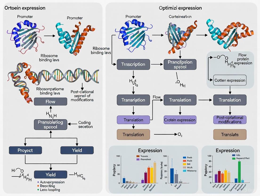
The Escherichia coli (E. coli) protein expression system stands as a cornerstone of modern biotechnology, enabling the production of recombinant proteins for research, industrial, and therapeutic applications [9]. Its popularity stems from a powerful combination of rapid growth, facile genetic manipulation, and cost-effective cultivation [10] [9]. A typical expression experiment requires four key elements: the gene encoding the protein of interest, a specialized bacterial expression vector, a compatible expression cell line, and equipment for bacterial cell culture [10]. The core of this system lies in the precise and coordinated function of its components—the vectors, promoters, and host strains. Optimizing the interplay between these parts is crucial for maximizing the yield of soluble, active protein, which is the central theme of this technical overview [10]. This guide provides a detailed resource for researchers to understand these critical components, troubleshoot common issues, and implement optimized protocols for successful protein production.
Core Component I: Expression Vectors and Promoter Systems
Expression vectors are engineered plasmids designed to drive the transcription and translation of a recombinant gene in E. coli. They serve as the vehicle that carries your gene of interest and provides the necessary genetic instructions for its high-level production [9].
Key Elements of an Expression Vector
- Promoter: A strong, regulatable DNA sequence where RNA polymerase binds to initiate transcription. The choice of promoter is one of the most critical factors determining expression levels [9].
- Ribosome Binding Site (RBS): A sequence upstream of the start codon that facilitates the binding of the ribosome to the mRNA for efficient translation initiation.
- Multiple Cloning Site (MCS): A short DNA sequence containing multiple restriction enzyme cleavage sites, allowing for the insertion of the target gene.
- Selectable Marker: A gene (e.g., for antibiotic resistance) that allows for the selection of bacteria that have successfully taken up the plasmid.
- Origin of Replication (ori): Determines the copy number of the plasmid within each cell, thereby influencing the potential yield of the target protein.
- Fusion Tags: Optional sequences that encode for tags (e.g., His-tag, GST, MBP) to facilitate protein purification, improve solubility, or enable detection [10] [9].
Common Promoter Systems
The promoter is the primary control switch for protein expression. The table below summarizes the characteristics of widely used promoter systems.
Table 1: Common Promoter Systems in E. coli Protein Expression
| Promoter System | Inducer | Mechanism of Action | Key Features | Best For |
|---|---|---|---|---|
| T7 (e.g., in pET vectors) | IPTG | The host strain (e.g., BL21(DE3)) contains a chromosomal copy of the T7 RNA polymerase gene under control of the lacUV5 promoter. IPTG inactivates the Lac repressor, allowing T7 RNA polymerase expression, which then transcribes the target gene from the T7 promoter on the plasmid with high efficiency [10] [11] [9]. | Very strong, high yields, but can have "leaky" basal expression [11] [12]. | High-level production of non-toxic proteins [9]. |
| T5/lac | IPTG | A hybrid promoter that is directly repressed by the Lac repressor protein. Adding IPTG derepresses the promoter, allowing transcription by the host's RNA polymerase [11]. | Tight regulation, less basal expression than some T7 systems. | General protein expression, particularly where tight control is needed [11]. |
| pBAD (araBAD) | L-Arabinose | The target gene is under the control of the arabinose-inducible araBAD promoter. This system is tightly repressed in the absence of arabinose and can be finely tuned by varying arabinose concentration [11] [13]. | Very tight regulation, tunable expression levels. | Expression of toxic proteins [11] [13]. |
| rhaBAD | L-Rhamnose | Similar to pBAD, this system uses the rhamnose-promoter and can be tightly regulated. In strains like Lemo21(DE3), it controls the expression of T7 lysozyme to tune the activity of the T7 RNA polymerase [11] [14]. | Tunable expression, reduces inclusion body formation. | Challenging proteins (toxic, insoluble, membrane proteins) [11] [14]. |
Core Component II: E. coli Expression Strains
Selecting the appropriate E. coli host strain is a key determinant of the success of a protein expression experiment. Strains are engineered to address specific challenges such as codon bias, protein toxicity, and insolubility [11] [9].
Strain Genotypes and Their Importance
Most protein expression strains share common genetic modifications to enhance protein production and stability:
- Protease Deficiencies (
lon,ompT): Mutations in genes encoding proteases reduce the degradation of the recombinant protein [10] [11] [14]. - DE3 Lysogen: This is a λ prophage integrated into the genome that carries the gene for T7 RNA polymerase, making the strain compatible with T7 promoter-based vectors (e.g., pET) [11] [14].
- hsdSB (
rB- mB-): This mutation inactivates the native restriction-modification system, preventing the degradation of unmethylated plasmid DNA [11].
Selecting a Specialized Expression Strain
The following table provides a guide to selecting a strain based on the specific characteristics of your target protein.
Table 2: Common E. coli Expression Strains and Their Applications
| Strain | Key Features | Primary Function | Mechanism |
|---|---|---|---|
| BL21(DE3) [11] [14] [9] | Deficient in Lon and OmpT proteases; contains DE3 lysogen for T7 RNA polymerase expression. | General-purpose protein expression. | Standard workhorse for non-toxic proteins. |
| BL21(DE3) pLysS/pLysE [11] [14] [13] | Contains a plasmid expressing T7 lysozyme, a natural inhibitor of T7 RNA polymerase. pLysE provides tighter control than pLysS. | Expression of toxic proteins. | Suppresses basal "leaky" expression before induction [11] [12]. |
| Rosetta2(DE3) [11] [14] [9] | Supplies tRNAs for rare codons (AGA, AGG, AUA, CUA, CCC, GGA) not commonly used in E. coli. | Expression of eukaryotic proteins or proteins with rare codons. | Prevents translation stalling and truncation, improving yield and integrity [15]. |
| Origami2(DE3) [11] [14] [9] | Mutations in thioredoxin reductase (trxB) and glutathione reductase (gor) genes. |
Enhancing disulfide bond formation in the cytoplasm. | Creates a more oxidizing cytoplasm, promoting correct folding of disulfide-bonded proteins. |
| SHuffle T7 Express [14] [16] | Combines trxB/gor mutations with cytoplasmic expression of disulfide bond isomerase (DsbC). |
Production of proteins with complex disulfide bonds. | Promotes both oxidation and isomerization of disulfide bonds for correct pairing in the cytoplasm. |
| ArcticExpress(DE3) [14] | Expresses cold-adapted chaperonins (Cpn10/Cpn60) from a psychrophilic bacterium. | Improving solubility of difficult-to-express proteins. | Chaperonins assist with proper protein folding at low temperatures (4-12°C). |
| Lemo21(DE3) [11] [14] [9] | Tunable expression of T7 lysozyme via the rhamnose-inducible promoter. | Expression of toxic, insoluble, or membrane proteins. | Allows fine-tuning of T7 RNA polymerase activity to find an expression level that balances yield and cell health. |
| Tuner(DE3) [11] [14] [9] | Contains a mutation in the lacY gene (lac permease). |
Tunable expression for toxic or insoluble proteins. | Allows uniform entry of IPTG into all cells, enabling precise, concentration-dependent induction across the entire culture. |
| C41(DE3) / C43(DE3) [14] [9] | Mutant derivatives of BL21(DE3) with mutations that reduce T7 RNA polymerase activity. | Expression of toxic and membrane proteins. | Genetic mutations prevent cell death associated with the expression of highly toxic proteins. |
The Integrated Workflow for Protein Expression
A generalized, optimized workflow for recombinant protein expression in E. coli involves multiple steps, from gene design to induction. The following diagram illustrates this process and the critical decision points.
Figure 1: A standard workflow for recombinant protein expression in E. coli.
Representative Experimental Protocol
The protocol below is a robust starting point for expressing a diverse range of proteins, incorporating strategies to enhance solubility [10].
- Construct Design: Clone the gene of interest into an expression vector (e.g., a pET vector with a T7 promoter). The vector should include an N-terminal purification tag, such as a hexahistidine (His₆)-tag, and a protease cleavage site (e.g., for TEV protease) for tag removal after purification [10].
- Transformation: Transform the expression vector into a suitable E. coli strain, such as BL21(DE3)-RIL. This strain supplies additional tRNAs for rare codons and is deficient in Lon and OmpT proteases [10].
- Starter Culture: Inoculate a single colony into a small volume of LB medium containing the appropriate antibiotic. Grow overnight at 37°C with shaking.
- Large-Scale Culture: Dilute the overnight culture 1:100 into a fresh, larger volume of LB medium with antibiotic in a baffled shaker flask to improve aeration. Grow at 37°C with vigorous shaking (200-250 rpm) until the culture reaches mid-log phase (OD₆₀₀ ~0.6 to 0.9) [10].
- Induction: Cool the culture to 18°C. Once the temperature has stabilized, induce protein expression by adding Isopropyl β-d-1-thiogalactopyranoside (IPTG) to a final concentration of 0.1 - 1.0 mM [10] [15].
- Post-Induction Incubation: Continue incubating the culture with shaking at 18°C overnight (~16-20 hours). The lower temperature slows the rate of protein synthesis, favoring correct folding and increasing soluble yield [10].
- Harvest: Centrifuge the culture to pellet the cells. The cell pellet can be processed immediately for protein purification or stored at -80°C.
Troubleshooting FAQs and Guide
Frequently Asked Questions
Q1: I get no colonies after transformation. What could be wrong?
- Toxic Gene Product: If your protein is toxic, even low-level basal expression can prevent cell growth. Use a tighter control strain like BL21(DE3) pLysS or BL21-AI [11] [13].
- Antibiotic & Plasmid Check: Verify the correct antibiotic is used for your plasmid. Check the quality of your plasmid and competent cells by transforming a control plasmid (e.g., pUC19) [13].
- Glucose Repression: For T7 systems, add 1% glucose to the plates and growth medium to further repress basal expression [12] [13].
Q2: My protein is not expressed, or the yield is very low.
- Verify Sequence and Cloning: Sequence your plasmid to ensure the gene is in-frame and free of mutations or premature stop codons [15] [13].
- Check for Rare Codons: Analyze your protein sequence for clusters of codons that are rare in E. coli (e.g., AGG, AGA, ATA for Arg, Ile). Use a codon-optimized synthetic gene or switch to a tRNA-supplemented strain like Rosetta2(DE3) [15] [14] [13].
- Check for Insolubility: The protein may be in the insoluble fraction as inclusion bodies. Analyze both the soluble supernatant and the insoluble pellet by SDS-PAGE [10] [13].
- Plasmid Instability: If using ampicillin, it can degrade during culture, leading to plasmid loss. Use carbenicillin (more stable) or a different antibiotic. Always use freshly transformed cells for expression cultures [13].
Q3: My protein is expressed but is insoluble. How can I improve solubility?
- Lower Induction Temperature: Induce at a lower temperature (e.g., 18-25°C) and express overnight. This slows down protein synthesis, giving the protein more time to fold correctly [10] [15] [13].
- Reduce Inducer Concentration: Use a lower concentration of IPTG (e.g., 0.1 mM or lower) to moderate the level of expression [15] [13].
- Use a Fusion Tag: Clone your gene downstream of a solubility-enhancing tag like Maltose-Binding Protein (MBP) or SUMO [12].
- Change the Strain: Use a chaperone-assisted strain like ArcticExpress(DE3) or a tunable strain like Lemo21(DE3) to find the optimal expression level [14].
- Test Different Media: Sometimes, using a minimal medium like M9 instead of rich LB can improve solubility [15] [13].
Q4: I see multiple bands or smearing on my gel, suggesting degradation.
- Use Protease-Deficient Strains: Ensure you are using a strain deficient in proteases like Lon and OmpT (e.g., BL21 derivatives) [10] [11].
- Add Protease Inhibitors: Include protease inhibitor cocktails, such as PMSF, in all lysis buffers. Note that PMSF is unstable in aqueous solution and must be added fresh [13].
- Work Quickly and Keep Samples Cold: Perform purifications quickly and keep lysates and extracts on ice to minimize protease activity.
Q5: How can I express a protein that requires disulfide bonds?
- Target to the Periplasm: Use a vector with a signal sequence (e.g., pelB, ompA) to export the protein to the oxidative periplasm, where disulfide bond formation naturally occurs [12] [16].
- Use Specialized Cytoplasmic Strains: For more complex disulfide bonds, use strains like SHuffle T7 Express, which provide an oxidizing cytoplasm and disulfide bond isomerase activity to correct mis-paired bonds [14] [16].
Troubleshooting Flowchart
The following diagram provides a logical pathway to diagnose and address the most common protein expression problems.
Figure 2: A troubleshooting guide for common protein expression issues.
The Scientist's Toolkit: Essential Research Reagents
Table 3: Key Reagents and Materials for E. coli Protein Expression
| Reagent / Material | Function / Purpose | Examples / Notes |
|---|---|---|
| Expression Vectors | Plasmid backbone containing promoter, tags, and selection marker for hosting the gene of interest. | pET (T7 promoter), pBAD (arabinose promoter), pMAL (MBP fusion) [10] [9]. |
| Competent Cells | Specialized E. coli cells treated to easily take up foreign DNA. | BL21(DE3), Rosetta2(DE3), SHuffle T7 Express [11] [14]. |
| Inducers | Chemical molecules that trigger transcription of the target gene. | IPTG (for T7/lac systems), L-Arabinose (for pBAD), L-Rhamnose (for rhaBAD/Lemo system) [10] [11]. |
| Antibiotics | Selectively maintain the plasmid within the bacterial population. | Ampicillin/Carbenicillin, Kanamycin, Chloramphenicol. Note: Carbenicillin is more stable than ampicillin for long cultures [13]. |
| Growth Media | Provide nutrients for bacterial growth and protein production. | LB (Luria-Bertani), TB (Terrific Broth), M9 Minimal Medium. Rich media like TB support high cell density [15]. |
| Purification Tags | Amino acid sequences fused to the protein to allow easy purification. | His-tag (Ni-NTA purification), GST (Glutathione resin), MBP (Amylose resin) [10] [9]. |
| Protease Inhibitors | Chemical compounds that inhibit cellular proteases, reducing protein degradation during purification. | PMSF, Commercially available cocktails. Must be added fresh to lysis buffers [13]. |
| Benzyldihydrochlorothiazide | Benzyldihydrochlorothiazide | Explore our high-purity Benzyldihydrochlorothiazide for research. This compound is for professional research use only and not for personal or human use. |
| Sulfo saed | Sulfo saed, MF:C21H21N5O9S3, MW:583.6 g/mol | Chemical Reagent |
Troubleshooting Guides
Troubleshooting Guide for Inclusion Body Formation
Problem: Target protein is expressed predominantly in insoluble inclusion bodies.
| Problem Cause | Diagnostic Signs | Recommended Solution | Key References |
|---|---|---|---|
| High Expression Rate | High expression levels but protein inactivity; aggregation. | Lower induction temperature (e.g., 16-25°C); reduce inducer concentration (e.g., 0.1-0.5 mM IPTG); use a weaker promoter. | [17] |
| Incorrect Folding due to Codon Bias | Protein insolubility in codon bias-adjusted strains; retarded cell growth. | Analyze codon usage; for sequences with >5% RIL codons, use standard strains like BL21(DE3)-pLysS instead of tRNA-enhanced strains. | [18] |
| Lack of PTMs / Disulfide Bonds | Common with eukaryotic proteins; improper folding. | Use E. coli strains with oxidizing periplasmic space (e.g., Origami); target protein to the periplasm; use chaperone co-expression vectors (e.g., GroEL/S, DnaK/DnaJ). | [17] |
| Non-optimal Physicochemical Conditions | Aggregation under specific culture conditions. | Adjust culture temperature (e.g., 16-30°C); optimize media pH (e.g., pH 7.5); supplement with additives (e.g., sugars, osmolytes). | [17] |
Troubleshooting Guide for Codon Bias and Translation Issues
Problem: Low or no protein yield, or production of truncated/insoluble products.
| Problem Cause | Diagnostic Signs | Recommended Solution | Key References |
|---|---|---|---|
| Rare Codons / Depleted tRNAs | Ribosome stalling; low protein yield; truncated products. | Use E. coli strains with plasmid-encoded rare tRNAs (e.g., CodonPlus(DE3)-RIL, Rosetta); perform whole-gene codon optimization. | [19] [18] |
| Non-optimal N-terminal sequence | Low yield regardless of overall codon optimization. | Use directed evolution libraries to optimize the first 5-10 N-terminal codons; employ tools like TISIGNER to minimize mRNA secondary structure. | [20] |
| Poor Translation Initiation | Low protein yield despite high mRNA levels. | Ensure a strong Shine-Dalgarno sequence; avoid strong secondary structures at the 5' end of the mRNA; verify the start codon is ATG. | [19] [20] |
| mRNA Secondary Structure | Reduced translation initiation and efficiency. | Use algorithms to predict and minimize secondary structure around the ribosome binding site and gene start. | [20] |
Frequently Asked Questions (FAQs)
Q1: My protein is expressed but is insoluble. What are my primary strategies to recover soluble, active protein?
A: Your strategy should involve both preventing aggregation and refolding existing aggregates.
- Prevention: Lower the growth temperature to 16-25°C immediately after induction, reduce the inducer concentration, and consider using a richer growth medium. If the protein is eukaryotic or requires disulfide bonds, use E. coli strains like Origami that facilitate disulfide bond formation in the cytoplasm and/or co-express molecular chaperones like GroEL/S [17].
- Recovery: You can attempt to solubilize inclusion bodies using denaturants like urea or guanidine hydrochloride, followed by a careful refolding process via dialysis or dilution. For proteins requiring disulfide bonds, supplement the refolding buffer with redox agents like reduced and oxidized glutathione [17].
Q2: I am expressing a eukaryotic protein in E. coli and it is inactive, likely due to missing post-translational modifications (PTMs). What can I do?
A: This is a common limitation of the bacterial system. Consider these approaches:
- Protein Engineering: If the PTM is not absolutely critical for activity, consider mutating the modification site (e.g., phosphorylation site) to a residue that mimics the modified or unmodified state.
- Alternative Hosts: If PTMs are essential, switch to a eukaryotic expression system such as yeast, insect, or mammalian cells, which possess the necessary machinery.
- Specialized E. coli Systems: For disulfide bonds, target your protein to the more oxidizing periplasm of E. coli or use strains like Origami that enhance disulfide bond formation [17].
Q3: What is the critical consideration when choosing an E. coli strain for expressing a gene with high rare codon content?
A: The primary consideration is the balance between translation speed and proper protein folding. While strains like BL21-CodonPlus(DE3)-RIL provide additional tRNAs for rare codons (AGA/AGG, AUA, CUA) and can prevent ribosome stalling, they can also cause too-rapid translation. This can lead to protein misfolding and aggregation into inclusion bodies, especially if the coding sequence has a high content (>5%) of these RIL codons [18]. For such genes, it is often better to use a standard strain like BL21(DE3)-pLysS, where slower translation at rare codons may facilitate correct co-translational folding.
Q4: How can I optimize the N-terminal sequence of my gene to improve expression yields?
A: The N-terminal sequence (first ~5-10 codons) significantly influences translation initiation and efficiency. Modern approaches include:
- Directed Evolution: Creating a DNA library with randomized N-terminal codons, fusing the gene to a fluorescent reporter (e.g., GFP), and using Fluorescence-Activated Cell Sorting (FACS) to select clones with the highest expression. This method has been shown to increase soluble yield by over 30-fold [20].
- Computational Tools: Using software like TISIGNER or CodonTransformer to design sequences that minimize mRNA secondary structure around the translation start site and optimize codon usage in a context-aware manner for the host organism [20] [21].
Experimental Protocols
Protocol 1: Testing the Impact of Codon Usage on Protein Solubility
This protocol systematically compares protein solubility between standard and codon-enhanced E. coli strains [18].
1. Principle To determine if the codon content of a target gene contributes to protein insolubility by expressing it in both a standard expression strain and a strain supplemented with rare tRNAs, then analyzing the solubility of the resulting protein.
2. Reagents and Equipment
- E. coli Strains: BL21(DE3)-pLysS (standard) and BL21(DE3)-CodonPlus(DE3)-RIL (codon-enhanced).
- Plasmid: Target gene in a T7 expression vector (e.g., pET series).
- Media: LB broth with appropriate antibiotics (e.g., Chloramphenicol for pLysS, Chloramphenicol and Streptomycin for CodonPlus-RIL).
- Inducer: Isopropyl β-d-1-thiogalactopyranoside (IPTG).
- Lysis Buffer: e.g., 50 mM Tris-HCl, pH 8.0, 100 mM NaCl, 1 mM EDTA, supplemented with lysozyme and protease inhibitors.
- Equipment: Sonicator, centrifuge, SDS-PAGE gel apparatus, Western blotting system.
3. Step-by-Step Procedure 1. Transform the plasmid containing your target gene into both the standard and codon-enhanced E. coli strains. 2. Inoculate starter cultures and grow overnight. 3. Dilute cultures into fresh, pre-warmed antibiotic media and grow at 37°C to an OD600 of ~0.5. 4. Induce protein expression by adding 0.5 mM IPTG. 5. Shift temperature to 25°C and continue shaking for 4-6 hours. 6. Harvest cells by centrifugation. 7. Resuspend cell pellet in Lysis Buffer and lyse cells by sonication on ice. 8. Centrifuge the lysate at high speed (e.g., 15,000 x g) for 20 minutes at 4°C to separate soluble (supernatant) and insoluble (pellet) fractions. 9. Analyze both the total lysate, soluble fraction, and insoluble fraction by SDS-PAGE and Western blotting.
4. Data Analysis Compare the Western blot signals between the two strains. A significant shift of the target protein from the soluble fraction in the standard strain to the insoluble fraction in the codon-enhanced strain indicates that overly rapid translation, facilitated by the supplemented tRNAs, is promoting misfolding and aggregation [18].
Protocol 2: Screening for Soluble Protein Expression Using FACS
This protocol uses a directed evolution approach to optimize the N-terminal sequence for high-yield soluble expression [20].
1. Principle A library of target genes with randomized N-terminal sequences is fused to a GFP reporter gene. E. coli cells expressing this library are sorted using FACS, where high fluorescence correlates with high levels of soluble target protein-GFP fusion. The optimized N-terminal sequences are then identified from the sorted population.
2. Reagents and Equipment
- Vector: pET22b or similar with a C-terminal GFP tag.
- Library: A DNA library of your target gene with randomized codons for the first 5-10 amino acids.
- E. coli Strain: BL21(DE3) or similar.
- Media: LB with appropriate antibiotics.
- Inducer: IPTG.
- Equipment: Fluorescence-Activated Cell Sorter (FACS), flow cytometer.
3. Step-by-Step Procedure 1. Clone the N-terminal library into the expression vector, creating in-frame fusions with the GFP gene. 2. Transform the library into an expression E. coli strain. 3. Induce protein expression with IPTG and grow cells, typically at a lower temperature (e.g., 18°C) to favor solubility. 4. Collect cells and resuspend in PBS or a suitable buffer for FACS analysis. 5. Sort Cells: Using FACS, gate the population based on the fluorescence of the positive control (cells expressing a GFP-only construct). Collect the top 1-5% of most fluorescent cells from the library population. 6. Recover and Plate the sorted cells to form single colonies. 7. Screen Colonies: Isolate plasmids from individual colonies and re-test for protein expression and solubility. 8. Sequence the plasmids from the best-performing clones to identify the optimized N-terminal sequence.
4. Data Analysis The primary data is the fluorescence histogram from the flow cytometer. A successful screen will show a broad distribution of fluorescence in the pre-sort library, with a distinct, highly fluorescent population post-sort. Sequencing multiple clones from this population will reveal consensus or preferred amino acids and codons at the N-terminus that confer high soluble yield [20].
Pathway and Workflow Visualizations
Protein Homeostasis and Inclusion Body Formation
This diagram illustrates the cellular equilibrium between proper protein folding and the formation of inclusion bodies in E. coli.
Codon Optimization and Solubility Screening Workflow
This flowchart outlines the experimental process for evaluating and improving protein solubility through codon and N-terminal optimization.
The Scientist's Toolkit: Research Reagent Solutions
| Reagent / Tool | Function & Application | Key Considerations |
|---|---|---|
| BL21(DE3)-CodonPlus(DE3)-RIL Strain | Supplies additional tRNAs for AGA/AGG (Arg), AUA (Ile), and CUA (Leu) codons. Prevents ribosome stalling on genes with high content of these "RIL" codons. | Can cause excessively fast translation leading to misfolding; use with caution if RIL codon content is high (>5%) [18]. |
| Origami E. coli Strains | Features mutations in the thioredoxin reductase (trxB) and glutathione reductase (gor) genes, creating a more oxidizing cytoplasm that promotes disulfide bond formation. |
Ideal for expressing eukaryotic proteins that require stable disulfide bonds for activity [17]. |
| Chaperone Plasmid Kits (e.g., GroEL/S, DnaK/DnaJ) | Plasmids for co-expressing molecular chaperones. Assist in the proper folding of recombinant proteins, reducing aggregation. | Different chaperone systems may be specific to different classes of proteins; may require testing multiple sets [17]. |
| pET Expression Vectors | A series of vectors utilizing the strong T7 lac promoter for high-level protein expression in E. coli. Offers various tags (His-tag, SUMO, etc.) and secretion signals. | The choice of vector (e.g., with solubility tags like Trx or MBP) can significantly impact yield and solubility. |
| PURExpress Disulfide Bond Enhancer | A commercial supplement added in vitro to the protein synthesis reaction to create an environment favorable for disulfide bond formation. | Useful in cell-free protein synthesis systems to produce complex proteins with multiple disulfide bonds [19]. |
| [1,2]Dioxino[4,3-b]pyridine | [1,2]Dioxino[4,3-b]pyridine, CAS:214490-52-5, MF:C7H5NO2, MW:135.12 g/mol | Chemical Reagent |
| Tantalum(IV) carbide | Tantalum(IV) Carbide|TaC Powder | High-purity Tantalum(IV) Carbide (TaC) powder for research. Used in UHTCs, cermets, and composites. For Research Use Only. Not for human use. |
This technical support center provides a focused resource for researchers optimizing recombinant protein expression in E. coli. The T7 expression system is the most widely used approach for producing high yields of recombinant proteins in this prokaryotic workhorse [22]. Within this system, a gene of interest is cloned downstream of a T7 promoter into an expression vector, which is then introduced into a specialized E. coli host strain containing a chromosomal copy of the T7 RNA polymerase gene [22]. Protein production is typically induced by the addition of an inducer like IPTG, leading to expression of the polymerase and subsequent transcription of the target gene [22] [9]. Despite the system's robustness, challenges such as low yield, poor solubility, and protein inactivity are common. The following guides and protocols are designed to help you troubleshoot these issues and refine your experimental workflow for successful protein production.
The journey from a gene of interest to a purified protein involves several critical stages, from initial vector design to final purification and analysis. The diagram below outlines this standard workflow.
Troubleshooting Guide: Common Protein Expression Issues
Problem 1: No or Low Yield of Target Protein
Possible Causes and Solutions:
| Possible Cause | Solution |
|---|---|
| RNase Contamination | Use nuclease-free tips and tubes. Add RNase Inhibitor to reactions [23]. |
| Non-optimal Template DNA Design | Verify the DNA sequence is correct and in-frame. Ensure the template includes a T7 terminator or UTR stem loop to stabilize mRNA [23]. |
| Rare Codons | Check for stretches of rare codons, especially near the 5' end. Use a host strain with supplemental tRNAs for rare codons (e.g., Rosetta series) or perform codon optimization [24] [9]. |
| Suboptimal Regulatory Sequences | Secondary structure or rare codons at the start of the mRNA can compromise initiation. Modify the 5' end via PCR or add a proven initiation region (e.g., first ten codons of Maltose-Binding Protein) [23]. |
| Incorrect Template DNA Concentration | The balance between transcription and translation is key. Use 250 ng of DNA per 50 µL reaction as a starting point and optimize from 25–1000 ng [23]. |
Problem 2: Target Protein is Insoluble or Inactive
Possible Causes and Solutions:
| Possible Cause | Solution |
|---|---|
| Incorrect Folding | Lower the incubation temperature (e.g., to 16-30°C) and extend the induction time (e.g., up to 24 hours) [23] [24]. |
| Lack of Disulfide Bonds | Use engineered strains that enhance disulfide bond formation in the cytoplasm, such as Origami or Shuffle strains [9]. Supplement the reaction with a disulfide bond enhancer system [23]. |
| Aggregation-Prone Protein | Fuse the target protein to a highly soluble partner tag, such as Maltose-Binding Protein (MBP) [25]. |
| Protein is Toxic to Host Cells | Use a tightly regulated expression system with very low background ("leaky") expression. For T7 systems, use hosts containing the pLysS plasmid, which produces T7 lysozyme to suppress basal polymerase activity [24]. |
Problem 3: Truncated Protein Products
Possible Causes and Solutions:
| Possible Cause | Solution |
|---|---|
| Premature Termination / Internal Initiation | Ensure proper translation initiation and termination sequences. Internal ribosome entry sites can produce truncated proteins [23]. |
| mRNA Instability | Verify the template DNA contains a T7 terminator or a UTR stem loop structure to increase mRNA stability and yield [23]. |
| Protease Degradation | Use host strains that are deficient in lon and ompT proteases, such as BL21 and its derivatives [9]. |
The following flowchart can help guide your troubleshooting process when you encounter a problem with protein expression.
Frequently Asked Questions (FAQs)
Q1: How do I choose the right E. coli expression strain? The choice of host strain is a critical determinant of success. BL21(DE3) is a common choice for non-toxic proteins. For toxic proteins, consider BL21(DE3)pLysS or C41(DE3)/C43(DE3) strains. If your protein contains rare codons, use a strain like Rosetta. For proteins requiring disulfide bonds, Shuffle or Origami strains are recommended [24] [9].
Q2: My protein is expressed but is insoluble. What are my options? You have several strategies: 1) Lower the growth temperature during induction (e.g., to 16-25°C); 2) Reduce the inducer concentration to slow down expression; 3) Fuse your protein to a solubility-enhancing tag like MBP; 4) Use a strain designed for enhanced disulfide bond formation if applicable; 5) If solubility cannot be achieved, purify from inclusion bodies and explore refolding protocols [23] [25] [26].
Q3: What can cause "leaky expression" (expression without induction) and how can I prevent it? Leaky expression occurs when the T7 RNA polymerase is active even before induction. This is a particular problem for proteins that are toxic to the host. To minimize leakiness, use expression hosts like BL21(DE3)pLysS, which contains the pLysS plasmid encoding T7 lysozyme, a natural inhibitor of T7 RNA polymerase [24] [9].
Q4: How can I verify that my cloned gene is correctly inserted in the expression vector? It is highly recommended to perform DNA sequencing on the cloned plasmid before proceeding with expression studies. This will confirm that the inserted sequence is correct, is in the proper reading frame, and has not acquired any unintended mutations during PCR or cloning [24].
The Scientist's Toolkit: Key Research Reagents
A successful protein expression experiment relies on the right combination of tools. The table below lists essential materials and their functions.
| Reagent / Material | Function & Application |
|---|---|
| pET Vectors (or similar) | Expression plasmids with a strong T7 promoter for high-level, inducible protein production [22] [9]. |
| BL21(DE3) E. coli Strain | A standard host strain deficient in lon and ompT proteases, containing a genomic DE3 lysogen for T7 RNA polymerase expression [9]. |
| Specialized E. coli Strains | Strains like Rosetta (supplies rare tRNAs), Shuffle (promotes disulfide bond formation), and BL21(DE3)pLysS (reduces leaky expression for toxic proteins) address specific expression challenges [24] [9]. |
| Isopropyl β-d-1-thiogalactopyranoside (IPTG) | A molecular mimic of allolactose that induces expression by binding to the lac repressor and activating transcription from the T7/lac promoter [9]. |
| Fusion Tags (His₆, MBP, GST) | His₆: Allows purification by immobilized metal affinity chromatography (IMAC). MBP: Often used as a powerful solubility enhancer. Tags can be removed post-purification using a specific protease site (e.g., TEV protease) [25]. |
| Ni-NTA Agarose | Resin for IMAC that chelates nickel ions, which bind with high affinity to polyhistidine tags, enabling rapid one-step purification of recombinant proteins [25]. |
| TEV Protease | A highly specific protease used to remove affinity tags from the purified protein of interest, leaving a minimal native sequence [25]. |
| Dodecyl 4-nitrobenzoate | Dodecyl 4-nitrobenzoate, CAS:35507-03-0, MF:C19H29NO4, MW:335.4 g/mol |
| Mercapto-propylsilane | Mercapto-propylsilane, MF:C3H8SSi, MW:104.25 g/mol |
Detailed Experimental Protocol: Evaluating Protein Solubility with His₆ and MBP Tags
This protocol describes how to construct and test the solubility of a protein with either a His₆ tag or a dual His₆-MBP tag, helping you choose the best strategy for large-scale production [25].
1. Cloning into Expression Vectors
- Use Gateway recombinational cloning to insert your target gene into two different destination vectors: pDEST527 (for a His₆-tag fusion) and pDEST-HisMBP or pDEST566 (for a dual His₆-MBP fusion) [25].
- Ensure the vectors are designed with a protease cleavage site (e.g., for Tobacco Etch Virus, TEV, protease) between the fusion tag and your protein of interest.
2. Small-Scale Pilot Expression
- Transform each expression vector into an appropriate expression host, such as Rosetta 2(DE3).
- Inoculate 5 mL cultures of LB medium containing the appropriate antibiotics (e.g., ampicillin, chloramphenicol) and grow overnight at 37°C.
- Dilute the overnight culture 1:100 into fresh medium and grow at 37°C with shaking until mid-log phase (OD600 ~0.5-0.7).
- Induce protein expression by adding IPTG to a final concentration of 0.1 - 1.0 mM.
- Continue incubation for 3-6 hours at 37°C, or alternatively, test lower temperatures (e.g., 25°C, 16°C) with overnight induction to improve solubility.
3. Analyzing Solubility via Lysis and Fractionation
- Harvest the cells by centrifugation.
- Resuspend the cell pellet in a suitable lysis buffer (e.g., 25 mM Tris-HCl, 200 mM NaCl, 25 mM imidazole, pH 7.2).
- Lyse the cells by sonication or lysozyme treatment.
- Centrifuge the lysate at high speed (e.g., >12,000 x g for 20 min) to separate the soluble fraction (supernatant) from the insoluble fraction (pellet, containing inclusion bodies).
- Analyze both the total lysate, soluble fraction, and insoluble fraction by SDS-PAGE to determine the distribution of your target protein.
4. Protease Cleavage Test for Solubility
- Purify the fusion protein from the soluble fraction using Ni-NTA Agarose.
- Incubate the purified, tagged protein with TEV protease to remove the His₆ or His₆-MBP tag.
- After cleavage, pass the mixture again over Ni-NTA Agarose. The cleaved tag will bind to the resin, while the untagged passenger protein will flow through.
- Analyze the flow-through by SDS-PAGE. If the passenger protein remains in the soluble fraction after tag removal, it is a good candidate for large-scale production without the solubility tag. If it precipitates, the MBP tag should be retained for subsequent work [25].
Practical Strategies for Enhancing Solubility and Protein Yield
Frequently Asked Questions (FAQs)
Q1: What are the primary factors I should consider when designing a vector for high-yield soluble protein expression in E. coli?
Achieving high yields of soluble protein requires a multi-factorial approach. Your primary considerations should be:
- Codon Optimization: The codon usage of your target gene should be optimized to match the tRNA pool of your E. coli expression strain. However, simple maximization of optimal codons is not always the best strategy, as it can lead to ribosomal congestion and increased metabolic burden. A balanced approach that matches the host's overall codon usage bias is often more effective [27].
- Fusion Tags: Incorporating an appropriate N-terminal fusion tag, such as a hexa-histidine tag, is a common first choice for improving solubility and enabling affinity purification [28].
- Signal Peptides (for secretion): If secreting the protein to the periplasm is desired, the selection of an efficient signal peptide is critical. There is no universal predictor, so empirical screening of a library of signal peptides is often necessary to find the optimal one for your protein of interest [29].
- Expression Conditions: Parameters like induction temperature, inducer concentration (e.g., IPTG), and media composition must be optimized. For example, lower temperatures (25°C–30°C) often favor correct folding and solubility, and lower IPTG concentrations can reduce metabolic stress [30].
Q2: My protein is expressed insolubly. What troubleshooting steps can I take?
When facing insoluble expression, follow this systematic troubleshooting guide:
- Verify Codon Usage: Re-analyze your gene sequence using multiple codon optimization tools. Consider having the gene synthesized with host-specific codon optimization [28] [31].
- Lower Expression Temperature: Induce protein expression at a lower temperature (e.g., 16°C–25°C) to slow down translation and facilitate proper folding [28].
- Reduce Inducer Concentration: Use a lower concentration of IPTG (e.g., 0.1–0.5 mM) to decrease the rate of protein synthesis and minimize aggregation [30].
- Test Different Fusion Partners: If a His-tag is insufficient, consider testing larger solubility-enhancing fusion partners like MBP or GST.
- Screen for Optimal Signal Peptide: If secretion is an option, use a signal peptide toolbox to screen for the most effective one for your protein [29].
- Use Specialized Strains: For proteins requiring disulfide bonds, switch to strains with oxidizing cytoplasms (e.g., Origami) or engineered strains that overexpress chaperones and foldases [32].
Q3: How does codon optimization truly affect my E. coli host, and can it be "over-optimized"?
Yes, "over-optimization" is a recognized phenomenon. While optimizing rare codons is crucial, simply maximizing the frequency of so-called optimal codons can be detrimental. If the host's native genes have a certain codon usage bias (e.g., 60-70% optimal codons), and you express a gene with 100% optimal codons, you can create an imbalance. This over-optimized gene may sequester a disproportionate share of specific tRNAs and ribosomes, exacerbating metabolic burden and potentially reducing the yield of both your target protein and essential host proteins [27]. The goal is to harmonize codon usage with the host's global tRNA availability, not just to maximize a single metric like the Codon Adaptation Index (CAI).
Troubleshooting Guides
Guide to Codon Optimization Tools and Parameters
Selecting the right tool and parameters is critical for successful gene design. The table below summarizes key characteristics of popular codon optimization tools.
Table 1: Comparative Analysis of Codon Optimization Tools and Key Parameters
| Tool Name | Optimization Strategy | Key Parameters Adjustable | Best Use Case |
|---|---|---|---|
| JCat [31] | Aligns with host genome codon usage. | CAI, GC content | Rapid, standard optimization for microbial hosts. |
| OPTIMIZER [31] | Matches codon usage to a reference set. | CAI, ICU, GC content | Custom optimization using user-defined reference genes. |
| ATGme [31] | Integrated design with multiple criteria. | CAI, GC content, restriction sites | One-stop solution for synthetic gene design. |
| GeneOptimizer [31] | Advanced algorithm using multiple parameters. | CAI, GC content, mRNA structure, CPB | High-performance optimization for difficult proteins. |
| TISIGNER [31] | Focuses on 5' sequence and translation initiation. | RBS strength, N-terminal codon context | Optimizing translation initiation efficiency. |
| IDT (Codon Optimization Tool) [31] | Proprietary algorithm for general use. | Limited user parameters | Quick design for standard gene synthesis orders. |
Optimizing Expression Conditions for Soluble Yield
The conditions during induction are as important as the vector design. The following table provides a guideline for key parameters.
Table 2: Experimental Conditions for Enhancing Soluble Protein Expression in E. coli
| Parameter | Typical Range | Effect & Rationale | Recommended Starting Point |
|---|---|---|---|
| Induction Temperature | 16°C - 30°C | Lower temperatures slow translation, reducing aggregation and favoring proper folding [28] [32]. | 25°C |
| IPTG Concentration | 0.01 - 1.0 mM | Low-level induction reduces metabolic burden and inclusion body formation [30]. | 0.1 - 0.5 mM |
| Induction Point (OD₆₀₀) | 0.4 - 0.8 | Induction during mid-exponential phase ensures healthy, metabolically active cells [30]. | 0.6 |
| Post-induction Duration | 4 - 16 hours | Shorter times (4-6h) for fast-growing; longer (o/n) for slow growth at low temp [28]. | 16 hours (o/n at 25°C) |
| Media | LB, TB, Auto-induction | Rich media (TB) supports higher cell density and protein yield. | Terrific Broth (TB) |
Experimental Protocols
Basic Protocol: High-Throughput Solubility Screening in a 96-Well Format
This protocol allows for the rapid parallel screening of up to 96 different protein constructs or expression conditions within one week [28].
Materials:
- Expression Vectors: Commercially synthesized, codon-optimized genes cloned into your expression vector (e.g., pMCSG53) [28].
- Host Strain: Chemically competent E. coli BL21(DE3) or equivalent.
- Growth Media: Luria-Bertani (LB) broth and agar plates with appropriate antibiotics.
- Inducer: 200 mM Isopropyl-β-D-thiogalactopyranoside (IPTG) stock solution.
- Equipment: 96-well deep-well plates, microplate shaker/incubator, spectrophotometer (for plate reading), microcentrifuge, and a liquid handling robot (optional, for automation).
Method:
- Transformation: Transform the commercially sourced plasmid into your expression host and plate on selective LB-agar plates. Incubate overnight at 37°C [28].
- Inoculation: Pick single colonies into a 96-deep-well plate containing 1 mL of LB media per well. Seal with a breathable membrane.
- Growth: Incubate at 37°C with shaking (~250 rpm) until the cultures reach mid-exponential phase (OD₆₀₀ ~0.6).
- Induction: Add IPTG to a final concentration of 200 µM. Lower the incubation temperature to 25°C and continue shaking for 16-20 hours (overnight) [28].
- Harvesting: Centrifuge the plate at 4,000 x g for 20 minutes to pellet the cells.
- Lysis & Solubility Check: Resuspend cell pellets in lysis buffer (e.g., with lysozyme). Perform a freeze-thaw cycle or use a chemical lysis reagent. Centrifuge the lysate at high speed (e.g., 4,000 x g for 30 min) to separate soluble (supernatant) and insoluble (pellet) fractions.
- Analysis: Analyze the total, soluble, and insoluble fractions by SDS-PAGE to determine the expression level and solubility of each construct.
Basic Protocol: Optimization of Induction Conditions
This protocol outlines a systematic approach to optimize IPTG concentration and induction time for maximizing yield of active enzyme [30].
Materials:
- Expression Strain: E. coli BL21(DE3) harboring your plasmid [30].
- Media: Terrific Broth (TB) [30].
- Inducer: 50 mM IPTG stock solution [30].
- Equipment: Shake flasks, incubator shaker.
Method:
- Cultivation: Inoculate TB medium with a fresh overnight culture and grow at 37°C with vigorous shaking (210 rpm) to ensure high oxygen transfer (kLa) [30].
- Induction Timing: When the culture reaches the desired optical density (e.g., OD₆₀₀ ~0.6), reduce the temperature to 25°C.
- IPTG Concentration Screening: Add IPTG to the culture at a range of final concentrations (e.g., from 0.045 mmol/L to 1.2 mmol/L) [30].
- Induction Duration: Continue incubation for varying periods (e.g., from 2 to 6 hours).
- Harvest and Analyze: Harvest cells by centrifugation. Measure the specific activity of your enzyme (e.g., using a functional assay) and total protein yield. The optimal condition is the combination of IPTG concentration and induction time that gives the highest specific activity, indicating a functional, well-folded protein rather than just high yield [30].
Visualization of Workflows and Pathways
HTP Protein Screening Workflow
This diagram illustrates the high-throughput pipeline for screening soluble protein expression [28].
Signal Peptide Function
This diagram shows the classical Sec/SPI pathway for signal peptide-mediated protein secretion in bacteria [33].
The Scientist's Toolkit: Research Reagent Solutions
Table 3: Essential Research Reagents for Protein Expression Optimization
| Reagent / Tool | Function / Purpose | Example & Notes |
|---|---|---|
| Commercial Gene Synthesis | Provides codon-optimized, sequence-verified genes cloned into a standard vector. Saves weeks of cloning time. | Twist Bioscience; ensures optimal gene sequence from the start [28]. |
| Expression Vectors | Plasmid backbone containing promoter, affinity tags, and origin of replication. | pMCSG53 vector (from dnasu.org) with cleavable N-terminal His-tag is a common workhorse [28]. |
| Specialized E. coli Strains | Engineered hosts for specific challenges like disulfide bond formation or rare tRNA supplementation. | Origami (disulfide bonds), BL21(DE3) pLysS (tight control), and Rosetta (rare tRNAs) [32]. |
| Codon Optimization Tools | Software to redesign gene sequences for improved translational efficiency in the host. | JCat, OPTIMIZER, GeneOptimizer; use multiple tools for comparison [31]. |
| Signal Peptide Toolbox | A pre-made collection of diverse signal peptides for empirical screening of secretion efficiency. | A library of 74 native B. subtilis SPs can be used to find the optimal SP for a given protein [29]. |
| 1-Nitropiperazine-d8 | 1-Nitropiperazine-d8 | |
| O-Methyl-talaporfin | O-Methyl-talaporfin, MF:C40H47N5O9, MW:741.8 g/mol | Chemical Reagent |
Frequently Asked Questions (FAQs)
1. What is the most critical first step if my recombinant protein is insoluble? Lowering the induction temperature is one of the most effective initial strategies. While 37°C is standard, temperatures between 10°C and 30°C can significantly improve solubility. Lower temperatures slow down transcription and translation, giving proteins more time to fold correctly and reducing aggregation into inclusion bodies [10] [34]. For proteins prone to misfolding, a lower temperature combined with reduced inducer concentration is often the best approach [35] [36].
2. I'm not getting any protein expression. What should I check? Start by verifying the compatibility between your plasmid and expression strain. Ensure that an IPTG-inducible T7 promoter plasmid is used in a DE3 lysogen strain, which supplies the T7 RNA polymerase [11]. If the protein is potentially toxic, use a strain with tighter control of basal expression, such as one carrying a pLysS plasmid [11]. Also, confirm that the culture medium contains the necessary antibiotics to maintain the plasmid [11].
3. How does IPTG concentration affect my protein yield and quality? The optimal IPTG concentration is often much lower than traditionally used. High IPTG concentrations (e.g., 1 mM) can overburden the host cell's metabolism, leading to excessive protein production that forms inclusion bodies [30] [36]. Studies show that concentrations between 0.05 mM and 0.2 mM are frequently sufficient for high yields of soluble protein and reduce metabolic stress on the E. coli cells [35] [30]. The ideal concentration can also depend on the cultivation temperature, with lower inducer concentrations being advantageous at higher temperatures [35].
4. My protein is soluble but the yield is low. How can I improve it? Optimize your culture medium. Rich media like Terrific Broth (TB) support high cell densities and can increase overall yield [30]. For more controlled growth or specific applications like isotope labeling, a defined minimal medium may be preferable [37] [35]. Furthermore, ensure adequate aeration during culture, as oxygen limitation can severely impair both cell growth and recombinant protein production [30].
Troubleshooting Guides
Problem: Low Solubility or Inclusion Body Formation
Potential Causes and Solutions:
Cause 1: Excessive expression rate and incorrect folding.
- Solution: Lower the induction temperature. Shift the culture from 37°C to a range of 16°C to 25°C immediately before adding IPTG [10] [34]. This slows down protein synthesis, allowing chaperones more time to assist with proper folding.
- Solution: Reduce the IPTG concentration. Use concentrations as low as 0.05 mM to 0.1 mM to moderate the expression level and favor the soluble fraction [35] [30].
- Solution: Use a specialized expression strain. Strains like Origami2 enhance disulfide bond formation in the cytoplasm, while Lemo21(DE3) allows tunable control of T7 RNA polymerase to find a balance between yield and solubility [11].
Cause 2: Lack of specific tRNAs or folding assistants.
Cause 3: Suboptimal growth medium.
- Solution: Experiment with different media. While LB is common, Terrific Broth (TB) often supports higher cell densities and better protein production [30]. For systematic optimization, use statistical design of experiments (DoE) to find the ideal concentrations of key components like yeast extract and peptone [37] [7].
Potential Causes and Solutions:
Cause 1: Protein degradation by host proteases.
Cause 2: Toxicity of the protein to the host cell.
- Solution: Use a tightly controlled expression system. Strains with pLysS or pLysE plasmids express T7 lysozyme, which inhibits basal T7 RNA polymerase activity and prevents protein expression before induction [11].
- Solution: Shorten the post-induction time. For some proteins, a short, high-intensity expression (3-6 hours) is more productive than overnight induction [36].
Cause 3: Insufficient aeration or incorrect induction point.
Experimental Protocols
Protocol 1: High-Throughput Screening of Expression Conditions
This protocol, adapted from high-throughput pipelines, allows for the rapid testing of multiple variables in a 96-well plate format [28] [35].
- Preparation: Transform your expression vector into a suitable E. coli strain (e.g., BL21(DE3)) and prepare an overnight culture.
- Inoculation: In a 96-deep well plate, inoculate 1 mL of auto-induction or LB medium per well with a small amount of the overnight culture.
- Cultivation and Induction: Grow the plate at 37°C with shaking until the OD600 reaches ~0.6.
- For temperature profiling: Move the plate to different shakers set at 16°C, 25°C, and 37°C. Add the same concentration of IPTG to all wells.
- For IPTG profiling: Keep the plate at a single temperature (e.g., 25°C) and add a gradient of IPTG concentrations (e.g., from 0.05 mM to 1.0 mM) to different wells.
- Expression: Continue shaking the plates for 16-20 hours (if at low temperature) or 3-6 hours (if at 37°C).
- Analysis: Harvest cells by centrifugation. Lyse the cells and separate the soluble and insoluble fractions by centrifugation. Analyze the fractions using SDS-PAGE to determine the optimal condition for soluble yield [28].
Protocol 2: Optimization of Induction Parameters using Response Surface Methodology (RSM)
RSM is a powerful statistical technique for optimizing multiple factors simultaneously with a minimal number of experiments [7] [36].
- Experimental Design: Select key factors (e.g., induction temperature, IPTG concentration, induction time) and define a range for each (e.g., Temperature: 18°C-37°C; IPTG: 0.1 mM-1.0 mM; Time: 3-8 hours). Use software to generate a Box-Behnken or Central Composite experimental design [36].
- Experimental Execution: Perform the cultivation and induction experiments as per the design matrix. For each run, measure the response variable (e.g., concentration of soluble protein in μg/mL).
- Model Fitting and Analysis: Input the experimental data into statistical software to build a mathematical model (e.g., a quadratic polynomial) that describes how the factors influence the response.
- Validation: The model will predict the optimal factor settings for maximum soluble yield. Perform a validation experiment using these predicted conditions to confirm the model's accuracy [7] [36].
Data Presentation
| Protein / Study Focus | Optimal Temperature (°C) | Optimal IPTG (mM) | Optimal Post-Induction Time | Key Medium Components | Reference |
|---|---|---|---|---|---|
| GFP | 35 | 0.5 | 4 hours | Yeast Extract (5 g/L), Galactose (5 g/L) | [7] |
| Fluorescent Protein (FbFP) | 28 - 37 | 0.05 - 0.1 | Varies with temperature | Wilms-MOPS Mineral Medium | [35] |
| Cyclohexanone Monooxygenase (CHMO) | 25 (induction) | 0.16 | 20 minutes | Terrific Broth (TB) | [30] |
| anti-MICA scFv (IB) | ~37 | ~0.55 | ~4.5 hours | Luria-Bertani (LB) Broth | [36] |
| General Guideline | 16 - 25 (for solubility) | 0.05 - 0.2 | 12-16 hrs (low temp); 3-6 hrs (high temp) | LB, TB, or Defined Media | [10] [34] [35] |
Table 2: Key Research Reagent Solutions
| Reagent / Material | Function & Application | Examples & Notes | |
|---|---|---|---|
| E. coli Expression Strains | Host organism for recombinant protein production. Different strains address specific issues. | BL21(DE3): General purpose, protease-deficient.Rosetta2: Supplies rare tRNAs for eukaryotic genes.Origami2: Enhances disulfide bond formation.Tuner/Lemo21: Allows precise, tunable expression levels. | [10] [11] |
| Expression Vectors | Plasmid carrying the gene of interest and regulatory elements. | pET/pRhotHi vectors: Use T7 promoter/lac operator system for strong, inducible expression. Often include affinity tags (e.g., His-tag) for purification. | [28] [10] |
| Inducers | Chemicals that trigger transcription of the target gene. | IPTG: Non-hydrolyzable lactose analog; most common inducer for lac/T7 systems. | [10] [35] |
| Culture Media | Provides nutrients for cell growth and protein production. | LB: Standard, low-cost.Terrific Broth (TB): High-density growth.Defined Media (e.g., Wilms-MOPS): Controlled conditions, ideal for labeling. | [37] [35] [30] |
Workflow Visualization
Optimization Workflow for E. coli Protein Expression
FAQs: Core Concepts and Applications
Q1: What are molecular chaperones and folding catalysts, and how do they differ in function?
Molecular chaperones and folding catalysts are two classes of folding modulators that assist in the correct folding of recombinant proteins in E. coli.
- Molecular Chaperones (e.g., DnaK-DnaJ-GrpE and GroEL-GroES systems) primarily function to suppress off-pathway aggregation reactions. They facilitate proper folding through ATP-coordinated cycles of binding and release of folding intermediates, thereby preventing the formation of inactive aggregates or inclusion bodies [38].
- Folding Catalysts (Foldases) accelerate specific, rate-limiting steps along the protein folding pathway. The main classes include:
Q2: Why should I consider co-expressing chaperones or foldases for my recombinant protein?
Co-expression is particularly beneficial when your target protein is prone to misfolding and deposition into inactive inclusion bodies, a common problem during high-level expression in E. coli [38]. This strategy aims to create a folding-enhancing environment inside the cell by increasing the concentration of these helper proteins. The key benefits include:
- Increased Solubility: Chaperones can bind to hydrophobic patches on folding intermediates, shielding them from inappropriate interactions that lead to aggregation [40].
- Improved Yield of Active Protein: By facilitating correct folding and, where applicable, correct disulfide bond formation, these modulators increase the fraction of your protein that is biologically active [40] [39].
- Rescue of Challenging Targets: This approach can be pivotal for producing complex proteins, such as antibody fragments (e.g., scFv, Fab), which often require extensive folding assistance [40].
Q3: What are the potential drawbacks or side effects of chaperone co-expression?
While powerful, chaperone co-expression is not a universal solution and can have unintended consequences:
- Proteolytic Degradation: Some chaperones, notably DnaK and GroEL, can also recruit proteases (e.g., Lon, ClpP) and actively target client proteins for degradation, potentially reducing your overall yield [41].
- Growth Inhibition: Overexpression of certain chaperones, like DnaK without its co-chaperones, can be toxic to the cell and inhibit growth, further impacting protein production [41].
- Imbalanced Proteostasis: Artificially elevating one chaperone system can disrupt the natural balance of the protein quality control network, potentially down-regulating other essential folding components [41].
- Variable Efficacy: The success of a chaperone set is highly protein-dependent. For instance, the GroEL/GroES system has a size limitation (~60 kDa) and may be ineffective for large proteins [41].
Troubleshooting Guides
Problem 1: Low Yield of Soluble Protein Despite Chaperone Co-expression
Potential Causes and Solutions:
| Cause | Diagnostic Check | Solution |
|---|---|---|
| Chaperone-induced proteolysis | Check protein levels over a time course; degradation may appear as a ladder of bands on SDS-PAGE [13] [41]. | Switch to a protease-deficient host strain (e.g., lon and ompT mutants) [10] [9]. Try a different chaperone set (e.g., try GroEL/ES if DnaK/DnaJ/GrpE is causing degradation, or vice versa) [41]. |
| Incompatible Chaperone System | Your target protein may be too large for GroEL (cavity size limit) or may not be a natural substrate for DnaK. | Research chaperone substrate specificity. Consider co-expressing a broader set of chaperones or use a chaperone plasmid set that provides multiple systems. |
| Suboptimal Growth Conditions | Chaperone function is energy-dependent and sensitive to physiological stress [38]. | Lower the induction temperature (e.g., to 18-25°C) and reduce inducer concentration (e.g., 0.1-0.5 mM IPTG) to slow down synthesis and match the folding capacity [13] [10]. |
Protocol: Testing Multiple Chaperone Systems
- Clone your target gene into a standard expression vector (e.g., pET series).
- Co-transform the expression plasmid with different "chaperone plasmids" (e.g., a plasmid carrying the groEL/groES operon, another with the dnaK/dnaJ/grpE operon, and an empty vector as a control).
- Grow and induce cultures in parallel under optimized conditions (e.g., in LB medium at 25°C overnight with 200 µM IPTG) [28].
- Lysethe cells and separate the soluble and insoluble fractions by centrifugation.
- Analyze the fractions by SDS-PAGE to determine which chaperone system, if any, increases the proportion of your target protein in the soluble fraction.
Problem 2: Chaperone Co-expression Causes Cell Growth Defects or Toxicity
Potential Causes and Solutions:
| Cause | Diagnostic Check | Solution |
|---|---|---|
| Toxic Overexpression of Chaperones | Observe reduced cell density (OD600) and elongated cell morphology in cultures co-expressing chaperones compared to control [41]. | Use a tightly regulated, inducible promoter (e.g., pBAD for arabinose induction) for the chaperone genes themselves. Titrate the inducer (e.g., L-arabinose concentration) to find a level that provides benefit without toxicity [13]. |
| Imbalanced Co-chaperone Expression | DnaK overexpression without its co-chaperone DnaJ can be toxic [41]. | Ensure that chaperone teams are co-expressed from the same operon or compatible plasmids to maintain stoichiometric balance (e.g., express DnaK with DnaJ and GrpE) [41]. |
| Metabolic Burden | General slowdown in growth after induction of both target and chaperones. | Use a high-copy number plasmid for the target protein and a low- or medium-copy number plasmid for the chaperones to reduce the metabolic load on the cell. |
Problem 3: My Protein Requires Disulfide Bonds, But Co-expression Isn't Helping
Potential Causes and Solutions:
| Cause | Diagnostic Check | Solution |
|---|---|---|
| Incorrect Cellular Compartment | The cytoplasm of standard E. coli strains is reducing, which prevents disulfide bond formation [10]. | Express your protein in the periplasm or use engineered cytoplasmic strains like SHuffle or Origami. These strains have mutations (trxB/gor) that promote disulfide bond formation in the cytoplasm [39] [9]. |
| Insufficient Disulfide Catalyst | Co-expressing a general chaperone may not address the specific need for disulfide isomerization. | Co-express disulfide bond isomerases like DsbC (for rearranging incorrect bonds) and DsbA (for initial bond formation) in strains engineered for this purpose [40] [39]. |
Research Reagent Solutions
The following table details key reagents essential for implementing advanced co-expression strategies.
| Reagent Name | Function/Benefit | Example Uses |
|---|---|---|
| Chaperone Plasmid Sets | Commercial plasmids encoding specific chaperone teams (e.g., GroEL/ES, DnaK/DnaJ/GrpE, TF). Allows systematic screening of different folding systems [38] [41]. | Identifying the optimal chaperone system for a new, difficult-to-express protein target. |
| Specialized E. coli Strains | Engineered host strains designed to overcome specific folding challenges. | |
| SHuffle T7 | Engineered for cytoplasmic disulfide bond formation; expresses disulfide isomerase DsbC. | Production of proteins requiring multiple or complex disulfide bonds (e.g., antibody fragments) [9]. |
| Origami B | Mutations in thioredoxin reductase (trxB) and glutathione reductase (gor) genes create an oxidizing cytoplasm favorable for disulfide bond formation [9]. | Enhancing the formation of cytoplasmic disulfide bonds. |
| Rosetta | Supplies tRNAs for rare codons (AGA, AGG, AUA, CUA, GGA, CCC). Prevents translational stalling and truncation [13] [9]. | Expression of eukaryotic proteins with codons that are rare in E. coli. |
| BL21 (DE3) pLysS | Contains T7 lysozyme to suppress basal expression of T7 RNA polymerase, enabling tighter regulation. | Expression of proteins toxic to E. coli by minimizing "leaky" expression before induction [13] [42]. |
| Fusion Tags | Tags fused to the target protein that can enhance solubility and provide a handle for purification. | |
| MBP (Maltose-Binding Protein) | A highly effective solubility enhancer; can be used in conjunction with chaperone co-expression [10]. | Dramatically improving the solubility of insoluble target proteins. Initial purification step via amylose resin. |
| SUMO (Small Ubiquitin-like Modifier) | Acts as a chaperone and is efficiently cleaved by specific proteases after purification. | High-yield production of native protein sequences without tags. |
| Artificial Chaperone Systems | Innovative approaches like mRNA engineering (CRAS, CLEX) to co-localize chaperones with the nascent protein chain [40]. | A novel strategy to further enhance the efficiency and specificity of chaperone-assisted folding. |
Experimental Workflows and Pathways
Chaperone Co-expression Screening Workflow
The following diagram illustrates a high-throughput pipeline for screening protein targets for expression and solubility under various conditions, including chaperone co-expression.
Chaperone-Mediated Folding and Degradation Pathways
This diagram outlines the dual role of major chaperone systems in E. coli, highlighting how they can facilitate both correct folding and proteolytic degradation.
Technical Support Center
Troubleshooting Guides
Low or No Protein Expression
Problem: The yield of the recombinant protein in the periplasm is low or undetectable.
Possible Causes and Solutions:
| Problem Cause | Solution | Additional Notes |
|---|---|---|
| Toxic protein causing host cell growth issues. | Use tighter regulation system: BL21(DE3)pLysS or BL21(DE3)pLysE strains [13]. | For T7 promoters, adding 0.1-1% glucose to medium represses basal expression [13]. |
| Codon usage bias: Gene contains codons rare for E. coli. | Check codon usage; replace rare codons (e.g., AGG, AGA for Arginine) with common ones [13]. | |
| Plasmid instability during culture, especially with ampicillin resistance. | Substitute carbenicillin for ampicillin in culture medium [13]. | Use fresh transformation and inoculate from fresh cultures for higher yields [13]. |
| Protein is insoluble and forms inclusion bodies. | Lower induction temperature (30°C, 25°C, or 18°C); try different IPTG concentrations (1 mM - 0.1 mM) [13]. | |
| Gene sequence errors like frame shifts or premature stop codons. | Check the DNA sequence of the construct [13]. |
Protein Solubility and Degradation Issues
Problem: The target protein is insoluble or appears to be degraded upon analysis.
Possible Causes and Solutions:
| Problem Cause | Solution | Additional Notes |
|---|---|---|
| Low solubility / Inclusion body formation. | Lower induction temperature and IPTG concentration [13]. Use BL21-AI strain with arabinose induction for tighter control [13]. | |
| Proteolytic degradation in the periplasm. | Add protease inhibitors (e.g., PMSF) to lysis buffers; use fresh PMSF [13]. | Periplasmic proteases Prc and DegP can target destabilized proteins [43]. |
| Premature translation termination due to codon bias. | Check for and replace rare codons in the sequence [13]. | Typically shows 1-2 dominant bands on a gel [13]. |
Frequently Asked Questions (FAQs)
Q: What are the key advantages of targeting my recombinant enzyme to the periplasm? A: Periplasmic secretion simplifies downstream purification, provides an oxidizing environment conducive for disulfide bond formation, and can shield the protein from cytoplasmic proteases. The periplasm also facilitates critical quality control checks, as demonstrated by the concerted action of proteases like Prc and DegP on misfolded proteins [43].
Q: How can I tell if my protein is being successfully secreted into the periplasm? A: Use cell fractionation techniques to separate the periplasmic fraction from the cytoplasm and membrane fractions. Then, analyze each fraction for the presence of your target protein via SDS-PAGE or Western blot. Confocal microscopy with specific labeling can also confirm periplasmic co-localization, as shown in studies tracking proteins like NDM-1 [43].
Q: My protein is functional but yields are low. What optimization strategies can I try? A: Focus on induction parameters: systematically test lower temperatures (18-30°C) and reduce inducer (IPTG or arabinose) concentrations. Ensure you are using a tightly regulated strain (e.g., BL21-AI for T7-based systems) and consider adding glucose to repress basal expression. Checking and optimizing codon usage can also significantly boost expression levels [13].
Q: What specific proteases should I be concerned about in the periplasm? A: Research has identified specific proteases responsible for quality control. For example, the protease Prc targets membrane-bound proteins like NDM-1 at specific residues (primarily Ala and Val), while DegP further degrades the released peptide fragments, showing a broader specificity (Ala, Val, Ile, Thr) [43]. Using protease-deficient strains or adding inhibitors can mitigate degradation.
Experimental Protocol: Analyzing Periplasmic Protein Quality Control
This protocol is adapted from recent research investigating the degradation of a destabilized metallo-β-lactamase (NDM-1) in the periplasm of live E. coli cells under zinc starvation, providing atomic-level insight into quality control mechanisms [43].
Key Research Reagent Solutions
| Reagent / Material | Function in the Experiment |
|---|---|
| Dual Plasmid System | Enables independent induction of labeled, membrane-anchored target protein (e.g., NDM-1) and unlabeled periplasmic proteases (e.g., Prc, DegP) [43]. |
| Dipicolinic Acid (DPA) | A chelator used to mimic zinc starvation, which destabilizes the native structure of zinc-dependent proteins like NDM-1 and triggers their quality control degradation [43]. |
| Isotope-labeled Nutrients (¹âµN, ¹³C) | Used in culture media to produce isotopically labeled proteins, allowing for detection and structural analysis via in-cell NMR spectroscopy [43]. |
| Protease-Deficient Strains (Δprc, ΔdegP) | Genetically modified E. coli strains used to dissect the individual contribution of specific proteases in the degradation process [43]. |
| BL21(DE3) pLysS/E Strains | Expression strains providing tighter control over basal protein expression, useful for managing toxic genes [13]. |
Detailed Methodology
Step 1: System Design and Transformation
- Employ a dual-plasmid system in E. coli to separately control the expression of your target protein (localized to the outer membrane) and the proteases of interest (localized to the periplasm) [43].
- Transform plasmids into appropriate, tightly regulated expression strains (e.g., BL21-AI) to minimize basal expression and toxicity [13].
Step 2: Cell Culture and Induction of Expression
- Grow cells in media containing suitable isotopes (e.g., ¹âµN-labeled ammonium sulfate) for NMR detection.
- Induce expression of the membrane-bound target protein first. The study achieved a concentration of ~190 µM of NDM-1 in the periplasm [43].
- Subsequently, induce expression of the proteases (Prc and/or DegP) to controlled levels.
Step 3: Triggering Quality Control and Degradation
- Add the chelator DPA to the culture to create zinc starvation conditions. This destabilizes the target protein (e.g., converts holoNDM-1 to apoNDM-1) and initiates its recognition by the periplasmic quality control machinery [43].
Step 4: Monitoring Degradation in Real-Time
- Use in-cell NMR spectroscopy to monitor the degradation process in live cells over time. The appearance of new NMR signals indicates the generation of soluble peptide fragments from the degraded membrane protein [43].
- The kinetics can be followed in real-time, with signals from degradation products typically plateauing around 6.5 hours post-induction [43].
- As a complementary method, use immunofluorescence confocal microscopy to confirm the periplasmic co-localization of the target protein and proteases and visualize degradation [43].
Step 5: Fragment Analysis and Protease Specificity
- Collect supernatant samples, which contain peptides released from the periplasm and reflect the internal degradation profile.
- Use triple-resonance NMR experiments (e.g., CBCACO, CACO) to identify the newly generated C-terminal residues of the peptide fragments. This reveals the cleavage specificity of the proteases. For example, Prc was shown to generate C-termini of Ala and Val, while DegP showed broader specificity (Ala, Val, Ile, Thr) [43].
- To pinpoint the exact cleavage sites, use NMR-based sequential assignment of the peptide fragments found in the supernatant [43].
Experimental Workflow and Pathway Visualization
Periplasmic Quality Control Workflow
Periplasmic Protease Specificity
Diagnosing and Solving Common Protein Expression Problems
Achieving high yields of recombinant protein in E. coli is a fundamental step in many research and drug development pipelines. However, two of the most frequent and interconnected obstacles scientists face are protein toxicity and plasmid instability. When your gene of interest (GOI) is toxic to the host cell, it can place selective pressure on the bacterial population, favoring cells that have either mutated the plasmid or lost it entirely. This often manifests as "no or low expression" in experimental results. This guide provides targeted, actionable strategies to diagnose and overcome these specific challenges, ensuring your protein expression experiments are successful and reproducible.
Troubleshooting Guides & FAQs
Diagnosis: Identifying Toxicity and Instability
Before implementing solutions, it's crucial to confirm that toxicity or plasmid instability is the root cause of your low expression.
FAQ: What are the classic symptoms of a toxic protein or an unstable plasmid in my culture?
- Slow Cell Growth: Cultures grow significantly slower after induction, or even during the pre-induction phase if basal expression is high [44].
- Culture Heterogeneity: The culture appears to have a mix of cells expressing the protein and those that are not, often visible as a "smeared" band on a gel rather than a sharp, defined band [45].
- Loss of Plasmid: Plasmid DNA extracted from the culture shows a low yield, or diagnostic digestion reveals incorrect banding patterns, including smaller, recombined plasmids [46] [47].
- Satellite Colonies: On your selection plates, you see many small colonies growing around a large central colony. This indicates the antibiotic has been degraded around the fast-growing colony, allowing non-transformants to survive [46].
- Varied Colony Sizes: When streaking out your plasmid, you observe a large difference in colony sizes. Smaller colonies are more likely to contain the intact, full-length plasmid, while larger, faster-growing colonies may harbor recombined or empty vectors [47].
Solutions: Targeted Strategies for Common Problems
Q: I suspect my protein is toxic, and I'm getting no colonies after transformation. What should I do?
A: Toxic proteins can prevent cell growth from the outset. Your strategy should focus on completely suppressing any expression before induction.
- Use Tighter Regulation: Switch to a more tightly regulated expression system.
- Choose the Right Strain: Use BL21 (DE3) pLysS or pLysE strains. These cells contain a plasmid that produces T7 lysozyme, a natural inhibitor of T7 RNA polymerase, which drastically reduces basal "leaky" expression [13] [44]. The BL21-AI strain offers an alternative, where the T7 RNA polymerase is under the control of a tightly regulated arabinose promoter, providing very low background expression [13].
- Repress with Glucose: Add 0.5-1% glucose to your growth medium and agar plates. Glucose represses the lac operon, which controls T7 RNA polymerase expression in DE3 strains, further silencing your gene before induction [13] [44].
- Propagate Plasmids Correctly: Always clone and propagate your expression plasmid in a strain that does not contain the T7 RNA polymerase gene (e.g., DH5α). Only transform into expression strains like BL21(DE3) immediately before the expression experiment [13].
Q: My transformed culture grows, but protein expression is low or non-existent after induction. Could plasmid instability be the cause?
A: Yes. Over time, especially with toxic genes, the plasmid can be lost or recombined. The cells growing in your culture may no longer contain the correct plasmid.
- Use Fresh Transformants: For each expression experiment, start from a freshly transformed colony. Avoid using glycerol stocks for inoculation, as the integrity of the plasmid can change over time in recA+ strains [13].
- Check Antibiotic Stability:
- If using ampicillin, consider switching to carbenicillin, which is more stable and less prone to degradation by β-lactamase in the culture. This maintains selective pressure throughout the growth phase, preventing cells from losing the plasmid [13] [46].
- Avoid over-growing cultures. Limit incubation time after plating to <16 hours to prevent satellite colony formation [46].
- Address Recombination Directly: If your plasmid has repetitive sequences (common in viral vectors), use recombination-deficient strains like Stbl2 or Stbl3 for cloning and propagation. These strains are engineered to minimize intramolecular recombination [46] [47].
Q: I see expression, but the protein is insoluble or degraded. How can I adjust conditions to improve yield and quality?
A: This is a classic issue where the protein is expressed but misfolds or is attacked by host proteases.
- Modulate Induction Conditions: Lowering the induction temperature slows down protein production, giving the protein more time to fold correctly.
- Use Protease-Deficient Strains: Ensure you are using strains like BL21, which are deficient in the lon and ompT proteases [9] [44].
- Add Protease Inhibitors: During cell lysis and purification, always use a fresh protease inhibitor cocktail (e.g., PMSF, which must be used within 30 minutes of preparation) to prevent degradation after the cells are broken open [13].
Research Reagent Solutions
The table below summarizes key reagents and their roles in combating toxicity and instability.
Table 1: Essential Reagents for Overcoming Expression Challenges
| Reagent Type | Specific Examples | Function & Application |
|---|---|---|
| Specialized E. coli Strains | BL21(DE3) pLysS / pLysE, BL21-AI, Lemo21(DE3), Stbl2/Stbl3 | Provides tighter regulation to minimize basal expression (pLysS, AI) or reduces recombination of unstable plasmids (Stbl) [13] [47] [44]. |
| Alternative Antibiotics | Carbenicillin | A more stable alternative to ampicillin for maintaining plasmid selection in long-term cultures [13] [46]. |
| Suppressors & Additives | Glucose (0.5-1%), L-Rhamnose (for Lemo21 strain) | Represses basal expression before induction. L-Rhamnose allows for tunable expression control [13] [44]. |
| Protease Inhibitors | PMSF, Commercial Inhibitor Cocktails | Prevents proteolytic degradation of the target protein during and after cell lysis [13] [48]. |
Optimized Experimental Protocols
Protocol: A Standard Workflow for Troubleshooting Low Expression
This workflow integrates solutions for toxicity and instability into a standard expression pipeline.
Table 2: Troubleshooting Workflow for Protein Expression
| Step | Standard Protocol | Troubleshooting Modifications |
|---|---|---|
| 1. Plasmid Propagation | Propagate in standard cloning strain (e.g., DH5α). | For unstable/viral plasmids: Use Stbl2 or Stbl3 strains. Grow at 30°C instead of 37°C [47]. |
| 2. Transformation | Transform into expression host (e.g., BL21(DE3)). | For toxic proteins: Use BL21(DE3) pLysS or BL21-AI. Plate on LB + antibiotic + 1% glucose [13] [44]. |
| 3. Starter Culture | Pick a single colony and grow overnight. | Always use a fresh colony. Do not start from an old glycerol stock. Inoculate directly into medium with antibiotic and glucose if needed [13]. |
| 4. Expression Culture | Grow to mid-log phase (OD600 ~0.6). | Monitor growth. Consistently slow growth may indicate toxicity. |
| 5. Induction | Add IPTG (e.g., 1 mM). Grow at 37°C for 3-4 hours. | Modulate conditions: Lower IPTG (0.1-0.5 mM), lower temperature (18-25°C), or induce for a longer duration (overnight) [13] [45]. |
| 6. Harvest & Analysis | Pellet cells and analyze by SDS-PAGE. | Run both soluble and insoluble fractions to check for inclusion bodies. Use fresh protease inhibitors in lysis buffer [13] [49]. |
Workflow Diagram
The following diagram visualizes the decision-making process for troubleshooting no or low protein expression.
Advanced Strategies: Codon Optimization and Solubility
If the above troubleshooting steps do not yield satisfactory results, consider these advanced strategies.
Codon Optimization: Check your gene sequence for rare codons. A cluster of rare codons (e.g., AGG, AGA for arginine) can cause ribosome stalling, leading to truncated proteins or low yield [13] [45]. Use online tools to analyze codon usage and consider having the gene synthesized with codons optimized for E. coli. Alternatively, use host strains like Rosetta, which supply tRNAs for these rare codons [9].
Enhancing Solubility: For persistently insoluble proteins, fuse your protein to a solubility tag like Maltose-Binding Protein (MBP) or SUMO using systems like the pMAL vectors [44]. These tags can dramatically improve folding and solubility. Additionally, you can co-express molecular chaperones (e.g., GroEL/GroES) to assist with the folding process.
FAQs and Troubleshooting Guides
Q1: What are inclusion bodies and why do they form in E. coli?
A: Inclusion bodies (IBs) are insoluble aggregates of misfolded protein that lack biological activity and are frequently formed when recombinant proteins, especially eukaryotic ones, are overexpressed in bacterial hosts like E. coli [50]. They are often the major component of these aggregates, which can also contain impurities from host cells such as nucleic acids, lipids, and other proteins [51]. Formation occurs when the overexpressed recombinant protein exceeds the host's folding capacity or encounters unfavorable conditions in the cell, leading to aggregation [52].
Q2: My protein is entirely in inclusion bodies. What initial strategies should I try to get soluble protein?
A: Your first approach should focus on preventing aggregation by optimizing expression conditions before moving to solubilization. The table below summarizes the key parameters to optimize [50] [13] [52].
Table 1: Initial Optimization Strategies to Prevent Inclusion Body Formation
| Parameter to Optimize | Strategy | Rationale |
|---|---|---|
| Growth Temperature | Lower induction temperature (e.g., 20°C - 30°C) [50]. | Reduces growth rate and protein synthesis speed, allowing more time for proper folding [50] [52]. |
| Induction Conditions | Induce at lower cell density (OD600 ~0.5), for a shorter time, or with a lower concentration of inducer (e.g., 0.1 mM IPTG) [50]. | Slows the rate of recombinant protein expression, reducing the burden on the folding machinery [50]. |
| Expression Strain | Use strains designed for solubility (e.g., ArcticExpress for low temps) or with chaperone co-expression [52]. | Chaperones assist in the folding and stabilization of newly synthesized proteins, preventing misfolding and aggregation [52]. |
| Fusion Tags | Use solubility-enhancing tags like Maltose-Binding Protein (MBP) or Glutathione S-transferase (GST) [50] [52]. | These tags can improve protein stability, prevent aggregation, and provide a handle for purification [52]. |
| Culture Additives | Add osmolytes (e.g., sorbitol, glycine-betaine) or 1% glucose [13] [52]. | Osmolytes ease osmotic stress and enhance folding; glucose exerts catabolite repression to reduce expression rate [52]. |
Q3: I have already optimized conditions but still have inclusion bodies. How can I solubilize them?
A: If prevention strategies are insufficient, you can solubilize the isolated inclusion bodies. A modern approach is to start with mild, non-denaturing methods before resorting to harsh denaturants. The following workflow outlines a strategic path for solubilization and refolding.
The table below provides experimental starting points for different solubilization methods.
Table 2: Comparison of Inclusion Body Solubilization Strategies
| Method | Typical Conditions | Advantages | Disadvantages |
|---|---|---|---|
| Spontaneous Solubilization | Incubation in optimal protein buffer (e.g., phosphate buffer) at 37°C for 16-48 hours [51]. | Simple, detergent-free; preserves biological activity; avoids need for refolding [51]. | Requires activity screening for each protein; not universally applicable [51]. |
| Mild Detergents | 1-2% N-lauroylsarcosine (NLS) [50] [51]. | Releases correctly folded proteins; avoids harsh denaturants [51]. | Detergent traces can be hard to remove and may impair protein activity [51]. |
| Chaotropic Agents (Denaturing) | 4-8 M Urea or 4-6 M Guanidine-HCl, often with reducing agents (e.g., 10-20 mM β-mercaptoethanol) [50]. | Highly effective at solubilizing robust aggregates. | Protein is fully denatured; requires complex and often inefficient refolding to regain activity [50] [51]. |
Q4: How do I refold a protein that was solubilized with strong denaturants?
A: Refolding requires careful removal of the denaturant to allow the protein to adopt its native conformation. Critical parameters include pH, redox conditions (using a mix of reduced/oxidized glutathione), the speed of denaturant removal, and protein purity [50]. A highly effective strategy is on-column refolding, especially for His-tagged proteins [50]. The protein is bound to an affinity column (e.g., Ni-NTA) in denaturing conditions. The column is then washed with a buffer lacking denaturants, promoting refolding while the protein is immobilized, which minimizes intermolecular aggregation. Finally, the refolded protein is eluted [50].
Q5: What if my protein is toxic to E. coli, leading to no expression?
A: Protein toxicity can cause cell death or plasmid loss, resulting in no expression. To address this [53] [13] [54]:
- Use tighter regulation: For T7 systems, use BL21(DE3)pLysS or pLysE strains. The T7 lysozyme expressed from the pLys plasmids inhibits basal T7 RNA polymerase activity, preventing leaky expression before induction [13] [54].
- Switch expression systems: Use the BL21-AI strain with the pBAD promoter, which is induced by arabinose and offers very tight, titratable control [13].
- Modify culture conditions: Add 1% glucose to the growth medium to repress basal expression (for lac-based promoters) and use lower induction temperatures [13].
Q6: How can high-throughput approaches help with solubility screening?
A: High-throughput (HTP) pipelines allow you to test many variables (e.g., constructs, expression strains, media, temperature) in parallel using 96-well plates [28]. This is efficient for identifying optimal conditions for soluble expression. The process involves:
- Target Optimization: Using computational tools (e.g., AlphaFold, BLAST) to identify and design constructs of structured, soluble domains [28].
- HTP Transformation & Expression: Transforming and expressing hundreds of constructs in parallel [28].
- Solubility Screening: Rapidly assessing which conditions yield soluble protein, enabling progression to large-scale purification [28].
The Scientist's Toolkit: Key Research Reagent Solutions
Table 3: Essential Reagents and Tools for Managing Protein Solubility
| Reagent / Tool | Function / Application |
|---|---|
| E. coli Strains | |
| BL21(DE3) | Standard workhorse for T7-based protein expression [53]. |
| BL21(DE3)pLysS/E | Suppresses basal expression for toxic proteins [13] [54]. |
| BL21-AI | Provides tight, arabinose-inducible control for toxic proteins [13]. |
| SHuffle | Engineered for cytoplasmic disulfide bond formation [1]. |
| ArcticExpress | Co-expresses cold-adapted chaperonins for low-temperature expression [52]. |
| Fusion Tags | |
| His-tag | Enables affinity purification and on-column refolding in denaturants [50] [52]. |
| MBP, GST | Enhances solubility of fused target proteins [50] [52]. |
| Solubilization Reagents | |
| Urea, Guanidine-HCl | Strong chaotropic agents for denaturing solubilization of IBs [50]. |
| N-Lauroylsarcosine | Mild detergent for non-denaturing solubilization of IBs [50] [51]. |
| β-Mercaptoethanol / DTT | Reducing agents to break disulfide bonds in IBs during solubilization [50]. |
| Chromatography | |
| HisTrap/ Ni-NTA | Affinity resin for purifying His-tagged proteins under denaturing or native conditions [50]. |
| Chaperone Plasmids | Plasmids for co-expressing GroEL/ES, DnaK/DnaJ, etc., to assist folding in vivo [52]. |
Addressing Protein Degradation and Truncated Products
Troubleshooting Guide
Why is my full-length protein not the major species in the product?
When your expressed protein shows multiple bands on a gel, with the full-length product being weak or absent, it is typically due to issues during the translation process or protein instability.
| Possible Cause | Explanation | Recommended Solution |
|---|---|---|
| Incorrect Initiation/Termination | Internal ribosome entry sites or issues with start/stop codons can lead to truncated proteins. [55] | Verify DNA template sequence is correct and in-frame; ensure presence of stable T7 terminator or UTR stem loop. [55] |
| Protein Degradation by Proteases | Cellular proteases may degrade your protein after synthesis. [56] | Use protease-deficient strains (e.g., BL21); add a fresh aliquot of protease inhibitors during cell lysis; induce expression at a high cell density (OD) and use shorter induction times. [56] |
| Premature Translation Termination | Ribosomes may fall off the mRNA template prematurely, producing incomplete peptides. [55] | Address mRNA secondary structures or rare codons at the beginning of the gene; consider using cell strains that supplement rare tRNAs (e.g., BL21(DE3)-RIL). [55] [10] |
How can I prevent my protein from being degraded?
Protein degradation is a common issue that can be mitigated by controlling both the cellular environment and the expression strategy.
| Possible Cause | Explanation | Recommended Solution |
|---|---|---|
| Protease Activity | Host cell proteases recognize and cleave the recombinant protein. [56] | Use protease-deficient expression strains; lower induction temperature (e.g., to 16-30°C); shorten induction time. [56] [7] [10] |
| Toxic Protein Expression | High-level expression of some proteins can stress cells, triggering protease activity. [46] | Use a tightly regulated promoter and a low-copy number plasmid; lower the growth temperature to slow expression and favor correct folding. [46] |
| Incorrect Folding | Misfolded proteins are more susceptible to degradation. [55] | Co-express molecular chaperones; reduce induction temperature; for disulfide bond-containing proteins, use specialized strains like Origami or supplement with disulfide bond enhancers. [55] [56] |
I see a single band, but the size is wrong. Why?
If the protein band is at the expected size but you suspect it is inactive, or if the size itself is unexpected, consider the following.
| Possible Cause | Explanation | Recommended Solution |
|---|---|---|
| Post-Translational Modifications | Phosphorylation or glycosylation can alter the protein's apparent molecular weight. [56] | This is common in eukaryotic proteins; consider if your protein is known to be modified and whether a bacterial system is appropriate. |
| Protein Multimerization | Proteins may form dimers or higher-order multimers that do not fully dissociate in SDS-PAGE. [56] | Analyze samples under reducing conditions; use stronger denaturing agents in the gel loading buffer. |
| Errors in Template DNA | Mutations introduced during cloning (e.g., by PCR) can cause unexpected size changes. [46] | Sequence the plasmid DNA before and after protein induction; use high-fidelity polymerases for PCR. |
Frequently Asked Questions (FAQs)
What is the first thing I should check if my protein is degraded or truncated?
The DNA template is the most common source of issues. [55] Verify its sequence and concentration. Ensure it is clean from inhibitors like phenol, ethanol, or salts, and confirm it contains necessary stabilizing elements like a T7 terminator. [55] [46] Using 250 ng of template per 50 µL reaction is a good starting point for optimization. [55]
Which E. coli strain should I use to minimize degradation?
Protease-deficient strains are highly recommended. Strains like BL21(DE3) and its derivatives (e.g., BL21(DE3)-RIL) are engineered to lack the Lon and OmpT proteases, significantly reducing protein degradation. [10] For proteins requiring disulfide bonds, consider using Origami strains. [56]
How do expression conditions affect protein integrity?
Lowering the incubation temperature is one of the most effective strategies. [55] [56] [10] Inducing expression at 16-25°C instead of 37°C slows down protein synthesis, giving the protein more time to fold correctly and making it less susceptible to proteases and aggregation. Combining low temperature with shorter induction times can further protect the protein. [56]
My protein is soluble but inactive. Could degradation be the cause?
Yes, subtle degradation or incorrect folding that is not visible on a gel can lead to loss of activity. [55] [56] To address this, try:
- Lower temperature induction and co-expression of chaperones to improve folding. [56]
- For proteins requiring disulfide bonds, use specialized strains with more oxidizing cytosol or supplement the reaction with a Disulfide Bond Enhancer. [55]
- Always sequence your plasmid to rule out silent mutations that affect activity. [56]
Research Reagent Solutions
The following reagents are essential for diagnosing and solving problems related to protein degradation and truncation.
| Reagent / Tool | Function in Troubleshooting |
|---|---|
| Protease-Deficient Strains (e.g., BL21(DE3)) | Minimizes proteolytic cleavage of the expressed recombinant protein. [10] |
| Rare tRNA Supplementing Strains (e.g., BL21(DE3)-RIL) | Prevents premature termination and frameshifts caused by codon bias in heterologous genes. [10] |
| RNase Inhibitor | Protects mRNA templates from degradation, which is crucial in cell-free systems and if RNase is introduced during template prep. [55] |
| Disulfide Bond Enhancer (e.g., PURExpress E6820) | Promotes correct formation of disulfide bonds in the bacterial cytoplasm, improving stability and activity of certain proteins. [55] |
| Monarch Plasmid Miniprep Kit | Provides high-quality, contaminant-free plasmid DNA, removing inhibitors of transcription/translation. [55] |
Experimental Workflow for Diagnosis and Optimization
The following diagram outlines a systematic approach to address protein degradation and truncation issues.
Key Optimization Protocols
Detailed Methodology: Low-Temperature Induction to Enhance Solubility and Reduce Degradation
This protocol is effective for improving the solubility and integrity of proteins prone to degradation or misfolding. [55] [10]
- Transformation and Starter Culture: Transform your expression plasmid into a protease-deficient host strain (e.g., BL21(DE3)). Inoculate a single colony into a small volume of LB medium with appropriate antibiotic and grow overnight at 37°C with shaking.
- Large-Scale Culture: Dilute the overnight culture 1:100 into fresh, pre-warmed LB medium with antibiotic. Grow at 37°C with vigorous shaking (200-250 rpm) until the OD600 reaches 0.6-0.8.
- Temperature Reduction and Induction: Remove the culture from the shaker and immediately lower the temperature. This can be done by placing the flask in a chilled water bath or by moving it to an incubator shaker set to the target temperature (e.g., 16°C, 18°C, or 25°C). Allow the culture to equilibrate for 15-30 minutes.
- Induce Expression: Add IPTG to a final concentration optimal for your system (e.g., 0.1-1.0 mM). For high-throughput screening, a concentration of 200 µM IPTG is often effective. [28]
- Overnight Expression: Continue incubating the induced culture with shaking for 16-24 hours at the low temperature.
- Harvesting: Pellet the cells by centrifugation (e.g., 4,000-5,000 x g for 20 minutes at 4°C). The cell pellet can be processed immediately or stored at -80°C.
Detailed Methodology: Investigating DNA Template Quality and Integrity
This procedure helps rule out DNA-related causes for truncated products. [55] [46]
- Quantification and Purity Check: Measure the concentration of your plasmid DNA using a spectrophotometer. A 260/280 nm ratio of ~1.8 indicates pure DNA. A lower ratio may suggest protein or phenol contamination.
- Gel Electrophoresis: Run 100-200 ng of your uncut plasmid DNA on an agarose gel. A single, tight, high-molecular-weight band confirms the plasmid is intact and free of genomic DNA or RNA contamination. A smeared band suggests degradation.
- Diagnostic Restriction Digest: Use restriction enzymes that excise the entire insert. Run the digested DNA on a gel. A single band of the expected size confirms the insert is present and correctly sized. Multiple unexpected bands may indicate recombination or complex insertions.
- Sequencing Verification: Sequence the entire gene insert, paying close attention to the 5' and 3' ends, to verify the correct start/stop codons and the absence of point mutations or frameshifts.
- Repurification (if needed): If contaminants are suspected, repurify the DNA using a commercial plasmid miniprep or PCR cleanup kit. [55]
Frequently Asked Questions
What are the first steps to take when I see no protein expression? First, verify your plasmid construction by sequencing to check for mutations or frameshifts [57] [58]. Then, confirm you are using the correct expression host strain and that your antibiotic is still effective [13].
My protein is expressed but is insoluble (in inclusion bodies). What can I do? Lowering the induction temperature (e.g., to 18-25°C) and reducing inducer concentration (e.g., to 0.1-0.5 mM IPTG) are the most common and effective strategies [13] [58]. You can also try using a solubilizing fusion partner like MBP or co-expressing molecular chaperones [58].
How can I reduce "leaky" basal expression of my toxic protein? Use expression strains with tighter regulation, such as BL21(DE3) pLysS/pLysE or the BL21-AI strain for T7-based systems [57] [13]. Adding 0.1-1% glucose to the growth medium before induction can also help repress basal expression [13] [58].
I get a truncated protein product. What is the likely cause? This is often due to rare codons in your gene sequence that cause premature translation termination [58]. Use codon optimization software and express your protein in a host strain like Rosetta or CodonPlus that supplements rare tRNAs [57] [58].
What is a high-throughput (HTP) screening approach for expression? An HTP pipeline involves testing many clones and conditions in parallel using 96-well plates [28]. This allows for rapid screening of variables like expression strain, temperature, and media to identify the best conditions for soluble expression [28].
Troubleshooting Guide
The following tables summarize common issues, their potential causes, and solutions to optimize your recombinant protein expression in E. coli.
Table 1: Troubleshooting No or Low Protein Expression
| Problem & Symptoms | Possible Reasons | Recommended Solutions & Optimization Strategies |
|---|---|---|
| No/Low Expression• Protein undetectable by Western Blot• Very low yield (< µg/L) | Vector: Incorrect construction, toxic protein, rare codons, high GC content at 5' end [57] [58] | • Sequence verification of plasmid [57] [58]• Codon optimization and use of tRNA-supplementing strains (e.g., Rosetta) [57] [58]• Use low copy number plasmid or tighter promoter (e.g., pBAD) for toxic proteins [13] [58] |
| Host Strain: Leaky expression, incorrect strain [57] [13] | • Use BL21 (DE3) pLysS/pLysE or BL21-AI for tighter control of toxic proteins [57] [13] | |
| Growth Conditions: Insufficient induction, protein degradation [57] [13] | • Vary induction temperature (16°C, 25°C, 30°C, 37°C) and IPTG concentration (0.1 - 1.0 mM) [13] [58]• Use freshly transformed cells and add glucose to medium for lac-based systems [13] [58] |
Table 2: Troubleshooting Protein Solubility and Integrity
| Problem & Symptoms | Possible Reasons | Recommended Solutions & Optimization Strategies |
|---|---|---|
| Insoluble Protein / Inclusion Bodies• Protein in pellet fraction after lysis and centrifugation | Incorrect Folding: High hydrophobicity, lack of chaperones, incorrect disulfide bonds [58] | • Lower induction temperature and reduce IPTG concentration [13] [58]• Use solubility-enhancing fusion tags (e.g., MBP, GST, SUMO) [58]• Co-express chaperones or use strains with oxidative cytoplasm (for disulfide bonds) [58] |
| Truncated Protein• Shorter than expected band on SDS-PAGE | Rare Codons: Cause premature translation termination [57] [58]Protein Degradation: Protease activity in host [13] [58] | • Codon optimization and use of tRNA-supplementing strains [57] [58]• Use protease-deficient host strains and add protease inhibitors (e.g., PMSF) to lysis buffer [13] [58]• Shorten induction time and induce at lower temperature [58] |
| Inactive Protein• Soluble protein lacks expected activity | Incorrect Folding: Lack of essential cofactors or PTMs [58]Mutations: Errors in cDNA sequence [58] | • Co-express chaperones and add cofactors to media [58]• Sequence plasmid before and after induction to check for mutations [58]• Consider switching to a different expression system if PTMs are required [58] |
Detailed Experimental Protocols
Basic Protocol 1: Computational Target Optimization
Begin with bioinformatic analysis to select and optimize protein targets for a higher probability of soluble expression [28].
- pBLAST against PDB: Navigate to NCBI Protein BLAST. Enter your protein sequence in FASTA format and set the database to "Protein Data Bank proteins (pdb)." Run a PSI-BLAST. Identify homologs with ≥40% sequence identity and 75-80% query coverage to define globular domains for cloning [28].
- AlphaFold2 Modeling: For targets without PDB homologs, use the ColabFold: AlphaFold2 server. Submit your sequence to generate 3D models. Analyze the pLDDT scores; residues with high confidence (pLDDT > 70) are good candidates for construct design [28].
- Codon Optimization: Before gene synthesis, use software to optimize the gene's codon usage for E. coli. This replaces rare codons that can lead to translation errors, truncation, or low yield [57] [58].
Basic Protocol 2: High-Throughput Transformation and Small-Scale Expression Screening
This protocol is adapted for a 96-well plate format to screen multiple constructs or conditions in parallel [28].
Materials:
- Commercially synthesized, codon-optimized plasmid clones in a 96-well plate [28].
- Research Reagent Solutions: See Table 3 for essential materials.
- Appropriate E. coli expression strains (e.g., BL21(DE3), BL21(DE3) pLysS, Rosetta).
- LB broth and LB agar plates with appropriate antibiotic(s).
- IPTG stock solution (e.g., 1M), filter sterilized.
- 96-well deep-well plates and a plate centrifuge.
Method:
- Transformation: Resuspend the dry plasmid DNA from the commercial plate in TE buffer. Transform 1-10 ng of plasmid into chemically competent expression cells. Plate on selective LB agar plates and incubate overnight at 37°C [28].
- Starter Culture: The next day, pick single colonies into 200-500 µL of LB medium with antibiotic in a 96-deep well plate. Incubate at 37°C with shaking (~300 rpm) for 3-5 hours until turbid [28] [58].
- Expression Culture: Dilute the starter culture 1:100 into a fresh deep-well plate containing 1-2 mL of LB with antibiotic. Incubate at 37°C with shaking until the OD600 reaches 0.5-0.6 [28] [58].
- Induction: Induce protein expression by adding IPTG to a final concentration of 0.1-1.0 mM. A common starting condition is 200 µM IPTG at 25°C overnight [28]. For troubleshooting, set up parallel inductions at different temperatures (e.g., 18°C, 25°C, 30°C, 37°C) [13].
- Harvesting: Centrifuge the plate at 3,500-4,000 x g for 15-20 minutes. Discard the supernatant. Cell pellets can be processed immediately for solubility screening or stored at -80°C [28] [58].
Basic Protocol 3: Expression and Solubility Screening
This protocol details how to analyze the samples from Basic Protocol 2 to determine expression levels and solubility.
Materials:
- Lysis buffer (e.g., PBS or Tris buffer with protease inhibitors like PMSF).
- Lysozyme.
- Benzonase nuclease (optional, to reduce viscosity from nucleic acids).
- SDS-PAGE gel equipment.
Method:
- Cell Lysis: Resuspend cell pellets from Basic Protocol 2 in lysis buffer. Add lysozyme (e.g., 1 mg/mL) and incubate on ice for 30 minutes. Optionally, use a freeze-thaw cycle or sonication to complete lysis. Add Benzonase to digest DNA/RNA if needed [28] [58].
- Separation of Soluble and Insoluble Fractions: Centrifuge the lysate at >12,000 x g for 20-30 minutes at 4°C. The supernatant contains the soluble protein fraction. The pellet contains the insoluble fraction (inclusion bodies) [28].
- Analysis: Load samples of the total lysate (before centrifugation), soluble fraction (supernatant), and insoluble fraction (resuspended pellet) onto an SDS-PAGE gel. Compare the band intensity of your protein of interest across samples to assess total expression level and the proportion of soluble protein [28].
The Scientist's Toolkit
Table 3: Key Research Reagent Solutions for Protein Expression
| Reagent / Material | Function & Application in Optimization |
|---|---|
| pMCSG53 Vector | An example of an expression vector with an N-terminal, cleavable hexa-histidine tag for affinity purification [28]. |
| BL21(DE3) pLysS | An E. coli strain that provides tighter control for T7 promoter-based expression of toxic proteins by producing T7 lysozyme to inhibit basal polymerase activity [57] [13]. |
| Rosetta / CodonPlus | Expression strains designed to enhance expression of proteins with rare codons by providing tRNAs for codons rarely used in E. coli [57] [58]. |
| IPTG (Isopropyl β-D-1-thiogalactopyranoside) | A molecular biology reagent used to induce protein expression in lac-operated systems; concentration and timing are key optimization variables [13] [58]. |
| Protease Inhibitors (PMSF) | Added to lysis buffers to prevent proteolytic degradation of the target protein during and after cell disruption [13]. |
| Fusion Tags (MBP, GST, SUMO) | Tags fused to the target protein to improve solubility; can also simplify purification and be cleaved off after purification [58]. |
Protein Expression Optimization Workflow
The following diagram outlines the logical decision-making process for troubleshooting and optimizing your protein expression experiment, integrating the strategies detailed above.
Protein Expression Optimization Workflow
High-Throughput Screening Pipeline
For laboratories engaged in structural genomics or screening many proteins, the following high-throughput pipeline maximizes efficiency.
High-Throughput Screening Pipeline
Evaluating Success: Strain Screening and System Comparison
In the field of recombinant protein production, identifying the optimal microbial host is a critical bottleneck that directly impacts experimental success and scalability. Systematic strain screening represents a paradigm shift from traditional ad-hoc approaches, enabling researchers to efficiently navigate the complex landscape of host-pathway interactions through structured, high-throughput methodologies. Within the context of Escherichia coli protein expression research, this approach leverages quantitative data and automated technologies to match protein characteristics with host capabilities, significantly enhancing the probability of obtaining soluble, functional protein [59] [60].
The fundamental challenge in protein expression optimization stems from the intricate interplay between heterologous genes and host cellular machinery. Without systematic approaches, researchers often face unpredictable outcomes including low yields, protein insolubility, and inclusion body formation [61]. By implementing a structured screening framework that encompasses computational prediction, experimental validation, and data-driven decision making, scientists can transform protein expression from an art into a reproducible science, accelerating progress in therapeutic development and basic research.
Troubleshooting Guides & FAQs
Frequently Asked Questions
Q1: What are the primary advantages of systematic screening over traditional single-strain testing? Systematic screening enables researchers to simultaneously evaluate multiple host strains under varied conditions, dramatically reducing optimization time while providing comparative data for informed decision-making. This approach captures the complex interactions between genetic determinants and culture parameters that single-strain testing often misses, leading to more robust and reproducible expression systems [28] [60].
Q2: How many host strains should be included in an initial screening panel? For most applications, a panel of 4-8 well-characterized E. coli strains covering different genetic backgrounds (e.g., BL21 derivatives, Rosetta, Tuner, and specialized strains for disulfide bond formation or membrane protein expression) provides a balanced approach between comprehensiveness and practical resource allocation [60]. This can be expanded for challenging targets.
Q3: What is the typical timeline for a complete systematic screening process? A basic screening workflow from clone to initial solubility data can be completed within 1-2 weeks using established high-throughput protocols. However, timelines extend for more comprehensive optimization that includes purification testing and scale-up verification [28].
Q4: Can systematic screening approaches be applied to membrane proteins or toxic proteins? Yes, though these protein classes require specialized host strains and modified protocols. For membrane proteins, strains with engineered cytoplasmic membranes can enhance proper insertion, while for toxic proteins, tightly regulated expression systems with minimal basal leakage are essential [28].
Q5: What are the most common pitfalls in interpreting screening results? Common pitfalls include over-reliance on single parameters (e.g., focusing solely on total expression while ignoring solubility), insufficient replication leading to false positives/negatives, and failure to validate small-scale results at production scale. Multi-parameter assessment is crucial for accurate interpretation [61] [60].
Troubleshooting Common Experimental Issues
Problem: Consistently Low Protein Solubility Across Multiple Strains
Potential Causes:
- Aggregation-prone protein sequence
- Insufficient molecular chaperone support
- Rate of expression exceeding folding capacity
- Absence of required post-translational modifications or cofactors
Solutions:
- Implement computational solubility prediction tools during construct design to identify and modify problematic regions [61]
- Co-express molecular chaperones such as GroEL/GroES or DnaK/DnaJ to assist with folding [59]
- Reduce expression temperature (e.g., to 16-25°C) and use weaker promoters to slow translation and improve folding
- Test fusion tags such as MBP, GST, or Trx that enhance solubility [60]
- Screen for optimal inducer concentration using a concentration gradient rather than a single value
Validation Approach: Compare soluble fraction yields across modified conditions using small-scale purification and quantitative analysis (e.g., SDS-PAGE with densitometry)
Problem: High Inter-Clone Variability in Expression Levels
Potential Causes:
- Plasmid copy number variations
- Mutations or rearrangements in expression construct
- Position effects from genomic integration (for non-episomal systems)
- Differential metabolic burden across clones
Solutions:
- Verify plasmid integrity by restriction digest and sequencing for multiple clones
- Implement codon optimization to match host preferences and reduce translational stalling [61]
- Utilize vector systems with consistent copy number control
- Employ quantitative PCR to verify gene copy number in selected clones [62]
- Include internal expression standards in screening assays to normalize measurements
Validation Approach: Sequence analysis of expression constructs from high- and low-performing clones to identify consistent patterns
Problem: Discrepancy Between Small-Scale Screening and Production-Scale Results
Potential Causes:
- Differences in aeration and mixing conditions
- Variations in induction timing and culture density
- Scale-dependent metabolic changes
- Inadequate monitoring of parameters at different scales
Solutions:
- Maintain consistent culture conditions across scales through careful process control
- Implement advanced microbioreactors that better mimic production-scale environments
- Collect high-density time-course data during screening to identify optimal harvest windows
- Monitor metabolic byproducts that may accumulate differently at various scales
- Use predictive modeling to extrapolate small-scale results to production volumes
Validation Approach: Parallel expression studies at 50mL, 500mL, and production scales with comparative analytics
Essential Methodologies & Protocols
High-Throughput Transformation Protocol
This protocol enables parallel processing of multiple expression constructs into various host strains, forming the foundation for systematic comparison [28].
Materials:
- Chemically competent cells of selected host strains
- Expression vectors containing target genes
- Recovery media (SOC or LB)
- Selection plates with appropriate antibiotics
- 96-well plates and multichannel pipettes
Procedure:
- Preparation: Aliquot 50μL of competent cells into 96-well PCR plates on ice
- Transformation: Add 1-5ng plasmid DNA to each well, mix gently without pipetting
- Incubation: Heat shock at 42°C for 30 seconds, then return to ice
- Recovery: Add 150μL recovery media, incubate with shaking at 37°C for 1 hour
- Plating: Spread appropriate dilutions on selection plates, incubate overnight
- Archive: Pick 2-4 colonies per transformation for glycerol stock preparation
Critical Parameters:
- Maintain consistent cell competence across all strains through quality control testing
- Include empty vector controls to assess background expression
- Use fresh transformation batches for entire screening panels to minimize batch effects
Analytical-Scale Expression and Solubility Screening
This core methodology enables parallel assessment of protein expression and solubility across multiple host strains and culture conditions [28] [60].
Materials:
- 96-deep well plates with gas-permeable seals
- Auto-induction or IPTG-inducible media
- Lysis buffer (e.g., FastBreak ) with benzonase or lysozyme
- Centrifuge capable of handling 96-well plates
- SDS-PAGE equipment or automated electrophoresis systems
Procedure:
- Inoculation: Transfer transformed clones to 96-deep well plates containing 1mL media
- Growth: Incubate with shaking (900-1000rpm) at 37°C to mid-log phase
- Induction: Add IPTG (typically 0.1-1.0mM) or shift to auto-induction media
- Expression: Continue incubation for 4-16 hours at temperature optimized for target (16-37°C)
- Harvest: Centrifuge at 4,000×g for 15 minutes, discard supernatant
- Lysis: Resuspend pellets in lysis buffer, incubate with shaking for 30-60 minutes
- Fractionation: Centrifuge at 10,000×g for 20 minutes to separate soluble and insoluble fractions
- Analysis: Load equivalent volumes of total lysate, soluble, and insoluble fractions for SDS-PAGE
Critical Parameters:
- Maintain consistent culture volume-to-flask area ratios for proper aeration
- Include expression controls (known good and poor expressing constructs)
- Normalize samples by cell density rather than volume for accurate comparison
- Document growth curves for correlation with expression data
Advanced Screening: Single-Cell Laser Raman Spectroscopy
For applications requiring ultra-high-throughput screening without cell disruption, Single-Cell Laser Raman Spectroscopy (SCLRS) provides a non-destructive analytical method [62].
- Principle: Measures intrinsic molecular vibrations that create unique spectral fingerprints of cellular composition
- Application: Rapid identification of high-producing strains based on characteristic peaks for recombinant proteins
Implementation:
- Sample Preparation: Concentrate cells to OD600 ~10-20 in compatible buffer
- Measurement: Place 5-10μL on aluminum-coated slides, acquire spectra (typically 10-30 seconds per cell)
- Analysis: Identify characteristic peaks (e.g., 1447 cmâ»Â¹, 1658 cmâ»Â¹, 2929-2943 cmâ»Â¹ for recombinant gelatin) [62]
- Sorting: Use spectral features to select high-producing clones for expansion
Advantages: Non-destructive, label-free, single-cell resolution, minimal sample preparation
- Limitations: Requires specialized equipment, spectral interpretation expertise, may need validation with traditional methods
Data Presentation & Analysis
Quantitative Comparison of E. coli Strain Performance
Table 1: Statistical analysis of soluble expression success rates across different E. coli host strains and culture conditions based on high-throughput screening of over 1,000 proteins [60]
| Host Strain | Primary Application | Success Rate Prokaryotic Targets (%) | Success Rate Eukaryotic Targets (%) | Optimal Temperature Range (°C) |
|---|---|---|---|---|
| BL21(DE3) | Standard expression | 68 | 45 | 25-37 |
| Rosetta(DE3) | Rare codon supplementation | 72 | 58 | 20-30 |
| Origami(DE3) | Disulfide bond formation | 51 | 62 | 20-25 |
| C41(DE3) | Toxic protein expression | 59 | 52 | 25-30 |
| Lemo21(DE3) | Toxic protein tuning | 63 | 55 | 20-28 |
Culture Condition Optimization Data
Table 2: Impact of culture parameters on soluble protein yield based on systematic screening of 12 different conditions across multiple protein targets [60]
| Culture Parameter | Options Tested | Recommended Default | Soluble Yield Improvement Over Baseline (%) |
|---|---|---|---|
| Induction Temperature | 16, 20, 25, 30, 37°C | 25°C | 45% |
| Induction OD600 | 0.4, 0.6, 0.8, 1.0 | 0.6 | 18% |
| Induction Duration | 2, 4, 6, 16, 20h | 16h | 32% |
| IPTG Concentration | 0.1, 0.4, 1.0 mM | 0.4 mM | 22% |
| Media Composition | LB, TB, Autoinduction | TB | 28% |
In Silico Screening Performance Metrics
Table 3: Success rates of systematic in silico screening for soluble expression of dimethyl sulfide monooxygenase (DMS) components in E. coli [61]
| Screening Component | Number of Genes Tested | Success Rate (%) | Key Performance Metric |
|---|---|---|---|
| DmoA subunit | 7 | 71% (5/7) | Soluble expression achieved |
| DmoB subunit | 7 | 57% (4/7) | Soluble expression achieved |
| Computational solubility prediction | 14 | 64% | Correlation with experimental results |
| Codon optimization | 14 | 79% | Improved expression over wild-type |
Visualization of Systematic Workflows
The Scientist's Toolkit: Research Reagent Solutions
Table 4: Essential research reagents and materials for implementing systematic strain screening workflows [28] [62] [60]
| Reagent/Material | Function in Screening | Application Notes | Commercial Sources |
|---|---|---|---|
| pMCSG53 vector | Protein expression with cleavable His-tag | Enables standardized cloning and purification | DNASU.org |
| Rosetta(DE3) cells | Rare codon supplementation | Enhances expression of eukaryotic genes | MilliporeSigma |
| FastBreak lysis reagent | Rapid cell lysis in 96-well format | Compatible with high-throughput systems | Promega |
| Zeocin | Selection antibiotic | Used for transformant selection and copy number amplification | Thermo Fisher |
| Ni²âº-NTA resin | His-tagged protein purification | Standardized purification across targets | QIAGEN |
| His-tag HRP conjugate | Western blot detection | Quantitative expression analysis | Various suppliers |
| Autoinduction media | Simplified protein expression | Eliminates need for OD monitoring and induction timing | MilliporeSigma |
| 96-deep well plates | High-throughput culture format | Enables parallel processing of multiple strains | Various suppliers |
Selecting the optimal expression system is a critical first step in recombinant protein production in E. coli. The choice involves balancing key performance characteristics including expression level, tightness (low basal expression), and titratability (the ability to precisely control expression levels with inducer concentration). No single system excels in all categories; the best choice often depends on the specific protein target and application. For example, producing a non-toxic protein for structural studies might prioritize raw yield, whereas expressing a toxic protein for functional assays demands a tight, titratable system. This guide provides a comparative analysis of the most commonly used systems—T7-lac, pBAD (arabinose), pTrc, and others—to help you diagnose and troubleshoot issues, ultimately optimizing your protein expression outcomes.
System Comparison and Selection Guide
Understanding the inherent strengths and weaknesses of each promoter system is essential for making an informed choice. The table below summarizes the core characteristics of four widely used systems.
Table 1: Key Characteristics of Common E. coli Expression Systems
| Expression System | Expression Level | Leakiness (Basal Expression) | Titratability | Primary Inducer |
|---|---|---|---|---|
| Champion pET (T7-lac) | Very High (+++) [63] | Low (++) [63] | Low (+) [63] | IPTG |
| Standard T7 | High (++/+++) [63] | High (+++) [63] | Low (+) [63] | IPTG |
| pTrc | Moderate (++) [63] | Moderate (+++) [63] | Moderate (++) [63] | IPTG |
| pBAD (Arabinose) | Moderate (+) [63] | Low (+) [63] | Very High (+++) [63] | L-Arabinose |
A more detailed systematic study, which standardized vector backbones to ensure a fair comparison, revealed further nuances. It found that while the LacI/PT7lac system generates the highest amount of transcript, this does not always translate to the highest yield of functional protein. In many cases, the XylS/Pm ML1-17 and LacI/PT7lac systems produced the highest amounts of functional protein [64].
Table 2: Advanced Functional Comparison of Expression Systems (Standardized Backbone Study)
| Expression System | Regulator/ Promoter | Transcript Level | Functional Protein Yield | Key Features and Notes |
|---|---|---|---|---|
| T7-lac | LacI/PT7lac | Highest [64] | High (among the best) [64] | High transcription doesn't always equal most functional protein [64]. |
| pBAD | AraC/PBAD | Not Highest [64] | Good [64] | Very tight regulation; often has the most translation-efficient UTR [64]. |
| XylS/Pm ML1-17 | XylS/Pm ML1-17 | Not Highest [64] | High (among the best) [64] | Highly flexible; does not require specific host features [64]. |
| pTrc | LacI/Ptrc | Not Highest [64] | Variable [64] | A hybrid trp/lac promoter recognized by E. coli RNA polymerase [63]. |
Troubleshooting Common Problems
Leaky Expression and Poor Control
Problem: Unwanted basal expression of your recombinant protein before induction, which is a common issue with T7-based systems and can be detrimental when expressing toxic proteins [63].
Solutions:
- Use tighter promoter variants: Switch from a standard T7 promoter to a T7-lac promoter (e.g., in Champion pET vectors), which includes a lac operator sequence downstream of the T7 promoter for enhanced repression by LacI [63].
- Choose a tighter system: For highly toxic genes, use the pBAD system, which is known for its very low basal expression [64] [63].
- Repress with glucose: For pTrc and other LacI-regulated systems, adding 0.5% glucose to the growth medium reduces basal expression by lowering intracellular cAMP levels, which are required for full CAP-mediated activation [63].
- Avoid plant-derived peptones with T7-lac: Be aware that soy peptone and malt extract in culture media can contain galactosides (raffinose, stachyose) that act as inducers of the T7-lac promoter, causing uncontrolled "derepression." For toxic proteins, use media without plant-derived components or test them carefully [65].
Low Yield of Functional Protein
Problem: Despite high transcript levels or good overall protein yield, you obtain insufficient amounts of soluble, active protein.
Solutions:
- Modulate expression rate: High transcription rates can overwhelm the cell's folding machinery. Use titratable systems like pBAD or T7 in specialized strains (e.g., Tuner(DE3)) to slow down the rate of production, which can improve solubility and function [66].
- Optimize induction conditions: Reduce induction temperature (e.g., to 25-30°C) and use lower IPTG concentrations (e.g., 0.1-0.5 mM) to slow down transcription and translation, giving proteins more time to fold correctly [30].
- Evaluate the system: If using T7-lac, confirm that high transcript levels are the bottleneck. Switching to a system like XylS/Pm ML1-17 may yield more functional protein despite lower mRNA levels [64].
- Improve disulfide bond formation: For proteins requiring disulfide bonds, use strains like Origami with an oxidizing cytoplasm, or co-express sulfhydryl oxidase and isomerase (e.g., Erv1p and DsbC) to promote correct folding [32].
"All-or-Nothing" and Heterogeneous Expression
Problem: Within a culture, you have a mixed population of fully induced and uninduced cells, leading to unreliable and non-titratable expression. This is common with IPTG- and arabinose-inducible systems due to positive feedback in native inducer transport [67].
Solutions:
- Use engineered strains: For T7 systems, use the Tuner(DE3) strain, which has a mutation in the lacY gene (lactose permease), allowing uniform uptake of IPTG across the population and enabling dose-responsive induction [67] [66].
- Modify the transporter for pBAD: To achieve homogeneous arabinose-induced expression, use a system where the high-affinity, constitutive AraE transporter is expressed from the plasmid, bypassing the native regulatory loop [67].
- Consider dual-system vectors: New single-vector systems combine the homogeneous T7 expression (in Tuner(DE3)) with the homogeneous pBAD expression (with constitutive AraE), allowing tunable, unimodal co-expression of two genes [67].
Essential Experimental Protocols
Protocol: Testing System Tightness and Induction Efficiency
Objective: To quantify the leakiness and dynamic range of your expression construct.
- Clone your gene into your expression vectors (e.g., T7-lac, pBAD, pTrc).
- Transform the plasmids into an appropriate expression strain.
- Inoculate 5 mL of LB medium (with appropriate antibiotics and 0.5% glucose for pTrc systems) with a single colony. Grow overnight at 37°C.
- Dilute the overnight culture 1:100 into fresh, pre-warmed medium (at least two flasks per construct). Grow at 37°C with shaking.
- Induce: When the culture reaches mid-log phase (OD600 ≈ 0.5), add inducer to one flask (e.g., 1 mM IPTG for T7/pTrc; 0.2% L-arabinose for pBAD). Leave the other flask as an uninduced control.
- Continue cultivation for 3-4 hours (or your optimized time).
- Harvest cells by centrifugation. Analyze by SDS-PAGE and/or Western blot to compare protein levels in induced vs. uninduced samples [63].
Protocol: Optimizing Induction for Solubility
Objective: To find conditions that maximize the yield of soluble, functional protein.
- Prepare cultures as in Steps 1-4 of the previous protocol.
- Vary induction parameters:
- Induce at OD600 ≈ 0.5 and continue cultivation for a longer period (e.g., 6-16 hours) at the lower temperatures.
- Harvest cells and lyse using a method like sonication or BugBuster reagent.
- Separate soluble and insoluble fractions by centrifugation at high speed (e.g., 15,000 x g for 15 min).
- Analyze the total lysate, soluble supernatant, and insoluble pellet by SDS-PAGE to determine the distribution of your target protein [30].
The Scientist's Toolkit: Key Research Reagents
Table 3: Essential Reagents for E. coli Protein Expression
| Reagent / Material | Function / Description | Example Use Cases |
|---|---|---|
| BL21(DE3) | Standard T7 expression host; protease-deficient [63]. | General-purpose high-yield protein production. |
| Tuner(DE3) | BL21(DE3) derivative with lacY mutation for uniform IPTG uptake [67] [66]. | Achieving titratable, homogeneous expression with T7 systems. |
| BL21-AI | Host for pBAD expression; T7 RNAP is under control of the tight araBAD promoter [66]. | Expression of toxic genes using pBAD system or T7/pBAD hybrid systems. |
| Origami | Strain with mutations that create an oxidizing cytoplasm [32]. | Promoting disulfide bond formation in recombinant proteins. |
| pET Series (T7) | Vectors with strong T7 promoter for high-level expression [64] [63]. | Maximizing protein yield for non-toxic proteins. |
| Champion pET (T7-lac) | pET vectors with added lacO sequence for tighter repression [63]. | Expressing moderately toxic genes with less leakiness. |
| pBAD Series | Vectors with arabinose-inducible araBAD promoter for tight, titratable expression [64] [63]. | Expressing toxic proteins or fine-tuning expression levels. |
| pTrc Series | Vectors with hybrid trp/lac promoter; recognized by E. coli RNAP [64] [63]. | Expression in any E. coli strain without requiring T7 RNAP. |
| IPTG | Non-metabolizable lactose analog; induces LacI-regulated promoters [63]. | Standard inducer for T7, T7-lac, and pTrc systems. |
| L-Arabinose | Natural sugar; induces the pBAD system by altering AraC conformation [63]. | Inducer for pBAD vectors and BL21-AI strain. |
Visualizing Regulatory Pathways and Workflows
T7-lac and pBAD Regulatory Mechanisms
Troubleshooting Logic and Workflow
Core Concepts: Understanding Phenotypic Heterogeneity in Bioprocessing
What is phenotypic heterogeneity and why does it matter for my E. coli protein production system?
Phenotypic heterogeneity refers to the phenomenon where genetically identical bacterial cells within a clonal population exhibit diverse characteristics, growth behaviors, and metabolic activities. In the context of E. coli bioprocessing, this means that even with a genetically uniform production strain, individual cells may vary significantly in their protein production capacity, stress resistance, and growth rates. This heterogeneity provides a selective advantage for bacterial populations under environmental perturbation, increasing population-level fitness but creating challenges for consistent bioproduction output [68].
How does this heterogeneity directly impact my protein production yield?
Heterogeneity affects production yield through several mechanisms:
- Subpopulation formation: Spatial heterogeneities in large-scale bioreactors lead to phenotypic diversification, where subpopulations may exhibit different production capabilities [69]
- Resource allocation imbalance: Cells divert energy differently between growth and production pathways, creating non-producing subpopulations that consume resources without contributing to yield
- Differential stress responses: Variations in stress tolerance create subpopulations with reduced productivity under bioprocess conditions
- Lineage-based inheritance: Robust phenotypic traits can be selectively inherited, potentially enriching for non-producing lineages over time [70]
Table 1: Quantitative Impact of Heterogeneity on E. coli Production Metrics
| Parameter | Homogeneous Population | Heterogeneous Population | Impact on Yield |
|---|---|---|---|
| Product formation consistency | Uniform fluorescence distribution | Multimodal fluorescence distribution | ± 15-30% variance [69] |
| Biomass accumulation | Predictable growth curves | Divergent subpopulation growth | Delayed peak productivity by 2-3 hours [69] |
| Stress response synchronization | Coordinated stress adaptation | Fractional survival and adaptation | 20-40% reduced yield under scale-up [69] |
| Transcriptional response uniformity | Synchronized promoter activity | Desynchronized ribosomal expression | 25% lower specific productivity [69] |
Troubleshooting Guide: Addressing Heterogeneity-Related Production Issues
Why does my production yield decrease when scaling from laboratory to production bioreactors?
This common scaling issue primarily results from increased phenotypic heterogeneity triggered by environmental gradients in large-scale bioreactors. While laboratory-scale reactors maintain homogeneous conditions, production-scale systems develop nutrient, oxygen, and pH gradients that drive population diversification [69].
Recommended solutions:
- Implement scale-down bioreactor systems that simulate large-scale gradients during process development
- Optimize mixing parameters to minimize cycling between different environmental zones
- Use fed-batch strategies with controlled substrate delivery to reduce gradient formation
- Consider strain engineering to reduce heterogeneity in key pathway regulation
Table 2: Troubleshooting Heterogeneity-Related Production Problems
| Problem | Potential Root Cause | Diagnostic Methods | Solution Strategies |
|---|---|---|---|
| Decreasing yield over extended cultivation | Enrichment of low-producing subpopulations | Single-cell RNA sequencing, Flow cytometry | Optimize selection pressure, Implement inducible systems |
| High batch-to-batch variability | Fluctuating subpopulation ratios | Automated real-time flow cytometry, Reporter strains | Standardize pre-culture conditions, Control inoculation density |
| Reduced yield under stress conditions | Fractional survival of production cells | Live/dead staining with productivity assays | Adaptive laboratory evolution, Pre-adaptation to mild stress |
| Inconsistent product quality | Heterogeneous post-translational modifications | Proteomic analysis at single-cell level | Engineer uniform glycosylation pathways, Optimize folding chaperones |
Experimental Protocols: Monitoring and Controlling Heterogeneity
Protocol 1: Real-Time Monitoring of Population Heterogeneity Using Automated Flow Cytometry
Principle: Automated real-time flow cytometry (ART-FCM) enables high-frequency monitoring of phenotypic heterogeneity in production strains, capturing dynamic population changes that occur during bioprocesses [69].
Materials:
- E. coli production strain with appropriate reporter constructs
- Automated flow cytometer with sampling interface (e.g., onCyt OC-300)
- Bioreactor system with sampling port integration
- Appropriate fluorescence dyes or reporter proteins
Method:
- Strain Preparation: Engineer production strain with ribosomal (rrnB) promoter fused to fluorescent protein (e.g., mEmerald) to monitor growth heterogeneity, and pathway-specific promoters to monitor production status [69].
- System Calibration: Establish baseline fluorescence distributions during balanced growth in homogeneous conditions.
- Process Integration: Connect ART-FCM system for autonomous sampling every 20 minutes throughout cultivation.
- Data Analysis: Quantify population distributions using coefficient of variation (CV) and subpopulation ratios.
- Process Intervention: Implement control strategies based on heterogeneity metrics to maintain optimal production population structure.
Expected Outcomes: This protocol typically reveals that shorter mean residence times in simulated gradient conditions result in pronounced subpopulation formation, whereas longer exposure attenuates heterogeneity, indicating transcriptional adaptation [69].
Protocol 2: Assessing Outer Membrane Heterogeneity Impact on Production
Principle: Structural and chemical diversity of the outer membrane, primarily conferred by lipopolysaccharides (LPS), is a key determinant of phenotypic heterogeneity with implications for cellular adhesion, environmental adaptation, and stress responses [68].
Materials:
- E. coli ATCC 25922 or production strain
- Atomic force microscopy (AFM) with colloidal probes
- EDTA solution (100 mM, pH 8.0)
- Phosphate buffer (0.01 M, pH 7.0)
- Gelatin-coated glass surfaces for cell immobilization
Method:
- Culture Preparation: Grow E. coli in LB broth for 24 hours at 37°C with shaking at 150 rpm.
- LPS Perturbation: Centrifuge culture at 2151 × g for 5 minutes, wash with Milli-Q water, then resuspend in 100 mM EDTA solution and incubate at 37°C for 30 minutes with gentle shaking to partially remove LPS [68].
- Cell Immobilization: Wash treated cells and resuspend in phosphate buffer. Adjust suspension to 10ⶠCFU/ml and deposit on gelatin-coated slides for 30 minutes.
- AFM Analysis: Perform force spectroscopy with colloidal probes to evaluate cell stiffness and adhesion forces across multiple cells.
- Heterogeneity Quantification: Calculate heterogeneity index from adhesion force and elasticity measurements.
Expected Outcomes: EDTA-induced disorganization of the outer membrane diminishes both adhesion forces and cell elasticity, markedly reducing structural diversity and cell-to-cell heterogeneity, eliminating strongly adherent and stiff phenotypic subgroups [68].
Visualization: Mechanisms and Monitoring workflows
Phenotypic Heterogeneity Mechanisms
Heterogeneity Monitoring Workflow
Research Reagent Solutions: Essential Tools for Heterogeneity Studies
Table 3: Key Research Reagents for Single-Cell Analysis of E. coli Production Strains
| Reagent Category | Specific Examples | Function in Heterogeneity Research | Application Notes |
|---|---|---|---|
| Fluorescent Reporter Plasmids | rrnB-mEmerald (growth), aroFBL-mCardinal2 (production) [69] | Monitor growth and production heterogeneity simultaneously | Enables multi-parameter tracking without spectral overlap |
| Metabolic Biosensors | QUEEN ATP biosensor [71], c-di-GMP riboswitch biosensor [71] | Quantify energy status and second messenger heterogeneity | Critical for linking metabolic state to production capacity |
| Membrane Perturbation Agents | EDTA (LPS removal) [68] | Modulate outer membrane heterogeneity | Reduces cell-to-cell variability in adhesion and mechanics |
| Single-Cell Isolation Tools | Microfluidic devices, Gelatin-coated surfaces [68] [72] | Enable individual cell analysis and tracking | Maintains viability while allowing single-cell manipulation |
| Viability and Staining Probes | DCFDA (oxidative stress), SYTOX Green (membrane integrity) [71] [73] | Assess stress heterogeneity and cell fate | Compatible with mass spectrometry analysis |
| Antibiotic Selection Markers | Kanamycin, Ampicillin resistance cassettes | Maintain plasmid stability in production strains | Avoid tetracycline for unstable constructs [46] |
Frequently Asked Questions
How can I determine if heterogeneity is causing my yield problems versus other factors?
Diagnosing heterogeneity as the root cause requires specific experimental approaches:
- Implement automated real-time flow cytometry to track population distributions throughout cultivation - heterogeneity problems show increasing CV over time [69]
- Use multi-reporter strains to simultaneously monitor growth, stress, and production - heterogeneous populations show disconnected expression patterns
- Perform single-cell RNA sequencing at multiple process time points - genuine heterogeneity shows distinct transcriptional subpopulations
- Compare laboratory versus scale-down reactor performance - heterogeneity issues typically worsen with simulated large-scale conditions
What are the most effective strategies for reducing detrimental heterogeneity in production strains?
Effective heterogeneity reduction requires multi-faceted approaches:
- Strain engineering: Implement constitutive promoters for essential pathway genes to reduce expression noise
- Process optimization: Extend exposure to mild stress conditions to promote population adaptation and reduce diversification [69]
- Controlled cryopreservation: Use standardized pre-culture protocols from single colonies to minimize initial population variation
- Dynamic feeding strategies: Implement feedback control based on online heterogeneity monitoring to maintain population stability
- Selection pressure: Use production-linked essential genes to eliminate non-producing subpopulations
How does cellular age and lineage affect productivity heterogeneity?
Recent single-cell tracking reveals that phenotypic resistance and production traits can be strongly correlated among family members, driving selective enrichment of robust lineages [70]. Key findings include:
- Pole age inheritance: Older cell poles preferentially accumulate certain proteins like efflux pumps, enhancing fitness of aged cells [70]
- Lineage-based survival: Cell survival and productivity patterns show strong correlation between siblings and decrease with relationship distance
- Heritable phenotypic states: Robust production traits can be inherited for multiple generations without genetic changes
- Genealogical analysis: Tracking family trees reveals that certain lineages maintain higher productivity across generations
What single-cell technologies provide the most actionable data for process optimization?
The most valuable technologies balance comprehensiveness with practicality:
- Automated real-time flow cytometry (ART-FCM): Provides high-frequency population distribution data enabling real-time process interventions [69]
- Single-cell mass spectrometry with phenotypic imaging: Correlates metabolic heterogeneity with functional outputs like oxidative stress resistance [73]
- Microfluidic single-cell culturing: Enables lineage tracking while controlling microenvironment
- Atomic force microscopy: Quantifies mechanical heterogeneity and its relationship to productivity [68]
- Raman spectroscopy: Provides label-free metabolic fingerprinting of individual cells
For researchers designing new implementation projects, ART-FCM currently offers the best balance of actionable data, temporal resolution, and practical implementation for industrial bioprocess optimization [69].
FAQs
What are the primary methods for assessing protein quality and conformation?
Protein quality is fundamentally assessed by analyzing the protein's amino acid composition, its digestibility, and its functional activity. For confirming native conformation, especially for complex proteins like membrane proteins, techniques such as planar lipid bilayer conductance studies, Western blot analysis, and NMR spectroscopy are used to verify proper folding and post-translational modifications. These methods can reveal whether a protein has achieved its correct three-dimensional structure and is functionally active [74] [75].
Why does my recombinant protein fromE. coliform inclusion bodies and lack activity?
The highly reductive environment of the E. coli cytosol and its inability to perform many eukaryotic post-translational modifications often result in the insoluble expression of heterologous proteins, leading to inactive aggregates or inclusion bodies. Proteins that require specific modifications (such as disulfide bond formation or the attachment of co-factors) for proper folding and activity are particularly susceptible. This rapid expression frequently results in misfolded proteins that lack biological activity [76].
How can I improve the solubility and native conformation of my expressed protein?
Several strategies can be employed to enhance soluble expression and correct folding:
- Lower Induction Temperature: Reduce the induction temperature to 30°C, 25°C, or even 18°C. Lower temperatures slow down protein production, allowing more time for proper folding and reducing aggregation [13].
- Use Specialized Cell Strains: Employ strains like BL21 (DE3) pLysS or BL21 (AI) for tighter regulation of expression, which is crucial if the protein is toxic to the cells [13].
- Address Codon Usage: Check the gene sequence for codons that are rare in E. coli and consider using cell strains that are enhanced for expressing proteins with such codons [13].
- Co-factor Supplementation: If the protein requires a metal ion or other cofactor, add it to the growth medium to assist in proper folding and activity [13].
My protein assay results are inconsistent. What could be interfering?
Inconsistent protein assay results are often due to interfering substances in your sample buffer. Different assay methods have specific sensitivities:
- BCA and Micro BCA Assays: are interfered with by reducing agents and chelators.
- Bradford Protein Assay: is sensitive to detergents.
- 660 nm Assay: is affected by ionic detergents [77]. The simplest solution is to dilute your sample in a compatible buffer to reduce the concentration of the interfering substance to a level that no longer affects the assay, provided your protein concentration is high enough to still be detected. Alternatively, you can dialyze or desalt the sample into a compatible buffer, or precipitate the protein to remove interferents [77] [78].
What does it mean if my purified protein shows low activity in functional assays?
Low activity can stem from several issues:
- Incorrect Buffer Conditions: The pH or ionic strength of your assay buffer may not be optimal for your specific protein's activity [77].
- Protein Misfolding: The protein may not be in its native, active conformation.
- Missing Modifications: The protein might lack essential post-translational modifications that occur in its native host but not in your expression system [74] [76].
- Protease Degradation: The protein may have been degraded during purification. Adding fresh protease inhibitors to your lysis and purification buffers is recommended [13].
Troubleshooting Guides
Protein Quantification Assays
| Problem | Possible Cause | Solution |
|---|---|---|
| Low Absorbance in Samples | Protein concentration is below the assay's detection limit. | Concentrate your sample or use a more sensitive assay (e.g., fluorescent assay) [78]. |
| Presence of interfering substances. | Dilute the sample, dialyze it, or precipitate the protein to remove interferents [77]. | |
| High Background/Inaccurate Readings | Contamination from dirty cuvettes or pipettes. | Use clean plastic or glass cuvettes (the Bradford dye can react with quartz) and ensure all equipment is clean [78]. |
| High concentration of detergents or other interfering substances in the buffer. | Check the assay's compatibility table for your buffer components and their acceptable concentrations. Dilute or change the buffer as needed [77] [78]. | |
| Precipitates in Sample | Detergents in the protein buffer are precipitating. | Dialyze or dilute the sample to reduce the detergent concentration [78]. |
| Inconsistent Standard Curve | Old or improperly stored dye reagent. | Use fresh reagents and store them as recommended by the manufacturer (e.g., Bradford reagent at 4°C) [78]. |
| Inaccurate pipetting of standards. | Prepare standard dilutions precisely according to the protocol and ensure pipettes are calibrated [78]. |
Protein Expression and Solubility inE. coli
| Problem | Possible Cause | Solution |
|---|---|---|
| No Protein Expression | Toxic gene product. | Use a tighter regulation system like BL21 (DE3) pLysS or BL21 (AI) strains [13]. |
| Incorrect cell strain or plasmid instability. | Use freshly transformed cells and check the plasmid sequence for errors [13]. | |
| Protein Expressed as Inclusion Bodies | Rapid expression in the reductive bacterial cytosol. | Lower the induction temperature and/or reduce the amount of inducer (IPTG) [76] [13]. |
| Lack of necessary chaperones or co-factors. | Co-express with molecular chaperones or add required co-factors to the medium [76]. | |
| Degraded Protein | Protease activity in the lysate. | Add protease inhibitors (e.g., PMSF) to all buffers during purification. Keep samples on ice [13]. |
| Low Activity in Soluble Fraction | Protein is misfolded or lacks disulfide bonds. | Use Origami or SHuffle strains that facilitate disulfide bond formation in the cytoplasm [76]. |
| Protein is not properly modified (e.g., lacking cOHB). | Ensure your expression system can perform the necessary post-translational modifications [74]. |
Experimental Protocols & Case Studies
Case Study: Resolving the Native Conformation ofE. coliOmpA
The outer membrane protein A (OmpA) of E. coli has been a model for studying membrane protein folding. Its native conformation was contentious, with evidence for both a narrow-pore and a large-pore structure. Research demonstrated that achieving the native large-pore conformation depends critically on specific post-translational modifications and environmental conditions [74] [79].
Key Findings:
- Cytoplasmic Modifications: Serines 163 and 167 in the N-terminal domain are modified in the cytoplasm by covalent attachment of oligo-(R)-3-hydroxybutyrates (cOHB). These modifications are essential for the initial integration of the protein into the membrane as a narrow pore [74] [79].
- Periplasmic Modifications: The C-terminal segment (residues 264-325) of OmpA isolated from outer membranes (M-OmpA) is also modified by cOHB. Furthermore, a disulfide bond between Cys290 and Cys302 is formed by the periplasmic enzyme DsbA. These periplasmic modifications are absent in OmpA isolated from cytoplasmic inclusion bodies (I-OmpA) [74].
- Temperature Dependence: The narrow pore formed by M-OmpA undergoes a temperature-induced transition to a stable large-pore conformation. This transition does not occur with I-OmpA or with M-OmpA treated with reducing agents, highlighting the decisive roles of cOHB modification, disulfide bond formation, and temperature in achieving the native state [74] [79].
Protocol: Planar Lipid Bilayer Conductance to Study OmpA Pore Conformation
This protocol is used to characterize the channel properties and conformational states of OmpA.
- Protein Preparation: Purify OmpA from E. coli outer membranes (M-OmpA) or inclusion bodies (I-OmpA) using the detergent LDS.
- Micelle Incorporation: Incorporate the purified protein into C8E4 detergent micelles.
- Bilayer Formation: Form a planar lipid bilayer from diphytanoylphosphatidylcholine (DPhPC) across a small aperture, separating two chambers containing an electrolyte solution (e.g., 1 M KCl, 20 mM HEPES, pH 7.4).
- Protein Insertion: Add the micellar protein solution to one chamber (the cis side). The protein will spontaneously insert into the bilayer.
- Current Recording: Apply a constant voltage (e.g., +50 mV to the cis chamber) and measure the ionic current flowing through the bilayer using a patch-clamp amplifier.
- Data Analysis: Analyze the current traces for discrete conductance steps. A conductance of ~80 pS indicates a narrow pore, while a conductance of ~450 pS indicates a large pore conformation.
- Temperature Transition: To induce the large-pore conformation, incubate the M-OmpA micellar solution at 40°C for 2 hours before incorporating it into the bilayer and measuring at room temperature [74].
The following workflow diagram summarizes the key steps and findings from the OmpA case study:
General Protocol: Troubleshooting Protein Activity Assays
When faced with low activity in a protease or other enzymatic assay, follow this methodological approach:
- Verify Buffer Conditions: Ensure that the pH, salt concentration, and presence of any required cofactors (e.g., Ca²âº, Mg²âº) are optimal for your specific enzyme, not just the standard used in the assay.
- Optimize Assay Time: The digestion or reaction time may be insufficient. If using a standard curve developed with one protease (e.g., trypsin), your enzyme of interest may act more slowly. Extend the incubation time (e.g., from 20 to 40 minutes or longer).
- Check Substrate Specificity: Confirm that the provided substrate is appropriate for your protease. If activity remains low, consider using a specific substrate designed for your protease.
- Ensure Proper Instrument Settings:
- For fluorescence-based assays, ensure the instrument gain is set low enough to avoid detector saturation.
- Confirm that the correct excitation/emission filter settings and positions (e.g., "top/top" for reading from above) are selected.
- Use reverse-pipetting techniques to prevent the introduction of air bubbles, which can cause significant error in low-volume fluorescent assays [77].
The Scientist's Toolkit: Key Research Reagent Solutions
The following table details essential materials and reagents frequently used in protein quality assessment and troubleshooting.
| Reagent / Material | Function in Experiment |
|---|---|
| BL21 (DE3) pLysS Cells | T7 RNA polymerase-containing E. coli strain for protein expression; the pLysS plasmid provides tighter regulation of basal expression, ideal for toxic proteins [13]. |
| C8E4 Detergent | A mild, non-ionic detergent used to solubilize membrane proteins like OmpA and maintain them in a soluble state for functional studies in planar lipid bilayers [74]. |
| DPhPC (Diphytanoylphosphatidylcholine) | A synthetic lipid with branched chains used to form highly stable planar lipid bilayers for single-channel conductance measurements [74]. |
| SOC Medium | A nutrient-rich bacterial growth medium used for the recovery of transformed cells after heat-shock or electroporation, improving cell viability and transformation efficiency [80]. |
| Protease Inhibitor Cocktails | A mixture of inhibitors (e.g., PMSF) added to lysis and purification buffers to prevent proteolytic degradation of the target protein during extraction and purification [13]. |
| Reducing Agents (DTT, β-mercaptoethanol) | Added to purification buffers to prevent oxidation of cysteine residues and the formation of unwanted disulfide bonds, which can lead to protein aggregation and loss of activity [81]. |
| Affinity Chromatography Resins | Resins (e.g., Ni-NTA for His-tagged proteins) used to purify recombinant proteins with high specificity and yield from complex cell lysates [81]. |
| Planar Lipid Bilayer Setup | An electrophysiology apparatus consisting of two chambers, electrodes, and an amplifier to measure the ionic current flowing through single protein channels inserted into an artificial lipid membrane [74]. |
Conclusion
Optimizing protein expression in E. coli is not a one-size-fits-all process but requires a tailored, systematic approach that integrates genetic design, host strain selection, and cultivation conditions. The key to success lies in understanding the fundamental principles, methodically applying a suite of optimization strategies, and rigorously validating outcomes through comparative analysis. As the field advances, the development of novel strains with enhanced folding machinery and the refinement of high-throughput screening methodologies will continue to push the boundaries of what is possible with this versatile microbial host. For biomedical research, these optimized pipelines are crucial for reliably producing the high-quality proteins needed for structural studies, functional assays, and the development of new biotherapeutics, ultimately accelerating discovery and innovation.
References
- 1. Advances and challenges in recombinant protein production [https://www.sciencedirect.com/science/article/abs/pii/S1046592824000354]
- 2. E. coli as a Model Organism: Advantages & Limitations [https://www.bosterbio.com/blog/post/e-coli-as-a-model-organism?srsltid=AfmBOop9Bw4n-WXd4UkZMd04KmBKv_8grDno7gQrKzewanVJ_VPmlmll]
- 3. Advantages and Disadvantages of Various Protein ... [https://www.betalifesci.com/blogs/articles/advantages-and-disadvantages-of-various-protein-expression-systems?srsltid=AfmBOormqrEBInc5NCQS-0R3suJB7PNM0vp4pK0efY6BJZXkyIewsFhD]
- 4. Protein Expression Systems: Advantages & Limitations [https://53biologics.com/blog/advantages-limitations-protein-expression/]
- 5. Troubleshooting: Bacterial Transformation [https://www.goldbio.com/blogs/articles/troubleshooting-bacterial-transformation?srsltid=AfmBOophWAjt7g9gkYejIVCDa1FefTVd4o-naVxQj4XZkLpdsHh5v5tc]
- 6. The Pros and Cons of Commonly Used Expression Systems [https://www.phenotypeca.com/guides/guide-to-selecting-expression-systems-for-biologics-manufacturing/the-pros-and-cons-of-commonly-used-expression-systems-escherichia-coli/]
- 7. Optimisation of GFP Recombinant Protein Expression in E. ... [https://www.atlantis-press.com/proceedings/cebs-25/126015137]
- 8. Troubleshooting Guide for NEBExpressâ„¢ Cell-free E. coli ... [https://www.neb.com/en-us/tools-and-resources/troubleshooting-guides/troubleshooting-guide-for-nebexpress-cell-free-e-coli-protein-synthesis-system-neb-e5360?srsltid=AfmBOorTUFLh7NTgwTTsfVIM8bx8Zi2hywdwZq17O_MxoCGDcOMvPLcV]
- 9. Protein Expression in E. coli: A Powerful System ... [https://www.sinobiological.com/resource/protein-review/ecoli-protein-expression-system]
- 10. Strategies to Optimize Protein Expression in E. coli - PMC [https://pmc.ncbi.nlm.nih.gov/articles/PMC7162232/]
- 11. Plasmids 101: E. coli Strains for Protein Expression [https://blog.addgene.org/plasmids-101-e-coli-strains-for-protein-expression]
- 12. Avoid Common Obstacles in Protein Expression [https://www.neb.com/en-us/tools-and-resources/feature-articles/bypassing-common-obstacles-in-protein-expression?srsltid=AfmBOoq34ITYLQ7SaK3JmIT53f7duz9AHTP6zlkf1tTsNwwMWorOEohB]
- 13. Bacteria Expression Support—Troubleshooting [https://www.thermofisher.com/us/en/home/technical-resources/technical-reference-library/protein-expression-support-center/bacterial-expression-support/bacterial-expression-support-troubleshooting.html]
- 14. E. coli expression strains – Protein ... [https://www.embl.org/groups/protein-expression-purification/services/protein-expression/e-coli-expression-strains/]
- 15. Troubleshooting: Protein Expression [https://www.goldbio.com/blogs/articles/troubleshooting-protein-expression?srsltid=AfmBOopZZE7qYeT5oKwmgvqEZfF4Ja4Q-4_yH_C0Z1Fk1GAoP867u_JB]
- 16. Protein Expression in E. Coli [https://www.neb.com/en-us/applications/protein-expression/protein-expression-in-e-coli?srsltid=AfmBOooThkQD4pugOvDcuck-Kd9LdhXGnPSSPQChKWknjVwZdxkh8lWJ]
- 17. Challenges Associated With the Formation of Recombinant ... [https://www.frontiersin.org/journals/bioengineering-and-biotechnology/articles/10.3389/fbioe.2021.630551/full]
- 18. Rare codon content affects the solubility of recombinant ... [https://microbialcellfactories.biomedcentral.com/articles/10.1186/1475-2859-8-41]
- 19. Troubleshooting Guide for NEBExpressâ„¢ Cell-free E. coli ... [https://www.neb.com/en-us/tools-and-resources/troubleshooting-guides/troubleshooting-guide-for-nebexpress-cell-free-e-coli-protein-synthesis-system-neb-e5360?srsltid=AfmBOop-k_tEoy9H6nBoTQJye_Shy-5qPZyopaYIDnGttXJYxvemtOZp]
- 20. Increasing recombinant protein production in E. coli via ... [https://pmc.ncbi.nlm.nih.gov/articles/PMC11880969/]
- 21. CodonTransformer: a multispecies codon optimizer using ... [https://www.nature.com/articles/s41467-025-58588-7]
- 22. Protein Expression in E. Coli [https://www.neb.com/en-us/applications/protein-expression/protein-expression-in-e-coli?srsltid=AfmBOoriGtyJMtMbycAijXBsBPvYSWw4xCafe6vt3AfG3XGGJ_gDstSU]
- 23. Troubleshooting Guide for NEBExpressâ„¢ Cell-free E. coli ... [https://www.neb.com/en-us/tools-and-resources/troubleshooting-guides/troubleshooting-guide-for-nebexpress-cell-free-e-coli-protein-synthesis-system-neb-e5360?srsltid=AfmBOoo0cAHYzEFobtc6_GoBaZ4jHWtUmJdzVetVDEVGqbrcdOd46blf]
- 24. Troubleshooting: Protein Expression [https://www.goldbio.com/blogs/articles/troubleshooting-protein-expression?srsltid=AfmBOor5W8ItenThhjdC8TXVxvwt01Jwi1nsk7zK6YLh9injYg31Zz2B]
- 25. Expression and Purification of Recombinant Proteins in ... [https://pmc.ncbi.nlm.nih.gov/articles/PMC7122414/]
- 26. Customized Bacterial Expression Services [https://www.genscript.com/bacterial-protein-customized-service.html]
- 27. Codon usage modulates the relationship between the burden ... [https://jbioleng.biomedcentral.com/articles/10.1186/s13036-025-00521-z]
- 28. Highâ€Throughput Pipeline for Protein Expression and ... [https://pmc.ncbi.nlm.nih.gov/articles/PMC12306927/]
- 29. A novel signal peptide toolbox for optimized heterologous ... [https://www.sciencedirect.com/science/article/pii/S1687157X25000952]
- 30. Optimization of growth and induction conditions for the ... [https://www.nature.com/articles/s41598-025-99461-3]
- 31. Comparative Analysis of Codon Optimization Tools [https://pmc.ncbi.nlm.nih.gov/articles/PMC12010093/]
- 32. Recent advances in recombinant production of soluble ... [https://microbialcellfactories.biomedcentral.com/articles/10.1186/s12934-025-02646-8]
- 33. Signal Peptides: From Molecular Mechanisms to Applications ... [https://pmc.ncbi.nlm.nih.gov/articles/PMC12190922/]
- 34. Determining the Optimal Temperature of Protein Expression [https://arvysproteins.com/blog/2021/04/19/determining-the-optimal-temperature-of-protein-expression/]
- 35. Optimizing recombinant protein expression via automated ... [https://microbialcellfactories.biomedcentral.com/articles/10.1186/s12934-017-0832-4]
- 36. Optimization of culture conditions for the expression ... [https://www.nature.com/articles/s41598-019-53200-7]
- 37. Smart culture medium optimization for recombinant protein ... [https://www.sciencedirect.com/science/article/abs/pii/S0734975025002241]
- 38. Molecular chaperones, folding catalysts, and the recovery ... [https://pubmed.ncbi.nlm.nih.gov/9276922/]
- 39. Protein Expression in E. Coli [https://www.neb.com/en-us/applications/protein-expression/protein-expression-in-e-coli?srsltid=AfmBOoqmj8SlQZ7luKHnoFRw4-uPyvtcPVBLmFakEKM5smgC04jEXNi9]
- 40. Molecular chaperones: A revolutionary approach for ... [https://www.sciencedirect.com/science/article/abs/pii/S1046592825001068]
- 41. Side effects of chaperone gene co-expression in recombinant ... [https://microbialcellfactories.biomedcentral.com/articles/10.1186/1475-2859-9-64]
- 42. Troubleshooting: Protein Expression [https://www.goldbio.com/blogs/articles/troubleshooting-protein-expression?srsltid=AfmBOorK15mZx3-hBW4J8I975OERr3IOvQ8ibN3ug9PT4x2soyhAaXPY]
- 43. Periplasmic protein quality control at atomic level in live cells [https://www.nature.com/articles/s41467-025-62340-6]
- 44. Avoid Common Obstacles in Protein Expression [https://www.neb.com/en-us/tools-and-resources/feature-articles/bypassing-common-obstacles-in-protein-expression?srsltid=AfmBOor8v008q-DoR5pDhBsL6wgwTHWT9bP9Dy97ELtcZ1ZshruwU5DB]
- 45. Troubleshooting: Protein Expression [https://www.goldbio.com/blogs/articles/troubleshooting-protein-expression?srsltid=AfmBOoricZj0dpLoFPE3Gguf57W5DZBkG0wKO20KHRxX2ctrYYOGGy8A]
- 46. Bacterial Transformation Troubleshooting Guide [https://www.thermofisher.com/us/en/home/life-science/cloning/cloning-learning-center/invitrogen-school-of-molecular-biology/molecular-cloning/transformation/bacterial-transformation-troubleshooting-guide.html]
- 47. Three Tips for Preventing Viral Plasmid Recombination in ... [https://blog.addgene.org/preventing-viral-plasmid-recombination]
- 48. Western blotting guide: Part 1, Introduction and Sample ... [https://www.jacksonimmuno.com/secondary-antibody-resource/technical-tips/western-blotting-guide-part-1/]
- 49. Western blot protocol [https://www.abcam.com/en-us/technical-resources/protocols/western-blot]
- 50. Handling Inclusion Bodies in Recombinant Protein ... [https://www.sigmaaldrich.com/US/en/technical-documents/protocol/protein-biology/protein-lysis-and-extraction/handling-inclusion-bodies?srsltid=AfmBOoobNiOc3BBYaft3u9GiLjKgRz6TnvZcDMcnM6D_xdV0WEDkdsp6]
- 51. Time to consider ruling out inclusion bodies denaturing ... [https://www.nature.com/articles/s41598-024-77899-1]
- 52. Additivities for Soluble Recombinant Protein Expression in ... [https://www.mdpi.com/2311-5637/10/3/120]
- 53. Strategies to overcome the challenges of low or no ... [https://www.sciencedirect.com/science/article/abs/pii/S0734975024001113]
- 54. Troubleshooting: Protein Expression [https://www.goldbio.com/blogs/articles/troubleshooting-protein-expression?srsltid=AfmBOopqug6_cs6WFp-e7wu5LYp6pYPnfM_48HnIWzcSLx88SsEUSFp9]
- 55. Troubleshooting Guide for NEBExpressâ„¢ Cell-free E. coli ... [https://www.neb.com/en-us/tools-and-resources/troubleshooting-guides/troubleshooting-guide-for-nebexpress-cell-free-e-coli-protein-synthesis-system-neb-e5360?srsltid=AfmBOopg9EQTFuT1xidUD5MwTJ2Tu0en6Jdp1Zl8xzWWhyd1YNmilovM]
- 56. General Troubleshooting Guide for Protein Synthesis [https://www.cusabio.com/m-306.html?srsltid=AfmBOop1P0kmi8sF1oQ1CVnFYOnuzRG4D7gu9qnZtf2Y5NxRyr6y35D8]
- 57. Troubleshooting: Protein Expression [https://www.goldbio.com/blogs/articles/troubleshooting-protein-expression?srsltid=AfmBOop2dq5kOwlzE8_YwWVcx6aI1tPvv3NfHys6kncpJ9yx8-lXJreo]
- 58. Protein Expression Protocol & Troubleshooting in E. coli [https://www.biologicscorp.com/protein-expression-protocol-troubleshooting-in-e-coli/]
- 59. Advancing recombinant protein production by bacteria [https://www.sciencedirect.com/science/article/pii/S2590262825000735]
- 60. High-throughput protein expression screening and ... [https://www.sciencedirect.com/science/article/abs/pii/S104620231100154X]
- 61. In silico screening and heterologous expression of soluble ... [https://pmc.ncbi.nlm.nih.gov/articles/PMC9259527/]
- 62. Efficient extracellular expression and rapid screening of ... [https://www.nature.com/articles/s41598-025-14855-7]
- 63. Bacterial Expression Support—Getting Started [https://www.thermofisher.com/us/en/home/technical-resources/technical-reference-library/protein-expression-support-center/bacterial-expression-support/bacterial-expression-support-getting-started.html]
- 64. A comparative analysis of the properties of regulated ... [https://pmc.ncbi.nlm.nih.gov/articles/PMC3621392/]
- 65. T7-lac promoter vectors spontaneous derepression caused ... [https://pmc.ncbi.nlm.nih.gov/articles/PMC8796431/]
- 66. Tunable expression rate control of a growth-decoupled T7 ... [https://microbialcellfactories.biomedcentral.com/articles/10.1186/s12934-021-01512-7]
- 67. A single vector system for tunable and homogeneous dual ... [https://www.nature.com/articles/s41598-024-83628-5]
- 68. Single-cell analysis of lipopolysaccharide-mediated ... [https://pmc.ncbi.nlm.nih.gov/articles/PMC12535489/]
- 69. Monitoring of the Single-Cell Behavior of an Escherichia ... [https://www.mdpi.com/2673-6284/14/3/54]
- 70. Lineage-dependent variations in single-cell antibiotic ... [https://www.nature.com/articles/s41467-025-59807-x]
- 71. Single-Cell Technologies to Study Phenotypic ... [https://www.mdpi.com/2076-2607/9/11/2277]
- 72. Single-cell pathogen diagnostics for combating antibiotic ... [https://pmc.ncbi.nlm.nih.gov/articles/PMC11800871/]
- 73. Integrative single-cell metabolomics and phenotypic ... [https://www.nature.com/articles/s41467-025-57992-3]
- 74. Resolving the native conformation of Escherichia coli OmpA [https://pmc.ncbi.nlm.nih.gov/articles/PMC3059720/]
- 75. A Review of Protein Quality Metrics and Their Applications [https://pmc.ncbi.nlm.nih.gov/articles/PMC8912699/]
- 76. Strategies to optimize protein expression in E. coli [https://pubmed.ncbi.nlm.nih.gov/20814932/]
- 77. Protein Assays and Quantitation Support—Troubleshooting [https://www.thermofisher.com/us/en/home/technical-resources/technical-reference-library/protein-assays-analysis-support-center/protein-assays-quantitation-support/protein-assays-quantitation-support-troubleshooting.html]
- 78. Bradford Assay Troubleshooting Guide Common Errors ... [https://go.zageno.com/blog/bradford-assay-troubleshooting]
- 79. Resolving the Native Conformation of Escherichia Coli OmpA [https://pubmed.ncbi.nlm.nih.gov/21069910/]
- 80. Troubleshooting: Bacterial Transformation [https://www.goldbio.com/blogs/articles/troubleshooting-bacterial-transformation?srsltid=AfmBOor1RyCxh-QtJDFp8Z7ofvEcteHruyuPNWPQ2cbpGZiIdA4UMR1s]
- 81. Protein Purificaiton Troubleshooting & Best Practices [https://www.bostonind.com/blog/protein-purificaiton-troubleshooting-and-best-practices?srsltid=AfmBOoq1PF-fLyZdH8aHCOrDLN7nen4NFu_OxCoxU51NTD_XNK-oABaj]