In Silico Validation for Computational Protein Assessment: Methods, Challenges, and Future Directions
This article provides a comprehensive overview of in silico validation for computational protein assessment, a field revolutionizing therapeutic discovery and biotechnology.
In Silico Validation for Computational Protein Assessment: Methods, Challenges, and Future Directions
Abstract
This article provides a comprehensive overview of in silico validation for computational protein assessment, a field revolutionizing therapeutic discovery and biotechnology. It explores the foundational principles of computational protein design, detailing key methodological shifts from energy-based to AI-driven approaches. The content covers practical applications in drug discovery and antibody engineering, addresses common troubleshooting and optimization challenges, and examines rigorous validation frameworks and performance comparisons of various tools. Aimed at researchers, scientists, and drug development professionals, this review synthesizes current capabilities and limitations, offering insights into the future of computationally accelerated protein design and its impact on biomedical research.
The Foundations of Computational Protein Design: From Energy Functions to AI
The computational design of proteins represents a frontier in biotechnology, enabling the creation of novel biomolecules for therapeutic, catalytic, and synthetic biology applications. This field is structured around three core methodological paradigms: template-based modeling, which leverages evolutionary information from known structures; sequence optimization, which identifies amino acid sequences that stabilize a given backbone; and de novo design, which generates entirely new protein structures and folds not found in nature. These approaches operate across a spectrum from evolutionary conservation to novel creation, collectively expanding our access to the protein functional universe—the vast theoretical space of all possible protein sequences, structures, and activities. Advances in artificial intelligence and machine learning are now revolutionizing all three paradigms, accelerating the exploration of previously inaccessible regions of the protein sequence-structure landscape and enabling the systematic engineering of proteins with customized functions [1].
Template-Based Protein Structure Modeling
Conceptual Foundation and Applications
Template-based protein structure modeling, also known as comparative modeling, operates on the paradigm that proteins with similar sequences and/or structures form similar complexes [2]. This approach leverages the rich evolutionary information contained within experimentally determined structures in the Protein Data Bank (PDB) to predict the structure of a target protein based on its similarity to known template structures. The methodology significantly expands structural coverage of the interactome and performs particularly well when good templates for the target complex are available. Template-based docking is less sensitive to the quality of individual protein structures compared to free docking methods, making it robust for docking protein models that may contain inherent inaccuracies [2]. This approach has proven valuable for predicting protein-protein interactions, modeling multi-domain proteins, and providing initial structural hypotheses for proteins with limited characterization.
Protocol: Template-Based Modeling with Multiple Templates
Objective: Generate an accurate structural model of a target protein sequence using multiple template structures to improve model quality and coverage.
Materials and Software Requirements:
- Target protein sequence in FASTA format
- TASSER(VMT) software (available from http://cssb.biology.gatech.edu/)
- Threading algorithms (e.g., SP3, HHsearch) for initial template identification
- Structural alignment tool (e.g., TM-align) for comparing target and template structures
- Computational resources: Cluster or high-performance computing node recommended for larger targets
Methodology:
Template Identification and Initial Alignment:
- Perform sequence-based searches against protein structure databases (e.g., PDB) using iterative hidden Markov models (HMMs) as implemented in HH-suite [2].
- Generate multiple target-template alignments using complementary methods including SP3 threading, HHsearch, and MUSTER to maximize template coverage [3].
- Select templates based on significance scores (E-value, probability scores) and structural coverage of the target sequence.
Alignment Refinement through Short Simulations:
- For each promising template, generate an alternative alignment (SP3 alternative alignment) using a parametric alignment method coupled with short TASSER refinement [3].
- Select refined models using knowledge-based scores and structurally align the top model to the template to produce improved alignments.
- Combine all generated alignments (SP3 alternative, HHsearch, and original threading alignments) to create a comprehensive set of target-template alignments.
Multiple Template Integration and Model Generation:
- Group templates into sets containing variable numbers of template/alignment combinations (VMT approach) [3].
- For each template set, run short TASSER simulations to build full-length models using the assembly of template fragments.
- Pool models from all template sets and select the top 20-50 models using the FTCOM ranking method, which evaluates model quality based on structural features and knowledge-based potentials.
Final Model Refinement:
- Subject the selected top models to a single longer TASSER refinement run for final prediction.
- Validate the final model using statistical potential scores, stereochemical quality checks, and consensus among alternative models.
- For protein-protein complexes, use interface-based structural alignment when conformational changes upon binding are suspected [2].
Table 1: Performance Metrics of TASSER(VMT) on Benchmark Datasets
| Target Difficulty | Number of Targets | Average GDT-TS Improvement | Comparison to Pro-sp3-TASSER |
|---|---|---|---|
| Easy Targets | 874 | 3.5% | Outperforms |
| Hard Targets | 318 | 4.3% | Outperforms |
| CASP9 Easy | 80 | 8.2% | Outperforms |
| CASP9 Hard | 32 | 9.3% | Outperforms |
Workflow Visualization
Sequence Optimization for Fixed Backbone Design
Principles and Energy Functions
Sequence optimization for fixed backbone design addresses the inverse protein folding problem: given a predetermined protein backbone structure, identify amino acid sequences that will fold into that specific conformation. This paradigm is central to nearly all rational protein engineering problems, enabling the design of therapeutics, biosensors, enzymes, and functional interfaces [4]. Conventional approaches employ carefully parameterized energy functions that combine physical force fields with knowledge-based statistical potentials to guide sequence selection. These energy functions typically include terms for van der Waals interactions, hydrogen bonding, electrostatics, and solvation effects, and they are used to score sequences during conformational sampling. The development of accurate energy functions represents a significant focus in computational protein design, with continual refinements improving their ability to distinguish stable, foldable sequences from non-functional ones [5].
Protocol: Learned Potential-Based Sequence Design
Objective: Design novel protein sequences for a fixed backbone structure using a deep learning approach that learns directly from structural data without human-specified priors.
Materials and Software Requirements:
- Target protein backbone (PDB format with N, Cα, C, O, and OXT atoms)
- 3D convolutional neural network model trained on CATH 4.2 S95 domains
- Computational resources: GPU acceleration recommended for network inference
- Analysis tools: Rosetta or similar package for structural validation
Methodology:
Backbone Preparation and Environment Encoding:
- Input the target backbone structure with defined atomic positions for N, Cα, C, O, and the C-terminal oxygen atom.
- For each residue position i, encode the local chemical environment (envi) comprising neighboring backbone atoms and adjacent residues.
- Represent the environment using 3D voxelization or geometric graphs capturing spatial relationships and chemical features.
Autoregressive Sequence and Rotamer Sampling:
- Iteratively sample sequences and side-chain conformations residue-by-reside using autoregressive conditional sampling:
- For each position i, predict the amino acid distribution pθ(ri∣envi) conditioned on the local environment.
- Sample an amino acid type from the predicted distribution.
- For the selected amino acid, sequentially predict side-chain torsion angles χi1 through χi4 using the conditional distributions pθ(χij∣χi1:j-1,ri,envi).
- Repeat the process across all positions to generate complete sequences with full-atom side-chain placements.
- Iteratively sample sequences and side-chain conformations residue-by-reside using autoregressive conditional sampling:
Sequence Evaluation and Optimization:
- Calculate the pseudo-log-likelihood (PLL) for designed sequences using: PLL(Y∣X) = Σi log pθ(yi∣envi)
- Optimize sequences by maximizing the PLL through iterative sampling and residue replacement.
- Generate multiple design trajectories (typically 5 per backbone) to explore sequence diversity while maintaining structural compatibility.
Validation and Selection:
- Assess designed sequences using structural quality metrics including:
- Packing quality (core hydrophobic residues, void volumes)
- Hydrogen bonding satisfaction (buried unsatisfied donors/acceptors)
- Secondary structure propensity (agreement with predicted secondary structure)
- Rotamer recovery statistics compared to native structures
- Select top designs for experimental characterization based on these quality metrics.
- Assess designed sequences using structural quality metrics including:
Table 2: Performance Metrics of Learned Potential Design on Test Cases
| Metric | All Alpha | Alpha-Beta | All Beta | Core Regions |
|---|---|---|---|---|
| Native Rotamer Recovery | 72.6% | 70.8% | 74.1% | 90.0% |
| Native Sequence Recovery | 25-45% | 28-42% | 26-44% | 45-60% |
| Secondary Structure Prediction Accuracy | Comparable to native | Comparable to native | Comparable to native | Comparable to native |
| Buried Unsatisfied H-Bonds | Matches native | Matches native | Matches native | Matches native |
Workflow Visualization
De Novo Protein Design
RFdiffusion for De Novo Structure Generation
De novo protein design seeks to generate proteins with specified structural and functional properties that are not based on existing natural templates. The RFdiffusion method represents a breakthrough in this area by adapting the RoseTTAFold structure prediction network for protein structure denoising tasks, creating a generative model of protein backbones that achieves outstanding performance on de novo protein monomer design, protein binder design, symmetric oligomer design, and functional site scaffolding [6]. Unlike previous approaches that struggled with generating realistic and designable protein backbones, RFdiffusion employs a diffusion model framework that progressively builds protein structures through iterative denoising steps. Starting from random noise, the method generates elaborate protein structures with minimal overall structural similarity to proteins in the training set, demonstrating considerable generalization beyond the PDB [6]. This approach has enabled the creation of diverse alpha, beta, and mixed alpha-beta topologies with high experimental success rates.
Protocol: De Novo Backbone Generation with RFdiffusion
Objective: Generate novel protein backbone structures conditioned on functional specifications using diffusion-based generative modeling.
Materials and Software Requirements:
- RFdiffusion software (available from public repositories)
- ProteinMPNN for sequence design [6]
- AlphaFold2 or RoseTTAFold for in silico validation
- Computational resources: High-performance GPU cluster recommended
- Conditioning information (as needed): Partial structures, symmetry constraints, functional motifs
Methodology:
Model Initialization and Conditioning:
- Initialize random residue frames (Cα coordinates and N-Cα-C orientations) for the target protein length.
- Provide conditioning information based on design objectives:
- For unconditional generation: No additional conditioning
- For symmetric oligomers: Symmetry operators and interface specifications
- For functional sites: Fixed coordinates of active site residues or binding motifs
- For binder design: Target protein surface for interface conditioning
Iterative Denoising Process:
- For each diffusion step (up to 200 steps):
- RFdiffusion makes a denoised prediction of the protein structure from the noised input.
- Update each residue frame by taking a step toward the prediction with controlled noise addition.
- For conditional generation, apply constraints to maintain desired features throughout the process.
- Employ self-conditioning where the model conditions on previous predictions between timesteps, improving coherence and performance.
- For each diffusion step (up to 200 steps):
Backbone Selection and Validation:
- Generate multiple backbone candidates (typically 100-1000) through independent denoising trajectories.
- Filter backbones based on structural metrics (compactness, secondary structure content, packing quality).
- Validate backbones using structure prediction networks (AlphaFold2 or ESMFold):
- Process: Input designed sequences from ProteinMPNN into structure predictor
- Success criteria: High confidence (pAE < 5) and low RMSD (< 2Ã… global, < 1Ã… on functional sites) to design model
Sequence Design and Experimental Characterization:
- Use ProteinMPNN to design sequences for validated backbones, typically sampling 8 sequences per design.
- Screen designs in silico using structural validation metrics.
- Select top candidates for experimental testing (cryo-EM, X-ray crystallography, functional assays).
Table 3: RFdiffusion Performance on Diverse Design Challenges
| Design Challenge | Success Rate | Key Metrics | Experimental Validation |
|---|---|---|---|
| Unconditional Monomer Design | High | AF2/ESMFold confidence, structural diversity | 6/6 characterized designs had correct structures |
| Symmetric Oligomers | High | Interface geometry, symmetry accuracy | Hundreds of symmetric assemblies characterized |
| Protein Binder Design | High | Interface complementarity, binding affinity | cryo-EM structure nearly identical to design model |
| Active Site Scaffolding | Moderate-High | Functional geometry preservation, stability | Metal-binding proteins and enzymes validated |
Protocol: Requirement-Driven Design with SEWING
Objective: Create novel protein structures that satisfy user-defined functional requirements by assembling fragments of naturally occurring proteins.
Materials and Software Requirements:
- Rosetta software with SEWING module (license required for academic use)
- Database of structural fragments from native proteins
- Cluster computing resources for large-scale sampling
- Custom RosettaScripts for specific design requirements
Methodology:
Requirement Specification and Starting Structure Selection:
- Define structural or functional requirements (e.g., binding pocket geometry, interface characteristics, metal coordination site).
- Select a starting substructure from the fragment database that contains some required features, or begin with a random fragment.
Monte Carlo Assembly Process:
- Perform Monte Carlo simulation with the following moves:
- Addition: Add a compatible substructure to a growing terminus (probability: 0.05-0.1)
- Deletion: Remove a substructure from a terminus (probability: 0.005-0.01)
- Switch: Replace a terminal substructure with an alternative (probability: balance)
- Use temperature cooling from starttemperature to endtemperature over min_cycles (typically 10,000 cycles).
- Continue assembly until requirements are satisfied or max_cycles (typically 20,000) is reached.
- Perform Monte Carlo simulation with the following moves:
Requirement Enforcement During Assembly:
- Incorporate custom score terms and filters to bias assembly toward requirement satisfaction.
- For ligand binding sites: Include geometric constraints for optimal ligand coordination.
- For protein interfaces: Enforce surface compatibility and interaction potential.
- For metal binding: Incorporate coordination geometry preferences.
Structure Selection and Refinement:
- Generate large assembly sets (>10,000 structures) and select top 10% by SEWING score.
- Perform rotamer-based sequence optimization using Rosetta's fixed-backbone design protocols.
- Refine selected designs with backbone flexibility to relieve clashes and improve packing.
- Filter final designs using quality metrics (packing statistics, hydrogen bonding, residue burial).
Workflow Visualization
Table 4: Key Research Reagents and Computational Tools for Protein Design
| Resource Name | Type | Function | Application Context |
|---|---|---|---|
| RFdiffusion | Software | De novo protein backbone generation | Creating novel protein folds and functional sites |
| RoseTTAFold | Software | Protein structure prediction | Validating designed structures and sequences |
| AlphaFold2 | Software | Protein structure prediction | In silico validation of design models |
| ProteinMPNN | Software | Protein sequence design | Optimizing sequences for fixed backbone structures |
| Rosetta SEWING | Software | Requirement-driven backbone assembly | Designing proteins with specific functional features |
| TASSER(VMT) | Software | Template-based structure modeling | Comparative modeling with multiple templates |
| 1-Step Human Coupled IVT Kit | Wet-bench reagent | In vitro protein expression | Rapid testing of designed proteins without cloning |
| CATH Database | Database | Protein structure classification | Template identification and fold analysis |
| PDB | Database | Experimental protein structures | Source of templates and fragment libraries |
The three core paradigms of computational protein design—template-based modeling, sequence optimization, and de novo design—provide complementary approaches for creating proteins with desired structures and functions. Template-based methods leverage evolutionary information to build reliable models, sequence optimization solves the inverse folding problem to stabilize designed structures, and de novo approaches enable the creation of entirely novel proteins not found in nature. The integration of artificial intelligence and machine learning across all three paradigms is dramatically accelerating the field, moving protein design from modification of natural proteins to the creation of custom biomolecules with tailor-made functions. As these methods continue to mature and integrate with experimental validation, they promise to unlock new possibilities in therapeutic development, synthetic biology, and biomaterials engineering, fundamentally expanding our ability to harness the protein functional universe for human benefit.
The field of computational protein design has undergone a revolutionary transformation through the integration of artificial intelligence, enabling researchers to predict and generate protein structures with unprecedented accuracy. This paradigm shift, catalyzed by DeepMind's AlphaFold system which effectively resolved the long-standing challenge of predicting a protein's 3D structure from its amino acid sequence, has created new frontiers in protein engineering and therapeutic development [7] [8]. The subsequent development of generative AI systems like RFdiffusion and sequence design tools like ProteinMPNN has established a comprehensive framework for de novo protein design, moving beyond prediction to creation of novel proteins with specified structural and functional properties [6] [9]. These technologies are significantly reshaping the landscape of drug discovery and development by enhancing the precision and speed at which drug targets are identified and drug candidates are designed and optimized [7]. Within the context of in silico validation computational protein assessment research, these tools provide robust platforms for generating and evaluating protein designs before experimental characterization, accelerating the entire protein engineering pipeline.
The integration of these systems has established a powerful workflow: RFdiffusion generates novel protein backbones conditioned on specific functional requirements, ProteinMPNN designs optimal sequences for these structural scaffolds, and AlphaFold provides critical validation of the resulting designs [6] [9]. This closed-loop design-validate cycle enables researchers to rapidly iterate and refine protein constructs computationally, significantly reducing the traditional reliance on expensive and time-consuming experimental screening. For research scientists and drug development professionals, understanding the capabilities, applications, and implementation requirements of these tools is essential for leveraging their full potential in therapeutic development, enzyme engineering, and basic biological research.
Table 1: Key AI Technologies in Protein Design
| Technology | Primary Function | Methodology | Key Applications |
|---|---|---|---|
| AlphaFold | Protein structure prediction | Deep learning with Evoformer architecture & structural modules | Predicting 3D structures from amino acid sequences [10] [8] |
| RFdiffusion | Protein structure generation | Diffusion model fine-tuned on RoseTTAFold structure prediction network | De novo protein design, motif scaffolding, binder design [6] [11] |
| ProteinMPNN | Protein sequence design | Message passing neural network with backbone conditioning | Sequence design for structural scaffolds, robust sequence recovery [9] |
| OpenFold3 | Open-source structure prediction | AlphaFold-inspired architecture | Academic alternative to AlphaFold with comparable performance [12] |
The core AI systems revolutionizing protein design employ complementary approaches that address different aspects of the protein design challenge. AlphaFold represents a breakthrough in structure prediction, utilizing a deep learning architecture that combines attention-based transformers with structural modeling to achieve accuracy competitive with experimental methods [10] [8]. The system has been made accessible through the AlphaFold Protein Structure Database, which provides open access to over 200 million protein structure predictions, dramatically expanding the structural coverage of known proteomes [10].
RFdiffusion builds upon this structural understanding by implementing a generative diffusion model that creates novel protein structures through a progressive denoising process [6] [11]. By fine-tuning the RoseTTAFold structure prediction network on protein structure denoising tasks, RFdiffusion obtains a generative model of protein backbones that achieves outstanding performance on unconditional and topology-constrained protein monomer design, protein binder design, symmetric oligomer design, and enzyme active site scaffolding [6]. The method demonstrates considerable generalization beyond structures seen during training, generating elaborate protein structures with little overall structural similarity to those in the Protein Data Bank [6].
ProteinMPNN addresses the inverse problem of designing amino acid sequences that fold into desired protein structures [9]. This deep learning-based protein sequence design method employs a message passing neural network architecture that takes protein backbone features—including distances between atoms and backbone dihedral angles—as input to predict optimal amino acid sequences. Unlike physically-based approaches like Rosetta, ProteinMPNN achieves significantly higher sequence recovery (52.4% versus 32.9% for Rosetta) while requiring only a fraction of the computational time [9].
Figure 1: Integrated AI Protein Design Workflow
Application Notes: Research Implementation Protocols
Protocol 1: De Novo Protein Monomer Design
Objective: Generate novel protein monomers with specified structural properties using RFdiffusion and ProteinMPNN.
Materials and Equipment:
- RFdiffusion software (available via GitHub repository)
- ProteinMPNN package
- AlphaFold2 or ESMFold for validation
- High-performance computing system with GPU acceleration
- Python 3.8+ environment with required dependencies
Procedure:
Environment Setup: Clone the RFdiffusion repository and install dependencies following the installation guide. Download pre-trained model weights for base RFdiffusion models.
Unconditional Generation: Execute RFdiffusion with contig parameters specifying desired protein length range. Example command for generating 150-residue proteins:
Structure Refinement: Select generated backbones with favorable structural characteristics (compactness, secondary structure composition). Filter out designs with irregular geometries or poor packing.
Sequence Design: Process selected backbones with ProteinMPNN to generate amino acid sequences. Use default parameters for initial design, with temperature setting of 0.1 for focused sampling.
In silico Validation:
- Process designed sequences through AlphaFold2 or ESMFold to verify they fold into intended structures.
- Calculate metrics: predicted aligned error (pAE), pLDDT confidence score, and TM-score relative to design model.
- Consider designs successful when AF2 structure has mean pAE < 5 and backbone RMSD < 2Ã… to designed structure [6].
Experimental Characterization: Express top-ranking designs recombinantly, purify proteins, and assess folding via circular dichroism spectroscopy and thermal stability assays [6].
Protocol 2: Protein Binder Design
Objective: Design novel proteins that bind to specific target molecules of therapeutic interest.
Materials and Equipment:
- RFdiffusion with complex base checkpoint
- Target protein structure in PDB format
- ProteinMPNN with symmetry-aware capabilities
- Molecular docking software (optional)
Procedure:
Target Preparation: Obtain 3D structure of target protein. If experimental structure unavailable, use AlphaFold-predicted structure from AlphaFold Database.
Conditional Generation: Configure RFdiffusion for binder design by specifying the target chain and desired interface regions in the contig string. Example for designing a binder to chain A of a target:
Interface Optimization: Generate multiple design variants focusing on complementary surface geometry and favorable interfacial interactions (hydrogen bonds, hydrophobic complementarity).
Sequence Design with Interface Constraints: Use ProteinMPNN with chain-aware decoding to design sequences that optimize binding interactions while maintaining fold stability.
Binding Validation:
- Use AlphaFold Multimer to predict complex structure between designed binder and target.
- Assess interface quality using computational metrics like interface RMSD, buried surface area, and predicted binding energy.
- Perform molecular dynamics simulations to evaluate binding stability.
Experimental Validation: Express and purify binders, measure binding affinity via surface plasmon resonance or isothermal titration calorimetry, and determine complex structure via cryo-EM or X-ray crystallography if possible [6].
Table 2: Performance Metrics for AI Protein Design Tools
| Validation Metric | Threshold for Success | Assessment Method | Typical Performance |
|---|---|---|---|
| Structure Accuracy | RMSD < 2.0 Ã… | AlphaFold prediction vs design model | 90% of designs for monomers [6] |
| Sequence Recovery | >50% native sequence | ProteinMPNN on native backbones | 52.4% vs 32.9% for Rosetta [9] |
| Binding Affinity | Kd < 100 nM | Experimental measurement | Picomolar binders achieved [11] |
| Design Robustness | pLDDT > 80 | AlphaFold confidence score | Improved with noise training [9] |
Protocol 3: Motif Scaffolding for Functional Sites
Objective: Scaffold functional protein motifs (e.g., enzyme active sites, protein-protein interaction interfaces) into stable protein structures.
Procedure:
Motif Definition: Identify critical functional residues and their spatial arrangement from structural data or evolutionary conservation.
Conditional Generation: Use RFdiffusion motif scaffolding capability by specifying fixed motif positions and variable scaffold regions in contig string:
Scaffold Diversity: Generate multiple scaffold architectures with varying secondary structure compositions and topological arrangements.
Sequence Design with Functional Constraints: Fix functional residue identities during ProteinMPNN sequence design while optimizing surrounding sequence for stability.
Functional Validation:
- Verify preservation of functional site geometry through structural alignment.
- For enzymatic motifs, use computational docking to assess substrate binding.
- For protein interaction motifs, assess interface preservation.
Figure 2: Motif Scaffolding Workflow
Table 3: Essential Resources for AI-Driven Protein Design
| Resource | Type | Function | Access |
|---|---|---|---|
| AlphaFold DB | Database | Pre-computed structures for 200+ million proteins | https://alphafold.ebi.ac.uk [10] |
| RFdiffusion Models | Software | Conditional generation of protein structures | RosettaCommons GitHub [13] |
| ProteinMPNN | Software | Neural network for sequence design | Public GitHub repository [9] |
| ESM Metagenomic Atlas | Database | 700+ million predicted structures from metagenomic data | https://esmatlas.com [7] |
| Protein Data Bank | Database | Experimentally determined protein structures | https://www.rcsb.org [7] |
| SE(3)-Transformer | Library | Equivariant neural network backbone | Conda/Pip install [13] |
Validation Framework: Computational Assessment Protocols
In silico Validation Pipeline: Establishing robust computational validation is essential for assessing design quality before experimental investment. The following multi-tiered approach provides comprehensive assessment:
Structural Quality Assessment:
- Calculate structural metrics: Ramachandran outliers, rotamer outliers, and clash scores.
- Assess structural plausibility using MolProbity or similar tools.
- Verify design novelty through structural comparison to PDB using FoldSeek or Dali.
Folding Confidence Validation:
- Process designs through AlphaFold2 with multiple sequence alignments to assess confidence (pLDDT and pAE).
- Use ESMFold for rapid folding confidence estimates.
- Consider designs with pLDDT > 70 and pAE < 10 as high confidence.
Stability Assessment:
- Perform molecular dynamics simulations (50-100 ns) to assess structural stability.
- Calculate RMSD, RMSF, and secondary structure persistence over simulation trajectory.
- Use AMBER or CHARMM force fields for physics-based assessment.
Functional Site Preservation:
- For motif scaffolding, ensure functional residue geometry is maintained within RMSD < 1.0 Ã….
- Verify accessibility of active sites or binding interfaces.
- Assess conservation of catalytic triads or binding residues.
This validation framework enables researchers to triage designs computationally, focusing experimental efforts on the most promising candidates and significantly increasing success rates [6] [14]. The integration of these computational assessments creates a robust pipeline for in silico protein design evaluation that aligns with the broader thesis of computational protein assessment research.
Implementation Considerations and Limitations
While AI-powered protein design tools have demonstrated remarkable capabilities, researchers should be aware of several practical considerations and limitations. Current approaches face inherent limitations in capturing the full dynamic reality of proteins in their native biological environments, as machine learning methods are trained on experimentally determined structures that may not fully represent thermodynamic environments controlling protein conformation at functional sites [14]. Performance can vary across different protein classes, with particular challenges in designing large proteins (>600 residues) where in silico validation becomes less reliable as they are generally beyond the single sequence prediction capabilities of AF2 and ESMFold [6]. Additionally, the accuracy of functional site design may be limited by the training data representation of specific motifs.
Successful implementation requires significant computational resources, including GPU acceleration for both RFdiffusion and ProteinMPNN, with adequate RAM for sequence searching during alignment and structure prediction [13] [15]. Researchers should incorporate noise during training and inference to improve robustness, as ProteinMPNN models trained with Gaussian noise (std=0.02Ã…) showed improved sequence recovery on AlphaFold protein backbone models [9]. For therapeutic applications, particular attention should be paid to potential immunogenicity and aggregation propensity of designed sequences, requiring additional computational assessment beyond structural accuracy alone.
The field continues to evolve rapidly, with new developments such as OpenFold3 emerging as open-source alternatives that aim to match AlphaFold's performance while providing greater accessibility and customization for the research community [12]. By understanding both the capabilities and current limitations of these AI protein design systems, researchers can more effectively leverage them in protein engineering pipelines and contribute to their continued refinement.
Computational protein design (CPD) represents a disruptive force in biotechnology, establishing a paradigm for engineering proteins with novel functions and properties that are unbound by known structural templates and evolutionary constraints [16] [17]. The overall goal of CPD is to specify a desired function, design a structure to execute this function, and find an amino acid sequence that folds into this structure [18]. This process is fundamentally an in silico exercise in reverse protein folding. The workflow is inherently cyclical, relying on iterative design, simulation, and validation steps to achieve a final, experimentally validated protein. Advances in artificial intelligence (AI) and machine learning have dramatically accelerated this field, enabling atom-level precision in the creation of synthetic proteins for applications ranging from therapeutic development to the creation of robust biomaterials [16] [19]. This document outlines the detailed workflow, protocols, and key reagents for conducting rigorous in silico validation within a computational protein assessment research framework.
Stage 1: Computational Design andIn SilicoModeling
Foundational Principles and Strategy
The design process begins with establishing a target protein backbone, which is typically an ideal combination of secondary structural elements like α-helices and β-strands [18]. The stability of this scaffold is a primary consideration, guided by the principle that native protein structures occupy the lowest free energy state [18]. Key stabilizing forces include the formation of a hydrophobic core, where non-polar residues are segregated from the solvent, and the optimization of hydrogen bonding networks, particularly within force-bearing β-sheets [18] [19].
Two predominant strategies are employed in this phase:
- Rational Design: This approach uses physical principles and energy functions to engineer proteins with specific topology and functional features [18].
- AI-Driven De Novo Design: This approach uses deep learning models to explore the entirety of possible sequence and structural space, generating proteins that are entirely novel and not based on natural templates [18] [16].
Key Algorithms and Tools for Design
The core of the design process involves identifying low-energy amino acid sequences for a given backbone through combinatorial rotamer optimization [18]. AI-based generative models have become central to this effort.
Table 1: Key Computational Tools for Protein Design and Sequence Optimization
| Tool Name | Function | Key Application |
|---|---|---|
| RFdiffusion [19] | De novo protein structure generation | Creates novel protein structures based on user-defined constraints. |
| ProteinMPNN [18] [19] | Protein sequence design | Rapidly generates amino acid sequences that fold into a given protein backbone. |
| LigandMPNN [18] | Protein sequence design | Specialized for designing protein sequences in the presence of ligands or other small molecules. |
| AlphaFold2 [20] [19] | Protein structure prediction | Validates that a designed sequence will fold into the intended structure. |
| AI2BMD [21] | Ab initio biomolecular dynamics | Simulates full-atom proteins with quantum chemistry accuracy to explore conformational space. |
Workflow Visualization: Core Design and Validation Loop
The following diagram illustrates the integrated workflow of the computational design and initial validation process:
Stage 2:In SilicoValidation and Stability Assessment
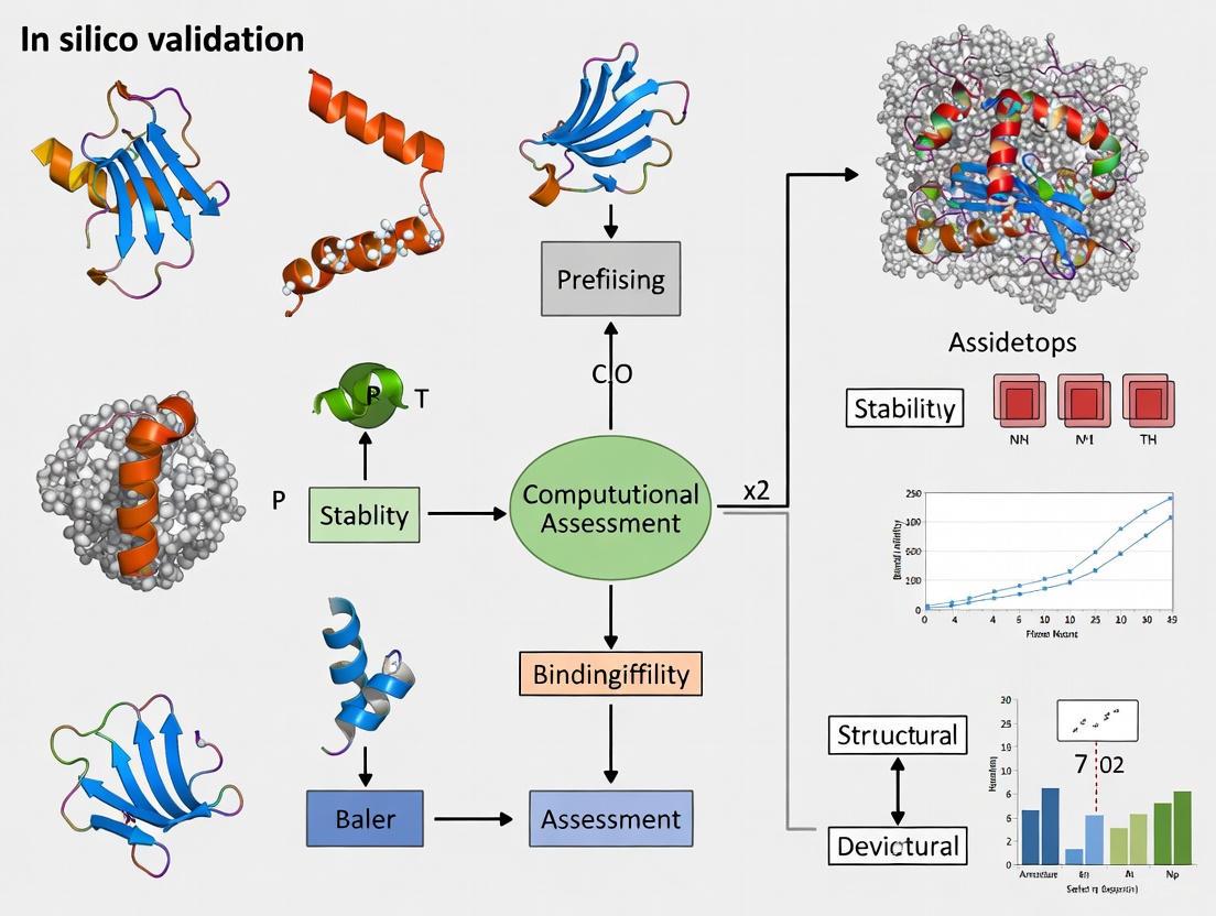
Once a protein is designed, comprehensive computational validation is critical to prioritize designs for costly and time-consuming experimental synthesis. This stage assesses the designed protein's stability, dynamics, and functional properties.
Molecular Dynamics (MD) Simulation Protocols
MD simulations serve as a "computational microscope" to observe protein behavior over time [21]. They are essential for probing conformational stability, folding pathways, and flexibility.
Protocol 3.1.1: Equilibrium Molecular Dynamics Simulation
This protocol is used to assess the structural stability and flexibility of a designed protein under simulated physiological conditions.
System Preparation:
- Obtain the initial atomistic coordinates from the design stage (e.g., from RFdiffusion or AlphaFold2 prediction).
- Solvate the protein in an explicit water box (e.g., TIP3P water model), ensuring a minimum distance of 1.0 nm between the protein and box edges.
- Add ions (e.g., Naâº, Clâ») to neutralize the system's charge and simulate a desired physiological salt concentration (e.g., 150 mM NaCl).
Energy Minimization:
- Use a steepest descent algorithm to minimize the energy of the solvated system, relieving any steric clashes. Run until the maximum force is below a threshold (e.g., 1000 kJ/mol/nm).
System Equilibration:
- Perform equilibration in two phases under NVT (constant Number of particles, Volume, and Temperature) and NPT (constant Number of particles, Pressure, and Temperature) ensembles.
- NVT Equilibration: Run for 100 ps while restraining protein heavy atoms, gradually heating the system to the target temperature (e.g., 310 K using a thermostat like Nosé-Hoover).
- NPT Equilibration: Run for 100 ps with restrained protein heavy atoms, coupling the system to a barostat (e.g., Parrinello-Rahman) to achieve the target pressure (e.g., 1 bar).
Production MD Run:
- Run an unrestrained simulation for a duration sufficient to observe relevant dynamics. For initial stability checks, 100 ns to 1 µs is typical. Use a time step of 2 fs.
- For ab initio accuracy, consider using AI-based MD systems like AI2BMD, which can simulate thousands of atoms with density functional theory (DFT)-level accuracy but are several orders of magnitude faster than conventional DFT [21].
Analysis:
- Calculate the root-mean-square deviation (RMSD) of the protein backbone to measure structural stability.
- Calculate the root-mean-square fluctuation (RMSF) of residues to identify flexible regions.
- Analyze the radius of gyration (Rg) to monitor compactness.
- Monitor the number and lifetime of intramolecular hydrogen bonds, a key metric for mechanical stability [19].
Protocol 3.1.2: Steered Molecular Dynamics (SMD) for Mechanical Strength
This protocol is used to quantitatively assess the mechanical unfolding resistance of a designed protein, which is particularly relevant for materials science applications [19].
- System Setup: Follow the steps in Protocol 3.1.1 for system preparation, energy minimization, and equilibration.
- Applying Force: Fix the C-terminal atom of the protein and apply a constant pulling force or a constant velocity to the N-terminal atom.
- Simulation Run: Run the simulation while the protein unfolds under the applied mechanical stress. Record the force and extension over time.
- Data Analysis: Plot a force-extension curve. The peak force observed before a major unfolding event is the unfolding force. Designs with maximized hydrogen-bond networks have demonstrated unfolding forces exceeding 1000 pN, about 400% stronger than natural titin domains [19].
AI-Based Ensemble and Trajectory Prediction
Static structures are insufficient to capture protein function. AI models are now being developed to predict the ensemble of conformations a protein can adopt, providing a more holistic view of dynamics [20].
Table 2: Computational Methods for Stability and Ensemble Validation
| Validation Method | Measured Property | Interpretation of Results |
|---|---|---|
| Equilibrium MD [21] | Root-mean-square deviation (RMSD), Radius of Gyration (Rg) | Low backbone RMSD (<0.2-0.3 nm) and stable Rg indicate a stable, folded design. |
| Steered MD [19] | Unfolding Force (picoNewtons, pN) | Higher forces indicate greater mechanical stability. >1000 pN is considered superstable. |
| Generative Models (e.g., AlphaFlow, DiG) [20] | Conformational Diversity & Root-mean-square fluctuation (RMSF) | Recovers flexible regions and alternative states; validates against experimental NMR data. |
| AI2BMD Folding/Unfolding [21] | Free Energy of Folding (ΔG) | A negative ΔG indicates a stable fold. Provides thermodynamic properties aligned with experiments. |
Advanced Workflow: Incorporating Ensemble Modeling
For a more thorough assessment, the core validation workflow can be enhanced with specialized ensemble and stability checks, as shown below:
The Scientist's Toolkit: Key Research Reagents and Computational Solutions
The following table details essential computational "reagents" – software, databases, and resources – required for executing the workflows described in this document.
Table 3: Essential Computational Reagents for Protein Design and Validation
| Resource Category & Name | Function in Workflow | Access Information |
|---|---|---|
| Design & Sequence Tools | ||
| ProteinMPNN [18] | Fast, robust protein sequence design for a fixed backbone. | Publicly available code repositories. |
| RFdiffusion [19] | De novo generation of novel protein structures from noise. | Publicly available code repositories. |
| Structure Prediction | ||
| AlphaFold2 [20] | Highly accurate protein structure prediction from sequence. | Publicly available; accessed via local installation or web APIs. |
| Simulation & Dynamics | ||
| AI2BMD [21] | Ab initio accuracy MD for large biomolecules; enables precise free-energy calculations. | Methodology described in literature; code availability may vary. |
| GROMACS [19] | High-performance classical MD simulation package. | Open-source software. |
| Data & Validation Resources | ||
| Protein Data Bank (PDB) [22] | Repository of experimentally determined 3D structures of proteins; used for training and validation. | Publicly accessible database (rcsb.org). |
| UniProt [22] | Comprehensive protein sequence and functional information. | Publicly accessible database (uniprot.org). |
| ATLAS / mdCATH [20] | Curated datasets of molecular dynamics trajectories; used for training and benchmarking ensemble models. | Publicly available datasets. |
| MS-Peg10-thp | MS-Peg10-thp, MF:C26H52O14S, MW:620.7 g/mol | Chemical Reagent |
| Amicoumacin B | Amicoumacin B, MF:C20H30N2O9, MW:442.5 g/mol | Chemical Reagent |
The integrated workflow of computational design, robust in silico validation, and experimental synthesis forms a powerful cycle for creating novel proteins with tailored functions. The protocols and tools outlined here provide a framework for researchers to rigorously assess the stability, dynamics, and functional potential of designed proteins before moving to the bench. As AI models for predicting ensemble properties and high-accuracy molecular dynamics simulations continue to mature, the reliability and precision of in silico validation will only increase, further accelerating the design-build-test cycle in synthetic biology and biotechnology.
Methodologies and Real-World Applications in Therapeutics and Engineering
The rapid expansion of protein sequence data has created a critical gap between known sequences and experimentally determined structures and functions. In silico computational methods have emerged as indispensable tools for bridging this gap, enabling researchers to predict protein properties, interactions, and functions with increasing accuracy. Among these methods, three deep learning architectures have demonstrated particular promise: Graph Neural Networks (GNNs), Convolutional Neural Networks (CNNs), and Transformer models. This article provides application notes and protocols for implementing these architectures within computational protein assessment research, framed specifically for drug development and protein engineering applications.
Architectural Foundations & Applications
Transformer Architectures in Protein Informatics
Transformer architectures, originally developed for natural language processing, have been successfully adapted for protein research due to their ability to process variable-length sequences and capture long-range dependencies through self-attention mechanisms [23]. The core innovation lies in the self-attention mechanism, which dynamically models pairwise relevance between elements in a protein sequence to explicitly capture intrasequence dependencies [23].
For protein sequences, the self-attention mechanism operates by defining three learnable weight matrices (Query, Key, and Value) that project input sequences into feature representations. The output is computed as a weighted sum of value vectors, with weights determined by compatibility between query and key vectors [23]. This architecture enables the model to learn complex relationships between amino acids that may be distant in the primary sequence but proximal in the folded structure.
Transformers have revolutionized multiple domains in protein science, including:
- Protein Structure Prediction: Models like ESMFold demonstrate near-experimental accuracy in predicting 3D structures from amino acid sequences [24] [25]
- Function Prediction: Self-supervised pre-training on large protein sequence databases enables accurate functional annotation without explicit supervision [23]
- Protein-Protein Interactions: Transformer-based models can predict interaction partners and binding affinities from sequence data alone [23]
Graph Neural Networks for Structure-Based Prediction
GNNs operate on graph-structured data, making them ideally suited for analyzing protein structures and interaction networks. In these representations, nodes typically correspond to amino acid residues or atoms, while edges represent spatial relationships or chemical bonds [26]. GNNs leverage message-passing algorithms to propagate information across the graph, enabling them to capture complex topological features essential for understanding protein function.
Key applications of GNNs in protein science include:
- Protein Function Prediction: GNNs learn representations from protein graphs at various granularity levels (atomic, residue, multi-scale) to predict Gene Ontology terms and interaction profiles [26]
- Protein-Protein Interaction Prediction: By modeling structural complexes as graphs, GNNs can predict binding interfaces and interaction strengths [26]
- Functional Annotation: GNN architectures leverage structural knowledge to improve the quality of protein function predictions beyond sequence-based methods [26]
Convolutional Neural Networks for Sequence-Pattern Recognition
CNNs employ hierarchical layers of filters that scan local regions of input data to detect spatially-localized patterns. For protein sequences, 1D-CNNs effectively identify conserved motifs, domain architectures, and sequence features that influence structure and function [27] [28].
Protocol implementations demonstrate CNNs applied to:
- Protein-Protein Interaction Prediction: Deep_PPI uses dual convolutional heads to process individual proteins before predicting interactions through fully connected layers [27]
- Protein Abundance Prediction: CNNs predict protein concentrations from mRNA levels by learning regulatory sequence motifs in untranslated and coding regions [28]
- Feature Extraction: Filter layers automatically detect biologically relevant patterns without manual feature engineering [28]
Table 1: Performance Comparison of Deep Learning Architectures on Key Protein Tasks
| Architecture | Application | Performance Metric | Value | Reference |
|---|---|---|---|---|
| Transformer (ESMFold) | Structure Prediction | Accuracy (Relative to Experimental) | Near-experimental | [24] [25] |
| 1D-CNN (Deep_PPI) | PPI Prediction (H. sapiens) | Accuracy | Superior to ML baselines | [27] |
| CNN | Protein Abundance Prediction (H. sapiens) | Coefficient of Determination (r²) | 0.30 | [28] |
| CNN | Protein Abundance Prediction (A. thaliana) | Coefficient of Determination (r²) | 0.32 | [28] |
| GNN | Gene Ontology Prediction | Quality Improvement | Promising | [26] |
Application Notes & Experimental Protocols
Protocol: Predicting Protein-Protein Interactions with Deep_PPI CNN
Objective: Predict binary protein-protein interactions from sequence information alone using a dual-branch convolutional neural network.
Materials:
- Protein sequences in FASTA format
- Known PPI pairs (positive and negative examples)
- Python 3.8+ with TensorFlow 2.8+ and Keras
- Hardware: GPU recommended for training
Methodology:
- Data Preparation:
- Curate balanced datasets of positive and negative interaction pairs
- Remove sequences with abnormal amino acids (B, J, O, U, X, Z)
- Filter out sequences shorter than 50 amino acids
- Apply PaddVal strategy to equalize sequence lengths (set to 90th percentile length)
Feature Engineering:
- Represent 20 native amino acids plus one special character using integers 1-21
- Apply one-hot encoding via Keras
one_hotfunction - Implement binary profile encoding with PaddVal strategy
- Generate 21-dimensional feature vectors for each residue position
Model Architecture:
- Implement two parallel 1D-CNN branches for each protein in a pair
- Each branch contains convolutional layers with ReLU activation
- Combine outputs from both branches via concatenation
- Add fully connected layers with decreasing dimensionality
- Final sigmoid activation for binary classification
Training Protocol:
- Initialize with He normal weight initialization
- Use Adam optimizer with learning rate 0.001
- Implement batch normalization between layers
- Apply five-fold cross-validation
- Monitor validation loss for early stopping
Validation:
- Evaluate on independent test sets for multiple species
- Compare against traditional machine learning baselines
- Assess generalization across organisms [27]
Protocol: Structure-Informed Function Prediction with GNNs
Objective: Predict protein function (Gene Ontology terms) from structural representations using graph neural networks.
Materials:
- Protein structures (PDB files or predicted structures)
- Gene Ontology annotations
- PyTorch Geometric or Deep Graph Library
- Hardware: GPU with ≥8GB memory
Methodology:
- Graph Construction:
- Represent proteins as graphs at atomic, residue, or multi-scale levels
- Nodes: amino acid residues with feature vectors (amino acid type, physicochemical properties)
- Edges: Spatial proximity (distance cutoffs 4-8Ã…) or chemical bonds
- Incorporate hierarchical relationships for multi-scale graphs
GNN Architecture Selection:
- Graph Attention Networks for weighted neighbor contributions
- Message Passing Neural Networks for information propagation
- Graph Convolutional Networks for hierarchical feature extraction
Model Implementation:
- Implement 3-6 graph convolution layers with skip connections
- Apply global pooling (attention-based or mean) for graph-level embeddings
- Add multi-layer perceptron head for GO term prediction
- Use multi-label loss function for simultaneous term prediction
Training Protocol:
- Use Adam optimizer with learning rate 0.001
- Implement class balancing for rare GO terms
- Apply gradient clipping to prevent explosion
- Regularize with dropout (0.2-0.5) between layers
Interpretation:
- Analyze attention weights to identify functionally important residues
- Visualize node contributions to predictions
- Map important regions to protein structures [26]
Protocol: Protein Abundance Prediction from Sequence with CNN
Objective: Predict protein abundance from mRNA expression levels and sequence features using a multi-input convolutional neural network.
Materials:
- Matched transcriptome-proteome datasets (mRNA TPM/FPKM, protein iBAQ)
- Sequence data (5' UTR, 3' UTR, CDS, protein sequence)
- TensorFlow 2.8+ with custom NaN-safe MSE loss function
- Hardware: GPU acceleration recommended
Methodology:
- Data Preprocessing:
- Convert all transcript abundances to TPM units
- Apply log2 transformation to iBAQ values
- Filter genes with <21 matched data points for robust regression
- Reserve genes with exactly 20 points as hold-out test set
Sequence Encoding:
- Extract 5' UTR, 3' UTR, CDS, and protein sequences
- Apply one-hot encoding (4 for nucleotides, 20 for amino acids)
- Pad sequences to uniform length for batch processing
- Implement custom generators for multi-input architecture
Multi-Input Architecture:
- Implement separate convolutional branches for each sequence type
- Each branch: 16 filters, kernel size 8-12, tanh activation, ReLU, sum pooling
- Process codon counts, nucleotide counts through dense layers
- Combine all features through intermediate dense layers (32→16 units)
Output Formulation:
- Final dense layer with 2 filters plus bias (a, b in: log2(iBAQ) = a * log2(TPM) + b)
- Expand for additional input genes: (2 + n genes) outputs
- Implement custom loss function handling varying valid data points per gene
Training Protocol:
- Use stochastic gradient descent without momentum
- Set learning rates based on input features (1e-3 to 1e-4)
- Train for 256-512 epochs with batch size 32
- Implement five independent repeats with tenfold cross-validation [28]
Visualization of Computational Workflows
CNN Protein-Protein Interaction Prediction Workflow
CNN PPI Prediction Flow
GNN Protein Function Prediction Workflow
GNN Function Prediction Flow
Transformer Protein Structure Prediction Workflow
Transformer Structure Prediction
Research Reagent Solutions
Table 2: Essential Computational Tools and Databases for Protein Informatics
| Resource | Type | Application | Access |
|---|---|---|---|
| ESM-2 | Transformer Model | Protein Structure & Function Prediction | GitHub |
| Deep_PPI | CNN Model | Protein-Protein Interaction Prediction | Research Code [27] |
| PyTorch Geometric | GNN Library | Protein Graph Representation Learning | Open Source |
| Protein Data Bank (PDB) | Structure Database | Experimental Structures for Training/Validation | Public Repository [25] |
| Swiss-Prot | Protein Database | Annotated Protein Sequences & Functions | Public Repository [27] |
| Gene Ontology Database | Functional Annotation | Protein Function Prediction Ground Truth | Public Repository [26] [28] |
| TensorFlow 2.8+ | Deep Learning Framework | Model Implementation & Training | Open Source [28] |
| TIM-1/GastroPlus | Physiological Modeling | GI Digestion Simulation & Validation | Commercial [29] |
The computational design of antibodies represents a frontier in modern biologics discovery, offering the potential to create novel therapeutics with precise target specificity. However, the accurate prediction of Complementarity-Determining Region (CDR) loop structures, particularly the hypervariable CDR-H3 loop, remains a primary challenge that directly impacts the developability of antibody-based therapeutics [30] [31]. CDR loops form the antigen-binding site and are critical for determining both affinity and specificity, yet their structural diversity and conformational flexibility present significant obstacles for computational modeling [30]. Recent advances in artificial intelligence (AI) and deep learning have revolutionized the field of antibody structure prediction, with specialized tools now achieving remarkable accuracy in CDR loop modeling [31] [32]. These improvements are essential for reliable developability assessment, which predicts the likelihood that an antibody candidate can be successfully developed into a manufacturable, stable, and efficacious drug [33]. This application note examines the current computational strategies for addressing CDR loop challenges and provides detailed protocols for incorporating developability assessment into early-stage antibody design workflows.
The CDR Loop Prediction Challenge
Structural Complexities of CDR Loops
Antibody binding specificity is primarily determined by six CDR loops - three each on the heavy (H1, H2, H3) and light (L1, L2, L3) chains [30]. While the antibody framework remains largely conserved, the CDR loops exhibit extraordinary structural diversity, with the CDR-H3 loop demonstrating the greatest variability in length, sequence, and structure [30] [31]. Five of the six loops typically adopt canonical cluster folds based on length and sequence composition, but the CDR-H3 loop largely defies such classification, making it the most challenging to predict accurately [30]. This challenge is compounded by the influence of relative VH-VL interdomain orientation on CDR-H3 conformation, as this loop is positioned directly at the interface between heavy and light chains [30].
Recent benchmarking studies reveal that even state-of-the-art prediction methods struggle with CDR-H3 accuracy. In comprehensive evaluations using high-quality crystal structures, current methods achieved average heavy atom RMSD values of 3.6-4.4 Ã… for CDR-H3 loops, significantly higher than errors for framework regions [31] [32]. These inaccuracies have direct consequences for downstream applications, including erroneous antibody-antigen docking results and unreliable biophysical property predictions such as surface hydrophobicity [30].
Critical Structural Inaccuracies and Their Impact
Computationally generated antibody models frequently contain structural inaccuracies that adversely affect developability assessments. Common issues include:
- Incorrect cis-amide bonds in CDR loops
- Wrong stereochemistry (D-amino acids instead of L-amino acids)
- Severe atomic clashes and non-physical bond lengths
- Inaccurate sidechain packing, particularly in CDR-H3 loops [30]
These errors significantly impact surface property predictions. Studies demonstrate that models containing cis-amide bonds and D-amino acids in CDR loops yield substantially different surface hydrophobicity profiles compared to experimental structures, potentially misleading developability assessments [30]. Since hydrophobicity is a conformation-dependent property, even small sidechain rearrangements can expose otherwise buried hydrophobic groups, altering perceived developability risk [30].
Table 1: Common Structural Inaccuracies in Antibody Models and Their Impact
| Structural Issue | Frequency in Models | Impact on Developability Assessment |
|---|---|---|
| Cis-amide bonds in CDRs | Up to 240 across 137 models [30] | Alters backbone conformation, affecting surface property predictions |
| D-amino acids | Up to 300 across 137 models [30] | Incorrect sidechain packing, misleading hydrophobicity estimates |
| Atomic clashes | Varies by modeling tool [30] | Physical implausibility, requires extensive refinement |
| Inaccurate CDR-H3 conformations | RMSD >2 Ã… in challenging cases [30] | Incorrect antigen-binding site characterization |
Computational Advances in Antibody Structure Prediction
AI-Driven Structure Prediction Tools
Recent years have witnessed transformative advances in antibody structure prediction, largely driven by deep learning approaches:
AlphaFold2 and Derivatives: While general protein structure predictors like AlphaFold2 (AF2) demonstrate remarkable accuracy for overall antibody structures (TM-scores >0.9) [31] [32], they show limitations for CDR-H3 loops, particularly for longer loops with limited sequence homologs [31]. This prompted the development of antibody-specific implementations.
Specialized Antibody Predictors: Tools such as ABlooper, DeepAb, IgFold, and Immunebuilder incorporate antibody-specific architectural adaptations to improve CDR loop modeling [30]. These tools typically achieve similar or better quality than general methods for antibody structures [30].
H3-OPT: This recently developed toolkit combines AF2 with a pre-trained protein language model, specifically targeting CDR-H3 accuracy [31] [32]. H3-OPT achieves a 2.24 Ã… average RMSD for CDR-H3 loops, outperforming other methods, particularly for challenging long loops [31]. The method employs a template module for high-confidence predictions and a PLM-based structure prediction module for difficult cases [34].
RFdiffusion for De Novo Design: Fine-tuned versions of RFdiffusion enable atomically accurate de novo design of antibodies by specifying target epitopes while maintaining stable framework regions [35]. This approach represents a paradigm shift from optimization to genuine de novo generation of epitope-specific binders.
Flow Matching for Antibody Design
FlowDesign represents an innovative approach that addresses limitations in current diffusion-based antibody design models [36]. By treating CDR design as a transport mapping problem, FlowDesign learns direct mapping from prior distributions to the target distribution, offering several advantages:
- Flexible prior distribution selection enabling integration of diverse knowledge
- Direct discrete distribution matching avoiding non-smooth generative processes
- High computational efficiency facilitating large-scale sampling [36]
In application to HIV-1 antibody design, FlowDesign successfully generated CDR-H3 variants with comparable or improved binding affinity and neutralization compared to the state-of-the-art HIV antibody ibalizumab [36].
Table 2: Performance Comparison of Antibody Structure Prediction Tools
| Tool | Methodology | CDR-H3 Accuracy (RMSD) | Strengths | Limitations |
|---|---|---|---|---|
| AlphaFold2 [31] | Deep learning with MSA | 3.79-3.92 Ã… [31] | High overall accuracy, excellent framework prediction | Limited CDR-H3 accuracy for long loops |
| ABlooper [30] | Antibody-specific deep learning | Similar to AF2 [30] | Fast prediction, antibody-optimized | May introduce structural inaccuracies |
| IgFold [31] | PLM-based | Comparable to AF2 [31] | Rapid prediction (seconds), high-throughput | Lower accuracy when templates available |
| H3-OPT [31] | AF2 + PLM | 2.24 Ã… (average) [31] | Superior CDR-H3 accuracy, template integration | Complex workflow, computational cost |
| RFdiffusion [35] | Diffusion-based de novo design | Atomic accuracy validated [35] | De novo design capability, epitope targeting | Requires experimental validation |
Experimental Protocols for Computational Developability Assessment
Protocol 1: Model Quality Validation with TopModel
Purpose: To identify and quantify structural inaccuracies in predicted antibody models that may affect developability assessments.
Materials:
- Predicted antibody 3D structures (PDB format)
- TopModel tool (https://github.com/liedllab/TopModel)
- PyMOL or similar visualization software [30]
Procedure:
- Input Preparation: Generate antibody structure models using preferred prediction tool(s). Save in PDB format.
- TopModel Analysis:
- Execute TopModel validation on each structure file
- Record counts of cis-amide bonds, D-amino acids, and atomic clashes
- Generate visualization output highlighting problematic regions
- Quality Assessment:
- Accept models with 0 D-amino acids and minimal cis-amide bonds (except cis-prolines)
- Flag models with severe clashes (>10% residues involved) for refinement
- Prioritize models passing all quality checks for further analysis
- Iterative Refinement: For failed models, consider alternative prediction tools or manual refinement of problematic regions.
Expected Results: Quality models should contain no D-amino acids, minimal non-proline cis-amide bonds, and fewer than 5% of residues involved in atomic clashes [30].
Protocol 2: Developability Assessment Using Therapeutic Antibody Profiler (TAP)
Purpose: To evaluate developability risk of antibody candidates based on surface physicochemical properties relative to clinical-stage therapeutics.
Materials:
- Antibody Fv sequences (VH and VL)
- ABodyBuilder2 for structure prediction (https://opig.stats.ox.ac.uk/webapps/abodybuilder2/)
- TAP implementation (https://github.com/oxpig/TAP)
- Reference dataset of clinical-stage therapeutic antibodies [37]
Procedure:
- Structure Modeling:
- Input VH and VL sequences into ABodyBuilder2
- Generate 3D structures for all candidates
- Validate model quality using Protocol 1
- TAP Analysis:
- Calculate five key developability metrics:
- Total CDR Length (Ltot)
- Patches of Surface Hydrophobicity (PSH)
- Positive Charge Patches (PPC)
- Negative Charge Patches (PNC)
- Spatial Aggregation Propensity (SAP)
- Compare metrics to distributions from clinical-stage therapeutics
- Calculate five key developability metrics:
- Risk Categorization:
- Assign "amber flags" to scores in 0th-5th or 95th-100th percentiles
- Assign "red flags" to scores beyond clinical-stage therapeutic ranges
- Calculate overall developability score for prioritization
- Visualization: Generate 3D surface representations colored by hydrophobicity/charge to identify problematic regions.
Expected Results: Developable candidates should show TAP metrics within the range of clinical-stage therapeutics, with minimal amber/red flags [37].
Figure 1: Computational Developability Assessment Workflow. This protocol integrates structure prediction, quality validation, and developability assessment in an iterative pipeline.
Protocol 3: De Novo Antibody Design with RFdiffusion
Purpose: To generate novel antibody binders targeting specific epitopes using diffusion-based generative models.
Materials:
- Target antigen structure (PDB format)
- Epitope specification (residue list)
- RFdiffusion implementation (https://github.com/RosettaCommons/RFdiffusion)
- ProteinMPNN for sequence design
- Fine-tuned RoseTTAFold2 for validation [35]
Procedure:
- Input Preparation:
- Prepare antigen structure with epitope residues specified
- Select antibody framework (e.g., humanized VHH for nanobodies)
- Define design regions (typically CDR loops)
- RFdiffusion Generation:
- Run fine-tuned RFdiffusion with epitope conditioning
- Generate 500-1000 backbone structures
- Filter based on interface quality and structural novelty
- Sequence Design with ProteinMPNN:
- Input selected backbones into ProteinMPNN
- Generate sequences optimized for stability and binding
- Filter sequences based on conservation and naturalness
- In Silico Validation:
- Use fine-tuned RoseTTAFold2 to predict complex structures
- Calculate interface metrics (ddG, buried surface area)
- Select top 50-100 designs for experimental testing
Expected Results: Initial designs typically exhibit modest affinity (tens to hundreds of nanomolar Kd), with potential for affinity maturation to single-digit nanomolar binders [35].
Table 3: Computational Tools for Antibody Design and Developability Assessment
| Tool Name | Type | Function | Access |
|---|---|---|---|
| TopModel [30] | Validation | Identifies structural inaccuracies (cis-amides, D-amino acids, clashes) | GitHub: liedllab/TopModel |
| ABodyBuilder2 [37] | Structure Prediction | Deep learning-based antibody modeling | Web server/API |
| H3-OPT [31] | Structure Prediction | Optimizes CDR-H3 loop prediction accuracy | Available upon request |
| RFdiffusion [35] | De Novo Design | Generates novel antibody binders to specified epitopes | GitHub: RosettaCommons/RFdiffusion |
| Therapeutic Antibody Profiler (TAP) [37] | Developability Assessment | Evaluates biophysical properties against clinical-stage therapeutics | GitHub: oxpig/TAP |
| FlowDesign [36] | CDR Design | Flow matching-based sequence-structure co-design | GitHub |
| IgFold [31] | Structure Prediction | PLM-based rapid antibody folding | GitHub |
The integration of advanced computational methods for antibody structure prediction and developability assessment represents a paradigm shift in biologics design. While challenges remain—particularly in accurate CDR-H3 loop prediction and structural validation—recent advances in AI-driven approaches now enable more reliable in silico profiling of antibody candidates. The protocols outlined in this application note provide a framework for systematic computational assessment, helping researchers identify developability risks early in the discovery process. As these methods continue to evolve, they promise to accelerate the development of novel antibody therapeutics with optimized properties for specialized administration routes and clinical applications.
The development of novel protein-based therapeutics represents a paradigm shift in modern medicine, rivaling and often surpassing traditional small-molecule drugs in treating complex diseases [38]. As of 2023, protein-based drugs are projected to constitute half of the top ten selling pharmaceuticals, with a global market approaching $400 billion [38]. This transformative growth has been catalyzed by advanced computational methodologies that enable researchers to preemptively address key development challenges including protein stability, immunogenicity, target specificity, and pharmacokinetic profiles.
In silico validation has emerged as a cornerstone of computational protein assessment, providing a critical framework for evaluating therapeutic potential before costly experimental work begins. These computational approaches allow researchers to simulate protein behavior under physiological conditions, predict interaction patterns with biological targets, and optimize structural characteristics for enhanced therapeutic efficacy. By integrating computational predictions with experimental validation, drug development professionals can accelerate the transition from candidate identification to clinical application while reducing development costs and failure rates.
The following application note details specific protocols and methodologies for leveraging in silico tools in the design and development of protein therapeutics and enzymes, with particular emphasis on practical implementation for research scientists.
Computational Assessment Protocols for Protein Therapeutics
In Silico Protein Digestibility Assessment
Protein digestibility represents a critical parameter in therapeutic development, directly influencing bioavailability and potential immunogenicity. Computational models can predict gastrointestinal stability, identifying sequences prone to enzymatic cleavage.
Protocol: In Silico Proteolytic Susceptibility Analysis
Purpose: To predict sites of proteolytic cleavage in simulated gastric and intestinal environments.
Methodology:
- Input Preparation: Obtain protein sequence in FASTA format and 3D structure (if available) in PDB format.
- Enzyme Selection: Configure computational digestion parameters for key proteases: pepsin (pH 2.0), trypsin, chymotrypsin, and pancreatin.
- Cleavage Simulation: Execute predictive algorithm based on:
- Known protease cleavage specificities
- Protein structural accessibility (surface exposure)
- Local flexibility parameters
- Output Analysis: Identify cleavage sites and generate semi-quantitative digestibility scores.
- Validation Correlation: Compare predictions with in vitro digestibility data when available.
Computational Tools: PEPSIM, ExPASy PeptideCutter, BIOVIA Discovery Studio
| Parameter | Implementation | Output Metrics |
|---|---|---|
| Protease Specificity | Position-specific scoring matrices | Cleavage probability scores |
| Structural Accessibility | Solvent-accessible surface area calculation | Relative susceptibility (0-1 scale) |
| Local Flexibility | B-factor analysis from PDB or molecular dynamics | Root mean square fluctuation (RMSF) |
| Digestibility Score | Composite algorithm weighting multiple factors | Predicted half-life, stability classification |
Interpretation Guidelines: Sequences with >80% predicted digestibility within 60 minutes are considered highly digestible; those with <20% digestibility are classified as resistant and may require further investigation for potential immunogenicity concerns [29].
Structure-Function Optimization Through Computational Engineering
Rational design of protein therapeutics employs computational tools to enhance stability, activity, and pharmacokinetic properties while reducing immunogenicity.
Protocol: Site-Directed Mutagenesis for Stability Enhancement
Purpose: To identify and validate amino acid substitutions that improve thermodynamic stability and reduce aggregation propensity.
Methodology:
- Structural Analysis: Load 3D protein structure and identify:
- Under-packed hydrophobic clusters
- Unsatisfied hydrogen bond donors/acceptors
- Unpaired charged residues
- Flexible regions with high B-factors
- Mutation Prediction: Utilize stability prediction algorithms (FoldX, RosettaDDG) to calculate ΔΔG values for potential mutations.
- Aggregation Propensity: Apply aggregation prediction algorithms (TANGO, AGGRESCAN) to screen stabilizing mutations for reduced aggregation potential.
- Structural Validation: Perform in silico structural analysis to confirm mutations do not disrupt active site or binding interfaces.
| Stabilization Strategy | Computational Approach | Therapeutic Example |
|---|---|---|
| Surface Charge Enhancement | Coulombic surface potential calculation | Supercharged GFP variants [38] |
| Hydrophobic Core Optimization | RosettaDesign packing quality assessment | Engineered antibody domains [38] |
| Disulfide Bridge Engineering | MODIP disulfide bond prediction | Engineered cytokines [38] |
| Glycosylation Site Addition | NetNGlyc/NetOGlyc prediction | Hyperglycosylated erythropoietin [38] |
Enzymatic Activity Optimization Through Computational Design
For enzyme therapeutics, computational methods can optimize catalytic efficiency, substrate specificity, and reaction conditions.
Protocol: Enzyme Assay Optimization Using Design of Experiments (DoE)
Purpose: To efficiently identify optimal assay conditions for enzymatic characterization using computational experimental design.
Methodology:
- Factor Identification: Select critical parameters for optimization (buffer pH, ionic strength, substrate concentration, enzyme concentration, cofactors, temperature).
- Experimental Design: Implement fractional factorial design to screen multiple factors simultaneously.
- Response Surface Methodology: Model the relationship between factors and enzymatic activity.
- Condition Prediction: Identify optimal assay conditions from modeled response surface.
- Validation: Experimentally verify predicted optimal conditions.
Implementation Note: This DoE approach can reduce optimization time from >12 weeks (traditional one-factor-at-a-time) to under 3 days for identifying significant factors and optimal conditions [39].
Diagram 1: In silico protein assessment workflow for therapeutic development.
Advanced Application: Targeted Delivery Systems
Computational approaches enable the design of protein therapeutics with enhanced tissue-specific targeting capabilities, particularly for challenging targets like intracellular sites and the blood-brain barrier.
Protocol: Ligand-Receptor Interaction Modeling for Targeted Delivery
Purpose: To design and optimize protein conjugates for tissue-specific targeting through computational prediction of ligand-receptor binding.
Methodology:
- Target Identification: Select target receptor based on tissue-specific expression (e.g., transferrin receptor for blood-brain barrier targeting).
- Molecular Docking: Perform rigid/flexible docking of potential targeting ligands against receptor structure.
- Binding Affinity Calculation: Calculate binding free energies using MM-PBSA/GBSA methods.
- Linker Optimization: Design optimal linkers between therapeutic protein and targeting moiety using molecular dynamics simulations.
- Validation: Predict cellular uptake efficiency through probabilistic models of receptor-mediated endocytosis.
Application Example: Proteins covalently conjugated to multiple copies of the transferrin aptamer show preferential accumulation in the brain relative to native proteins, as predicted through computational modeling and confirmed experimentally [38].
Diagram 2: Computational workflow for designing targeted protein therapeutics.
Essential Research Reagents and Computational Tools
The following table details key resources for implementing the described computational protocols in protein therapeutic development.
| Category | Specific Tools/Reagents | Application in Protein Therapeutic Development |
|---|---|---|
| Structure Prediction | AlphaFold2, RosettaFold, I-TASSER | De novo protein structure prediction for targets without experimental structures |
| Molecular Dynamics | GROMACS, AMBER, NAMD | Simulation of protein dynamics, stability, and binding events |
| Docking Software | AutoDock Vina, HADDOCK, SwissDock | Prediction of protein-ligand and protein-protein interactions |
| Stability Analysis | FoldX, CUPSAT, PoPMuSiC | Calculation of mutation effects on protein stability (ΔΔG) |
| Digestibility Prediction | PeptideCutter, PEPSIM | In silico simulation of gastrointestinal proteolysis |
| Immunogenicity Prediction | NetMHCIIpan, IEDB tools | Prediction of T-cell epitopes for reducing immunogenic potential |
| Expression Optimization | OPTIMIZER, GeneDesign | Codon optimization for recombinant expression in host systems |
Regulatory Considerations and Validation
The integration of computational assessments into regulatory frameworks is evolving, with agencies including the European Medicines Agency (EMA) and U.S. Food and Drug Administration (FDA) increasingly acknowledging the role of in silico approaches [29]. According to recent European Food Safety Authority (EFSA) guidance, "In silico tools aiming at predicting the behaviour of a protein in relation to gastrointestinal digestion can complement but not substitute in vitro digestibility experiments" [29]. This underscores the importance of coupled computational-experimental validation strategies.
Critical limitations of current computational approaches include simplified enzyme specificity modeling, exclusion of key physiological factors like protein folding and post-translational modifications, and lack of dynamic gastrointestinal conditions in digestibility models [29]. Future developments in digital twin methodology and more sophisticated physiologically based kinetic (PBK) models show promise for enhancing predictive accuracy [29].
Computational protein assessment represents a transformative approach in the design and development of novel protein therapeutics and enzymes. The protocols detailed in this application note provide a framework for leveraging in silico tools to address key development challenges including stability, activity, specificity, and delivery. While computational methods continue to evolve, their integration with experimental validation provides a powerful strategy for accelerating therapeutic development and enhancing success rates in clinical translation. As the field advances, increased sophistication in predictive modeling and broader regulatory acceptance will further solidify the role of in silico approaches in the protein therapeutic development pipeline.
Application Notes: The Role of Automation in Computational Protein Assessment
The integration of artificial intelligence (AI) and automated platforms is fundamentally reshaping computational protein science. These tools are transitioning from specialized assets to accessible resources that accelerate in silico validation, a process critical for modern drug development and nutritional assessment [29] [40]. This shift enables researchers to predict protein behavior, function, and interactions with a speed and scale previously unimaginable, supporting a paradigm where computational evidence is increasingly accepted in regulatory submissions [40].
Key Quantitative Metrics for Automated Protein Assessment
The performance of AI-driven tools for protein analysis is benchmarked using standardized quantitative metrics. The following table summarizes key performance indicators from recent studies.
Table 1: Performance Metrics of Selected Computational Protein Assessment Tools
| Tool Name | Primary Application | Key Performance Metric | Reported Value/Outcome | Context / Dataset |
|---|---|---|---|---|
| Deep_PPI [27] | Protein-Protein Interaction (PPI) prediction | Predictive Accuracy | Surpassed existing state-of-the-art PPI methods | Validation on multiple species datasets (Human, C. elegans, E. coli) |
| I-TASSER [41] | Protein structure prediction & design validation | RMSD to Target Structure | <2 Ã… in 62% of cases for top designed sequence | Tested on 52 non-homologous proteins |
| I-TASSER [41] | Protein structure prediction & design validation | RMSD to Target Structure | Increased to 77% when considering top 10 designed sequences | Tested on 52 non-homologous proteins |
| Clustering-based Protein Design [41] | Native sequence recapitulation | Average Sequence Identity to Native | 24% for first cluster tag | 52 non-homologous single-domain proteins |
| Clustering-based Protein Design [41] | Native sequence recapitulation | Average Core Identity to Native | 42% for highest-identity cluster tag | 52 non-homologous single-domain proteins |
AI Automation Platforms for Research Workflows
Beyond domain-specific protein tools, a new class of AI-native automation platforms is emerging. These platforms help orchestrate complex, multi-step computational and experimental workflows, making sophisticated in silico protocols more accessible and reproducible.
Table 2: AI Automation Platforms for Research Workflow Management
| Platform Name | Best For | Key AI Feature | Application in Research |
|---|---|---|---|
| Lindy [42] | General-purpose AI agents | No-code creation of custom AI agents ("Lindies") | Automating literature review, data synthesis, and routine analysis tasks |
| Gumloop [42] | Technical, developer-focused automation | Chrome extension for browser action recording | Web scraping public biological data, automating data entry into databases |
| Vellum.ai [42] | LLM-driven agent development | Natural language prompt building and orchestration | Designing multi-step AI agents for complex data analysis pipelines |
| Relevance AI [42] | Open-ended agentic workflows | Sub-agent creation for complex tasks | Building a "team" of AI agents where each specializes in a different analytical task |
| VectorShift [42] | Technical teams & multi-LLM workflows | Drag-and-drop Pipelines with Python SDK | Building and deploying complex simulation workflows that leverage multiple AI models |
Experimental Protocols
Protocol: Deep Learning-Based Prediction of Protein-Protein Interactions (PPIs)
This protocol describes the methodology for using the Deep_PPI model to predict interactions solely from protein sequences [27].
2.1.1 Background Protein-Protein Interactions (PPIs) are fundamental to most biological processes. Accurate computational prediction of PPIs accelerates the understanding of cellular mechanisms and the identification of novel drug targets. The Deep_PPI model employs a deep learning architecture to achieve high prediction accuracy across multiple species.
2.1.2 Materials
- Benchmark Datasets: Publicly available PPI datasets for the species of interest (e.g., Human, C. elegans, E. coli, H. sapiens). These datasets contain curated positive and negative protein interaction pairs [27].
- Computational Environment: A Python environment with deep learning libraries such as Keras and TensorFlow.
- DeepPPI Framework: The DeepPPI model, which utilizes a one-dimensional convolutional neural network (1D-CNN).
2.1.3 Procedure
- Data Preparation:
- Tokenization and Padding: Use the
PaddValstrategy to ensure all protein sequences in a pair have the same length. The value is typically set to the length of the 90th percentile of proteins in the dataset [27]. - Sequence Encoding: Represent each of the 20 native amino acids and one special character with an integer from 1 to 21. Subsequently, use the Keras
one-hotencoding function to convert each residue in the padded sequences into a binary vector [27].
- Tokenization and Padding: Use the
- Model Training and Prediction:
- Feature Extraction: Feed the encoded protein sequences into the Deep_PPI model.
- Convolutional Processing: The model processes each protein pair through two separate 1D-CNN "heads" to extract high-level features.
- Concatenation and Classification: The outputs from the two branches are concatenated and passed through a fully connected layer to generate the final prediction (interacting or non-interacting) [27].
2.1.4 Visualization of Workflow The following diagram illustrates the Deep_PPI prediction workflow, from sequence input to final classification.
Protocol: In Silico Assessment of Designed Protein Sequences
This protocol is used to validate whether a computationally designed amino acid sequence will fold into the intended target protein structure, a critical step in protein engineering [41].
2.2.1 Background Computational protein design aims to discover novel sequences that fold into a target structure. This protocol uses a combination of free-energy minimization, sequence clustering, and folding simulations to select and validate designed sequences.
2.2.2 Materials
- Target Backbone Structure: A Protein Data Bank (PDB) file containing the desired 3D structure.
- Computational Tools:
- Computational Resources: Workstation or computing cluster capable of running Monte Carlo simulations and homology modeling.
2.2.3 Procedure
- Sequence Generation via Monte Carlo Minimization:
- Perform multiple independent runs (e.g., 10) of Monte Carlo sampling to explore the sequence space. The FoldX force field is used to evaluate the free energy of each sequence decoy, with side chains modeled by SCWRL [41].
- Sequence Selection by Clustering:
- Cluster all generated sequence decoys based on "sequence distance" calculated using the BLOSUM62 matrix.
- Select the center sequence (cluster tag) from the largest cluster as the primary candidate. Additional sequences from other large clusters can also be selected for validation [41].
- Validation by Folding Simulation:
- Submit the selected designed sequences to the I-TASSER server for ab initio structure prediction.
- I-TASSER generates 3D models by iteratively assembling fragments from threading templates [41].
- Analysis of Results:
- Calculate the Root-Mean-Square Deviation (RMSD) between the I-TASSER-predicted structure and the original target backbone.
- A low RMSD (e.g., <2 Ã…) indicates that the designed sequence successfully folds into the target structure, validating the design [41].
2.2.4 Visualization of Workflow The following diagram outlines the multi-stage process for designing and validating a novel protein sequence.
The Scientist's Toolkit: Research Reagent Solutions
This section details essential computational tools and platforms that form the backbone of modern in silico protein assessment workflows.
Table 3: Essential Computational Tools for Automated Protein Research
| Tool / Platform Name | Type | Primary Function in Protein Assessment |
|---|---|---|
| I-TASSER/I-TASSER-MTD [43] | Structure Prediction Server | Predicts 3D protein structures and functions from amino acid sequences, including for multi-domain proteins. |
| AlphaFold/ColabFold [43] [40] | Structure Prediction Tool | Provides highly accurate protein structure predictions using deep learning; accessible via web or local installation. |
| trRosetta [43] | Structure Prediction Server | Web-based platform for fast and accurate protein structure prediction powered by deep learning and Rosetta. |
| FoldX [41] | Force Field / Algorithm | Calculates the free energy of protein structures and models, crucial for assessing stability and designing mutations. |
| SCWRL [41] | Modeling Tool | Predicts the optimal side-chain conformations for a given protein backbone and amino acid sequence. |
| RosettaAntibody & SnugDock [43] | Specialized Modeling Suite | Models antibody structures from sequence and docks them to protein antigens to predict immune complexes. |
| ClusPro [43] | Docking Server | Performs rigid-body docking of two proteins to generate models of protein-protein complexes. |
| AutoDock Suite [43] | Docking Software | Performs computational docking and virtual screening to study protein-ligand interactions for drug discovery. |
| HADDOCK [43] | Docking Server | Integrates experimental data to guide the 3D modeling of biomolecular complexes. |
| Phyre2 [43] | Protein Modeling Portal | Predicts protein structure, function, and ligand binding sites using remote homology detection. |
| Q11 peptide | Q11 peptide, MF:C70H99N19O20, MW:1526.6 g/mol | Chemical Reagent |
| BP Fluor 546 DBCO | BP Fluor 546 DBCO, MF:C52H47Cl3N4O11S3, MW:1106.5 g/mol | Chemical Reagent |
Navigating Pitfalls: Accuracy, Specificity, and Model Limitations
Common Failure Points in Protein-Ligand and Protein-Protein Interaction Modeling
In silico validation computational protein assessment research is a cornerstone of modern drug discovery and basic biological research. The accurate prediction of how proteins interact with small molecules (protein-ligand) and other proteins (protein-protein) is crucial for understanding disease mechanisms and developing new therapeutics. However, both modeling approaches face significant challenges that can compromise prediction reliability. This application note details the common failure points across these domains, provides structured experimental protocols for model validation, and offers visualization tools to guide researchers in avoiding these pitfalls. As deep learning (DL) continues to transform both molecular docking and PPI prediction, understanding these limitations becomes increasingly critical for translating computational predictions into biomedical reality [44] [45].
Protein-Ligand Docking Failure Points
Protein-ligand docking aims to predict the three-dimensional structure of a protein-ligand complex and estimate their binding affinity. Traditional physics-based docking tools face limitations due to their reliance on empirical rules and heuristic search algorithms, which result in computationally intensive processes and inherent inaccuracies [44]. While DL-based docking methods can overcome some limitations by extracting complex patterns from vast datasets, they introduce new challenges.
Performance and Physical Plausibility Issues
A comprehensive multidimensional evaluation of docking methods reveals a striking performance stratification across traditional, hybrid, and DL-based approaches [44]. The table below summarizes key failure metrics across different docking methodologies:
Table 1: Performance Comparison of Docking Methodologies Across Benchmark Datasets
| Method Category | Method | Pose Accuracy (RMSD ≤ 2 Å) | Physical Validity (PB-valid) | Combined Success Rate |
|---|---|---|---|---|
| Traditional | Glide SP | 85.88% (Astex) | 97.65% (Astex) | 84.71% (Astex) |
| Generative Diffusion | SurfDock | 91.76% (Astex) | 63.53% (Astex) | 61.18% (Astex) |
| Regression-based | KarmaDock | 22.35% (Astex) | 25.88% (Astex) | 5.88% (Astex) |
| Hybrid | Interformer | 68.24% (Astex) | 89.41% (Astex) | 62.35% (Astex) |
Generative diffusion models like SurfDock achieve exceptional pose accuracy with RMSD ≤ 2 Å success rates exceeding 70% across all datasets, yet exhibit suboptimal physical validity scores (as low as 40.21% on the DockGen dataset of novel protein binding pockets) [44]. This reveals deficiencies in modeling critical physicochemical interactions, such as steric clashes or hydrogen bonding, despite favorable RMSD scores. Regression-based models perform particularly poorly, often failing to produce physically valid poses, with combined success rates (RMSD ≤ 2 Å & PB-valid) as low as 5.88% on benchmark tests [44].
Generalization and Sampling Challenges
DL docking methods exhibit significant challenges in generalization, particularly when encountering novel protein binding pockets unseen during training [44] [46]. Performance degradation is pronounced in real-world scenarios such as:
- Apo-protein docking: Using predicted or unbound protein structures rather than holo-structures [47] [46]
- Cross-docking: Docking to alternative receptor conformations from different ligand complexes [46]
- Multi-ligand docking: Binding multiple ligands concurrently to a target protein [47]
The PoseBench benchmark reveals that DL co-folding methods generally outperform conventional and DL docking baselines, yet popular methods such as AlphaFold 3 still struggle with prediction targets featuring novel binding poses [47]. Furthermore, certain DL co-folding methods demonstrate high sensitivity to input multiple sequence alignments, while others struggle to balance structural accuracy with chemical specificity when predicting novel or multi-ligand targets [47].
Scoring Function Limitations
A critical failure point in practical docking applications is the inaccurate ranking of compounds by predicted binding affinity. Receiver operating characteristic (ROC) analysis of eight free-license docking programs revealed that most lack specificity, frequently misidentifying true negatives [48]. The use of convolutional neural network (CNN) scores, such as those implemented in GNINA, can improve true positive identification when applied as a filter before affinity ranking [48].
Protein-Protein Interaction Prediction Failure Points
Computational prediction of PPIs from amino acid sequences remains challenging despite advances in deep learning [49]. While high-throughput experimental methods exist, they remain costly, slow, and resource-intensive, creating dependence on computational approaches [27] [45].
Data and Generalization Limitations
Table 2: Common Failure Points in PPI Prediction Models
| Failure Category | Specific Issue | Impact on Prediction Accuracy |
|---|---|---|
| Data Limitations | Sparse experimental PPI data | Limited training examples, especially for non-model organisms |
| Class imbalance | Bias toward non-interacting pairs in many datasets | |
| Data leakage | Overestimation of performance due to similar sequences in training and test sets | |
| Generalization Issues | Cross-species prediction | Performance degradation with evolutionary distance from training data |
| Novel protein families | Poor performance on proteins with low similarity to training examples | |
| Mutation effects | Difficulty predicting how mutations alter existing interactions |
PLMs, while revolutionary for protein structure prediction, face inherent limitations for PPI prediction as they are primarily trained using single protein sequences and lack "awareness" of interaction partners [49]. In conventional PLM-based PPI predictors, a classification head must extrapolate signals of inter-protein interactions by grouping common patterns of intra-protein contacts, which has limited parameters to deal with complex interaction patterns [49].
Performance evaluation reveals significant degradation when models trained on human PPI data are tested on evolutionarily distant species. While PLM-interact achieves AUPR improvements of 2-28% over other methods, its performance on yeast and E. coli (AUPR of 0.706 and 0.722, respectively) remains substantially lower than on more closely related species [49].
Structural and Dynamic Considerations
Sequence-based PPI predictors face inherent limitations compared to structure-based approaches, including:
- Intrinsic disorder: Approximately 30-40% of the human proteome contains intrinsically disordered regions that lack clearly defined structure, challenging accurate prediction [50]
- Conformational dynamics: Proteins with dynamic conformations (e.g., switching between apo- and holo-states) are poorly modeled by current predictors [50]
- Structural quality dependence: Structure-based methods require high-quality structures, which are unavailable for many proteins [50]
Despite these limitations, sequence-based methods remain broadly applicable due to the relative scarcity of high-quality protein structures and can succeed where structure-based methods fail, as demonstrated by PepMLM's successful design of peptide binders where RFDiffusion (structure-based) failed [50].
Experimental Protocols for Validation
Protocol 1: Comprehensive Docking Validation
Purpose: To systematically evaluate protein-ligand docking method performance across multiple critical dimensions.
Materials:
- Benchmark datasets (Astex diverse set, PoseBusters set, DockGen)
- Docking software (traditional, DL-based, and hybrid methods)
- PoseBusters toolkit for chemical and geometric consistency checking
- RMSD calculation scripts
Procedure:
- Pose Prediction Accuracy Assessment:
- Dock each ligand in benchmark sets to its corresponding protein
- Calculate RMSD between predicted pose and experimental reference
- Determine success rate using RMSD ≤ 2.0 Å threshold
Physical Validity Check:
- Run PoseBusters validation on all predicted complexes
- Assess bond lengths, angles, stereochemistry, and steric clashes
- Calculate percentage of physically valid poses
Virtual Screening Evaluation:
- Perform docking with known binders and decoys
- Generate ROC curves and calculate AUC values
- Apply CNN score cutoff (0.9 recommended) before affinity ranking [48]
Generalization Testing:
- Evaluate performance on novel protein binding pockets (DockGen)
- Test cross-docking to alternative receptor conformations
- Assess apo-protein docking using unbound structures
Protocol 2: PPI Prediction Cross-Species Validation
Purpose: To rigorously assess PPI prediction model generalization across evolutionarily distant species.
Materials:
- Multi-species PPI datasets (human, mouse, fly, worm, yeast, E. coli)
- PPI prediction tools (PLM-interact, TUnA, TT3D, etc.)
- Computing resources capable of handling large protein language models
Procedure:
- Dataset Curation:
- Obtain experimentally verified positive PPIs from dedicated databases
- Generate negative pairs using non-interacting proteins from different cellular compartments
- Apply stringent filters to avoid data leakage (≤30% sequence similarity)
Model Training:
- Train models exclusively on human PPI data
- Use appropriate validation sets for hyperparameter tuning
- For PLM-interact, use 1:10 ratio between classification loss and mask loss [49]
Cross-Species Evaluation:
- Test trained models on held-out species (mouse, fly, worm, yeast, E. coli)
- Calculate AUPR and AUROC for each species
- Analyze performance degradation with evolutionary distance
Mutation Effect Analysis:
- Fine-tune model on wild-type and mutant protein pairs
- Predict interaction changes for known disruptive mutations
- Validate predictions against experimental mutation data from IntAct [49]
Visualization of Validation Workflows
Protein-Ligand Docking Validation
PPI Prediction Cross-Species Validation
Research Reagent Solutions
Table 3: Essential Research Reagents and Tools for Interaction Modeling
| Category | Tool/Resource | Primary Function | Application Context |
|---|---|---|---|
| Benchmark Datasets | Astex Diverse Set | Evaluate pose prediction accuracy | Protein-ligand docking validation |
| PoseBusters Benchmark | Assess physical plausibility of complexes | Steric clash and geometry validation | |
| DockGen | Test generalization to novel binding pockets | Method robustness assessment | |
| Validation Tools | PoseBusters Toolkit | Chemical and geometric consistency checking | Automated validation of predicted structures |
| PLM-interact | Protein-protein interaction prediction | Cross-species PPI forecasting | |
| Software Solutions | GNINA with CNN scoring | Improved true positive identification | Virtual screening specificity enhancement |
| DiffDock | Diffusion-based docking pose generation | Handling flexible ligand docking | |
| Data Resources | STRING Database | Known and predicted protein interactions | PPI prediction training and validation |
| PDBBind | Experimentally determined binding data | Docking method training and testing | |
| IntAct Mutation Data | Experimentally verified mutation effects | PPI mutation impact analysis |
This application note has detailed the common failure points in both protein-ligand and protein-protein interaction modeling, highlighting that while DL methods offer significant advances, they introduce new challenges including physical implausibility, generalization limitations, and scoring inaccuracies. The provided experimental protocols and visualization workflows offer structured approaches for rigorous model validation. As the field continues to evolve, researchers must maintain critical assessment of both traditional and DL-based methods, recognizing that each approach has distinct strengths and limitations. Systematic validation across multiple dimensions—pose accuracy, physical validity, interaction recovery, and generalization capability—remains essential for advancing robust computational protein assessment research.
In the realm of in silico validation for computational protein assessment, the performance of predictive algorithms is paramount. The metrics of sensitivity and specificity serve as critical indicators of algorithmic reliability, yet their relationship often exhibits a characteristic divergence where improving one can compromise the other. This application note delineates structured methodologies for quantitatively assessing this trade-off, providing researchers, scientists, and drug development professionals with standardized protocols for rigorous computational evaluation. The framework is contextualized within protein structure prediction and interaction analysis—a domain where accurate performance assessment directly impacts research validity and therapeutic development pipelines. Based on contemporary research, this document synthesizes evaluation strategies to guide the selection and optimization of computational tools for specific research objectives.
Theoretical Foundations and Key Metrics
Sensitivity (True Positive Rate) measures the proportion of actual positives correctly identified, while Specificity (True Negative Rate) measures the proportion of actual negatives correctly identified. In computational protein assessment, this often translates to correctly identifying interacting residues or accurate structural features (sensitivity) versus correctly excluding non-interacting residues or inaccurate features (specificity) [51] [52].
The inverse relationship between sensitivity and specificity defines the Receiver Operating Characteristic (ROC) curve. The Area Under the ROC Curve (AUC) provides a single scalar value measuring overall performance across all thresholds [52]. However, holistic AUC can mask critical performance in operationally relevant ranges, necessitating analysis of specific curve regions [52].
Solution Divergence is a related concept referring to the presence of multiple viable solutions or predictions for a single problem. Recent studies indicate that higher solution divergence correlates with enhanced problem-solving abilities in computational models, suggesting its value as a complementary metric for algorithm assessment [53].
Quantitative Metrics Table
Table 1: Key Performance Metrics for Algorithmic Assessment
| Metric | Calculation | Interpretation | Optimal Range |
|---|---|---|---|
| Sensitivity | TP / (TP + FN) | Proportion of true positives detected | Close to 1.0 |
| Specificity | TN / (TN + FP) | Proportion of true negatives correctly excluded | Close to 1.0 |
| AUC-ROC | Area under ROC curve | Overall discriminative ability | 0.9-1.0 (Excellent) |
| Solution Divergence | Spectral analysis of prediction variants [53] | Diversity of valid solutions | Context-dependent |
Experimental Protocols for Performance Assessment
Protocol 1: Threshold-Dependent Sensitivity-Specificity Analysis
This protocol evaluates algorithm performance across different classification thresholds, enabling identification of optimal operating points.
Materials and Reagents:
- Benchmark dataset with known positives and negatives
- Computational algorithm for assessment
- High-performance computing resources
Procedure:
- Dataset Preparation: Curate a gold-standard dataset with verified positive and negative instances. For protein disorder prediction, utilize datasets from Protein Data Bank with disorder residues annotated [51].
- Algorithm Prediction: Execute target algorithm across the benchmark dataset without applying a predefined threshold.
- Threshold Variation: Apply classification thresholds from 0.01 to 0.99 in increments of 0.01 [51].
- Performance Calculation: At each threshold, calculate:
- True Positives (TP), False Positives (FP), True Negatives (TN), False Negatives (FN)
- Sensitivity = TP / (TP + FN)
- Specificity = TN / (TN + FP)
- ROC Construction: Plot sensitivity against (1 - specificity) across all thresholds [51].
- AUC Calculation: Compute the area under the ROC curve using numerical integration methods.
Technical Notes:
- For large-scale datasets (e.g., >2000 sequences), automated scripting is essential [51].
- Account for potential class imbalance through stratified sampling or weighted metrics.
Protocol 2: Region of Interest (ROI) Performance Enhancement
Traditional AUC optimization may not guarantee performance in critical operational ranges. This protocol enhances sensitivity at high-specificity regions through targeted optimization [52].
Materials and Reagents:
- Pre-trained model
- Task-specific fine-tuning dataset
- AUCReshaping implementation [52]
Procedure:
- ROI Definition: Identify the critical specificity range based on application requirements (e.g., 90-98% specificity for abnormality detection) [52].
- Baseline Performance: Evaluate baseline model sensitivity at the target specificity.
- AUCReshaping Application:
- Identify positive class samples misclassified at high-specificity thresholds
- Iteratively boost weights for these samples during training
- Compute modified loss value and backpropagate [52]
- Validation: Apply the high-specificity threshold determined during validation to testing phase.
- Performance Assessment: Compare sensitivity at target specificity before and after reshaping.
Technical Notes:
- AUCReshaping has demonstrated sensitivity improvements of 2-40% at high-specificity levels in binary classification tasks [52].
- The method is particularly valuable for class-imbalanced datasets common in medical imaging and anomaly detection.
Protocol 3: Multi-Algorithm Comparative Assessment
This protocol enables direct comparison of multiple algorithmic approaches for protein structure prediction, assessing their relative strengths across different peptide characteristics [54].
Materials and Reagents:
- Set of target peptides with varied physicochemical properties
- Multiple prediction algorithms (AlphaFold, PEP-FOLD, Threading, Homology Modeling)
- Molecular dynamics simulation software
- Validation tools (Ramachandran plot, VADAR)
Procedure:
- Peptide Selection: Curate a diverse set of peptides (e.g., 10 peptides randomly selected from human gut AMPs) [54].
- Structure Prediction: Apply each algorithm to predict structures for all peptides.
- Quality Assessment: Analyze predicted structures using:
- Ramachandran plot analysis
- VADAR structural validation
- Molecular dynamics simulations (100ns each) [54]
- Performance Correlation: Correlate algorithm performance with peptide properties (hydrophobicity, charge, disorder propensity).
- Algorithm Suitability Mapping: Identify optimal algorithm choices for different peptide types.
Technical Notes:
- AlphaFold and Threading complement each other for hydrophobic peptides [54].
- PEP-FOLD and Homology Modeling show advantages for hydrophilic peptides [54].
- PEP-FOLD generally provides compact structures with stable dynamics [54].
Computational Workflows and Visualization
Performance Assessment Workflow
Diagram 1: Performance assessment workflow
Multi-Algorithm Comparison Logic
Diagram 2: Multi-algorithm comparison logic
Research Reagent Solutions
Table 2: Essential Computational Tools for Protein Assessment
| Tool/Resource | Type | Primary Function | Application Context |
|---|---|---|---|
| AlphaFold2 [55] [54] | Structure Prediction Algorithm | Predicts 3D protein structures from sequence | Protein complex prediction; interaction screening |
| DISpro [51] | Disorder Prediction Tool | Identifies protein disorder regions with adjustable sensitivity/specificity | Structural genomics; function annotation |
| PEP-FOLD3 [54] | De Novo Peptide Modeling | Predicts structures for short peptides (5-50 amino acids) | Antimicrobial peptide design |
| AUCReshaping [52] | Performance Optimization | Reshapes ROC curve to enhance sensitivity at high-specificity | Medical imaging; anomaly detection |
| RaptorX [54] | Property Prediction Server | Predicts secondary structure, solvent accessibility, and disorder regions | Structure-property analysis |
| Modeller [54] | Homology Modeling | Comparative protein structure modeling | Template-based structure prediction |
The systematic assessment of sensitivity-specificity divergence provides critical insights for selecting and optimizing computational algorithms in protein research. The protocols outlined herein enable researchers to move beyond singular metric optimization toward comprehensive algorithmic evaluation. By implementing threshold-dependent analysis, region-of-interest enhancement, and multi-algorithm comparison, scientists can make informed decisions aligned with specific research objectives. As computational methods continue to advance in structural bioinformatics, rigorous performance assessment remains fundamental to validating predictive models and ensuring research reproducibility in drug development pipelines.
The revolutionary advancements in AI-based protein structure prediction, acknowledged by the 2024 Nobel Prize in Chemistry, have created a paradigm shift in structural biology [56]. However, proteins are not static entities; their functions are fundamentally governed by dynamic transitions between multiple conformational states [56]. This dynamic behavior is crucial for understanding enzymatic catalysis, signal transduction, molecular transport, and allosteric regulation [56]. Molecular dynamics (MD) simulations bridge this critical gap by providing atomic-level insights into protein motion, conformational landscapes, and time-dependent functional mechanisms that static structures cannot capture.
The limitations of single-state structural representations are particularly evident in studying pathological conditions. Many diseases, including Alzheimer's disease and Parkinson's disease, stem from protein misfolding or abnormal dynamic conformations [56]. Similarly, in drug discovery, the effectiveness of covalent inhibitors depends on detailed static and dynamic multi-scale structures of both the target and the protein-ligand complex [57]. MD simulations enable researchers to move beyond these limitations by modeling the dynamic reality of proteins in their native biological environments, making them indispensable for modern in silico validation and computational assessment.
Essential Concepts and Biological Significance
The Energy Landscape of Protein Dynamics
Protein dynamic conformations encompass a process of structural change over time and space, involving both subtle fluctuations and significant conformational transitions [56]. As illustrated in the conceptual energy landscape, a protein samples multiple conformational states including stable states, metastable states, and the transition states between them [56]. The conformational ensemble—the collection of independent conformations under given conditions—reflects this structural diversity and captures the distribution of protein conformations under thermodynamic equilibrium [56].
Factors Driving Conformational Diversity
Protein dynamics arise from both intrinsic and extrinsic factors. Intrinsic factors include disordered regions lacking regular secondary structure, relative rotations between structural domains, and sequence-encoded conformational preferences [56]. Proteins such as G Protein-Coupled Receptors (GPCRs), transporters, and kinases undergo functionally essential conformational changes [56]. Extrinsic factors include ligand binding, interactions with other macromolecules, environmental conditions (temperature, pH, ion concentration), and mutations in the amino acid sequence [56].
Specialized MD Simulation Databases
High-quality datasets are fundamental for researching protein dynamic conformations and training deep learning models. The table below summarizes key specialized databases documenting protein dynamic conformations through MD simulations.
Table 1: Specialized Databases for Protein Dynamic Conformations
| Database Name | Data Content | Number of Trajectories | Time Scale | Specialization | Primary Applications |
|---|---|---|---|---|---|
| ATLAS (2023) | MD Data | 5,841 across 1,938 proteins | Nanoseconds | General proteins | Protein dynamics analysis [56] |
| GPCRmd (2020) | MD Data | 2,115 across 705 systems | Nanoseconds | GPCR proteins | GPCR functionality and drug discovery [56] |
| SARS-CoV-2 (2024) | MD Data | ~300 across 78 proteins | ns/μs | SARS-CoV-2 proteins | SARS-CoV-2 drug discovery [56] |
| MemProtMD | MD Data | 8,459 simulations | Microseconds | Membrane proteins | Membrane protein folding and stability [56] |
Research Reagent Solutions for MD Workflows
Table 2: Essential Research Reagents and Tools for Molecular Dynamics
| Research Tool | Type | Function | Key Applications |
|---|---|---|---|
| GROMACS | MD Software | High-performance molecular dynamics | Simulating Newton's equations of motion for systems with hundreds to millions of particles [56] |
| AMBER | MD Software | Molecular dynamics with force fields | Biomolecular simulations with specialized force fields [56] |
| CHARMM | MD Software | Molecular dynamics with force fields | All-atom empirical energy functions for biochemical systems [56] |
| AlphaFold2 | Structure Prediction | Deep learning for structure prediction | Providing initial structural models for MD simulations [56] [58] |
| DeepSCFold | Complex Modeling | Protein complex structure prediction | Modeling quaternary structures for multi-chain MD simulations [58] |
| VMD | Visualization & Analysis | Molecular visualization and analysis | Trajectory analysis, structure rebuilding, and interactive molecular dynamics [57] |
Integrated MD Protocol for Protein Conformational Assessment
Comprehensive Workflow for Dynamic Profiling
The following diagram outlines the integrated protocol for assessing protein dynamics through molecular dynamics simulations:
Detailed Protocol Steps
Initial Structure Acquisition and Validation
- AlphaFold2 Prediction: Generate initial structural models using AlphaFold2 for targets lacking experimental structures [54]. Validate predicted aligned error (PAE) and per-residue confidence estimates (pLDDT) to identify reliable regions.
- Experimental Structure Processing: Retrieve structures from PDB and prepare for simulation by adding missing residues using Modeller [54] or homologous grafting.
- Comparative Modeling: For challenging targets like short peptides, employ complementary approaches including threading and de novo folding (PEP-FOLD) to generate alternative starting conformations [54].
System Preparation and Solvation
- Force Field Selection: Choose appropriate force fields (CHARMM, AMBER, OPLS) based on system composition and simulation goals.
- Solvation and Ionization: Solvate the protein in explicit water models (TIP3P, TIP4P) using triclinic or rectangular boxes with minimum 1.2 nm distance between protein and box edges. Add ions to neutralize system charge and achieve physiological concentration (0.15 M NaCl).
- Membrane Protein Embedding: For membrane proteins, use specialized tools to embed within lipid bilayers matching native composition.
Energy Minimization and Equilibration
- Energy Minimization: Perform steepest descent minimization (maximum 50,000 steps) until maximum force <1000 kJ/mol/nm to relieve steric clashes.
- System Equilibration: Conduct multi-stage equilibration:
- NVT Ensemble: 100 ps with position restraints on protein heavy atoms (force constant 1000 kJ/mol/nm²), gradually heating from 0K to target temperature (310K).
- NPT Ensemble: 100-500 ps with position restraints, achieving stable density and pressure coupling.
Production MD Simulation
- Simulation Parameters: Run unrestrained production simulations using GROMACS [56] or OpenMM [56] with 2-fs integration time step. Apply LINCS constraints to all bonds involving hydrogen atoms.
- Simulation Duration: For local dynamics, 50-100 ns simulations may suffice. For large-scale conformational changes or folding studies, extend to microsecond timescales [54].
- Replica Simulations: Perform 3-5 independent replicas with different initial velocities to assess reproducibility and enhance conformational sampling.
Trajectory Analysis Framework
- Structural Stability Metrics: Calculate Root Mean Square Deviation (RMSD) to assess global stability and Root Mean Square Fluctuation (RMSF) for residue-level flexibility.
- Conformational Clustering: Use algorithms (GROMOS, k-means) to identify dominant conformational states and representative structures.
- Principal Component Analysis: Extract essential dynamics by diagonalizing the covariance matrix of atomic positional fluctuations to identify collective motions.
- Free Energy Calculations: Employ methods like MMPBSA/MMGBSA to estimate binding free energies or construct Markov State Models to map kinetic pathways.
Experimental Validation and Integration
- Cross-Validation with Experimental Data: Compare simulation results with experimental B-factors, NMR relaxation data, hydrogen-deuterium exchange, and cryo-EM density maps.
- Mutational Validation: Correlate simulation predictions with experimental mutational studies and functional assays to establish biological relevance.
Application in Drug Discovery: Case Study
Covalent Inhibitor Development for Lung Cancer Proteins
Integrated MD protocols have demonstrated significant utility in covalent inhibitor development for challenging targets like lung cancer proteins. Recent research applied advanced in silico techniques to identify and characterize novel covalent inhibitors of TFDP1, LCN2, and PCBP1—key proteins in lung cancer pathobiology [57].
The study employed a comprehensive computational workflow:
- Virtual Screening: 369 covalent inhibitors were screened against three protein targets, with 366 satisfying Lipinski's Rule of Five criteria for drug-likeness [57].
- Molecular Docking: Top-ranking compounds with favorable binding affinities and consistent RMSD values were selected for further analysis [57].
- MD Validation: 100-ns simulations confirmed binding stability and conformational stability of protein-ligand complexes, with stable RMSD profiles indicating robust interactions [57].
This integrated approach identified promising covalent inhibitors through rigorous dynamic assessment, demonstrating how MD simulations provide critical validation beyond static docking poses by evaluating complex stability and interaction persistence under dynamic conditions [57].
Molecular dynamics simulations represent an indispensable component of modern computational protein assessment, providing the critical dynamic dimension that static structures cannot capture. As the field progresses beyond the static structure paradigm, integrated protocols combining AI-based structure prediction with rigorous MD validation will increasingly drive advances in understanding biological mechanisms, disease pathogenesis, and therapeutic development. The standardized protocols outlined here provide researchers with a comprehensive framework for implementing dynamic assessment in protein engineering and drug discovery pipelines.
Ensuring Credibility: Validation Frameworks and Tool Performance
Computational modeling and simulation (CM&S) is increasingly used in the medical device industry and therapeutic development to accelerate the creation of next-generation therapies. A central challenge has been developing credible models that can support regulatory review. The ASME V&V 40 standard provides a risk-based framework for establishing the credibility of a computational model and is recognized by the US Food and Drug Administration (FDA) [59]. The core of this framework is the precise definition of the model's purpose through its Context of Use (COU).
The COU is a concise, structured description that clearly defines how a model will be used to inform a specific decision [60] [61]. For computational models, it precisely states the role of the simulation, the specific conditions under which it is applied, and the decisions it supports. A well-defined COU is the critical first step in the V&V 40 process, as it determines the specific credibility evidence required to build trust in the model's application [59].
The Interrelationship Between COU and Model Credibility
The ASME V&V 40 standard establishes a direct, proportional relationship between a model's COU and the level of evidence needed to demonstrate its credibility. The standard employs a risk-informed approach, where the consequence of a model error in the context of its intended use dictates the rigor of the Validation and Verification (V&V) activities [59].
This risk-based framework is flexible, requiring that "model credibility is commensurate with the risk associated with the model" [59]. A high-risk COU, such as using a Finite Element Analysis (FEA) model to predict the structural fatigue of an implantable transcatheter aortic valve for design verification, demands an extensive and rigorous validation plan [59]. Conversely, a model with a low-risk COU may require less comprehensive evidence. The COU directly shapes the entire V&V process, determining the necessary level of verification, the scope and extent of validation testing, and the need for uncertainty quantification.
Table: Credibility Evidence Requirements Based on Model Risk Level
| Credibility Element | Low-Risk COU | Medium-Risk COU | High-Risk COU |
|---|---|---|---|
| Verification | Code verification only | Partial solution verification | Full solution verification with mesh convergence |
| Validation | Comparison to limited data set | Comparison to multiple data sets | Comprehensive validation against relevant physics |
| Uncertainty Quantification | Not required | Input uncertainty propagation | Full uncertainty and sensitivity analysis |
| Documentation | Summary report | Detailed technical report | Extensive documentation for regulatory submission |
Practical Application: Implementing the V&V 40 Framework
Implementing the V&V 40 framework involves a structured process from defining the COU to executing a credibility plan. The following workflow outlines the key stages, with the COU as the foundational step that influences all subsequent activities.
Defining the Context of Use
A well-articulated COU follows a specific structure. For computational protein assessment, a COU might be: "A predictive model to estimate binding affinity for the prioritization of lead compounds during early-stage drug discovery." This statement includes the model's category, its specific function, the subject, and its role in the development process, providing clear boundaries for the credibility assessment [60].
Experimental and Computational Protocols for Validation
Validation is a core activity for establishing model credibility. It involves comparing model predictions to experimental or clinical data. The following protocol outlines a general approach for validating a computational protein assessment model, such as one predicting protein intake or binding affinity.
Table: Key Research Reagent Solutions for Protein Assessment Validation
| Reagent / Material | Function in Validation |
|---|---|
| Reference Standard (e.g., NIST-traceable BSA) | Provides an accurate baseline for calibrating protein quantification assays and validating model predictions against a known quantity [62]. |
| Cell Lysates or Biological Matrix | Serves as a complex, physiologically relevant sample to test the model's performance in a realistic environment [62]. |
| Validated Protein Quantification Assay (e.g., modified protein-amidoblack-complex) | An independent, validated method used to generate ground-truth data for comparison with model outputs [62]. |
| Placebo/Formulation Buffer | Used in specificity testing to prove the model's output is influenced by the protein and not by buffer components [62]. |
Protocol: Validation of a Computational Protein Assessment Model
1. Objective To validate the output of a computational protein assessment model against experimental data, ensuring its credibility for a specific COU.
2. Materials and Equipment
- Reference protein standard
- Relevant biological samples (e.g., tumor lysate, purified protein)
- Validated analytical method for protein quantification (e.g., enzymatic assay, chromatography)
- Computational model and required hardware/software
- Data analysis software (e.g., SPSS, validated Excel spreadsheet)
3. Experimental Procedure 3.1. Sample Preparation:
- Prepare a dilution series of the reference standard across the model's intended operating range.
- Prepare blinded test samples of known and unknown protein concentrations.
3.2. Data Generation:
- Analyze all samples using the validated analytical method. Follow a predefined plate layout that includes the standard curve in triplicate, test samples in triplicate, and negative controls [62].
- For protein quantification, ensure the acceptance criteria for the reference curve are met (e.g., correlation factor and slope of the linear fit) [62].
3.3. Model Prediction:
- Run the computational model to generate predictions for the test samples.
4. Data Analysis 4.1. Linearity and Accuracy:
- Plot model predictions against experimental results.
- Calculate the regression line and correlation coefficient (e.g., Pearson’s coefficient). A strong positive correlation (e.g., 0.92-0.96) indicates good agreement [63].
- Perform a recovery calculation by spiking samples with a known amount of protein. Acceptance criterion is typically 100 ±5% recovery [62].
4.2. Precision:
- Calculate the coefficient of variation (CV) for replicate analyses. A CV of less than 5% demonstrates good precision [62].
4.3. Agreement Assessment:
- Use a Bland-Altman plot to visualize the mean differences and limits of agreement between the two methods [63].
- Use an intraclass correlation coefficient (ICC) to assess reliability; an ICC above 0.9 indicates excellent reliability [63].
5. Acceptance Criteria Model validation is achieved if all pre-defined metrics—such as correlation, accuracy, precision, and clinical agreement—meet the thresholds established in the V&V plan based on the model's risk.
Case Studies in Medical Device and Therapeutic Development
The principles of the V&V 40 standard have been successfully applied across the medical product lifecycle.
Computational Heart Valve Modeling: An end-to-end example demonstrates the application of ASME V&V 40 for a Transcatheter Aortic Valve (TAV) FEA model. The model's COU was structural component stress/strain analysis for metal fatigue evaluation as part of Design Verification. The credibility activities were aligned with the high-risk nature of an implantable device and followed practices outlined in ISO5840-1:2021 [59].
Shoulder Arthroplasty Models: Case studies show how traditional benchtop validation was supplemented with clinical validation activities. This approach enhanced model credibility by ensuring the modeling approach was not only technically accurate but also clinically relevant, a key consideration for regulatory acceptance [59].
Validation of a Food Frequency Questionnaire (FFQ): In nutritional research, a new Korean Protein Assessment Tool (KPAT) was validated against an established FFQ. The study used Pearson correlation, Bland-Altman plots, and intraclass correlation coefficients to demonstrate agreement, following validation principles aligned with V&V 40. The high correlation (0.92-0.96) and excellent reliability (ICC=0.979) established credibility for the tool's COU: assessing dietary protein intake [63].
The ASME V&V 40 framework, anchored by a precisely defined Context of Use, provides a rigorous and flexible methodology for establishing credibility in computational models. For researchers in computational protein assessment and drug development, adopting this standard ensures a risk-informed, evidence-based approach to model development and validation. This not only strengthens scientific confidence in model predictions but also facilitates regulatory review, ultimately accelerating the development of safe and effective therapies.
In the evolving landscape of in silico computational protein assessment, the selection of appropriate experimental comparators forms the critical bridge between digital predictions and biological reality. As computational models increase in complexity, robust validation strategies integrating in vitro, in vivo, and clinical data become essential for verifying predictive accuracy and translational relevance. This framework is particularly crucial in drug development, where preclinical target validation significantly de-risks subsequent clinical development stages [64]. The convergence of these validation domains provides a multi-dimensional perspective on target engagement, biological impact, and therapeutic potential that no single approach can deliver independently.
This application note establishes structured protocols for designing validation workflows that effectively balance these complementary data types, with specific emphasis on their role in strengthening computational protein research for pharmaceutical applications.
Comparative Analysis of Validation Approaches
Each validation modality offers distinct advantages and limitations. Understanding these characteristics enables researchers to construct efficient, complementary experimental designs.
Table 1: Key Characteristics of Validation Approaches
| Parameter | In Vitro Validation | In Vivo Validation | Clinical Validation |
|---|---|---|---|
| Biological Complexity | Simplified, controlled systems | Whole-organism physiology | Human patient population context |
| Throughput | High | Moderate to low | Very low |
| Cost Factors | Lower cost per experiment | Significant facility and maintenance costs | Extremely high trial costs |
| Translational Value | Limited by reductionist nature | Moderate, species-dependent | Direct human relevance |
| Key Applications | Initial target screening, mechanism of action | Disease modeling, PK/PD relationships, toxicity | Diagnostic standards, therapeutic efficacy |
| Key Limitations | Lack of systemic context | Species-specific differences, ethical considerations | Regulatory constraints, population heterogeneity |
The hierarchical relationship between these approaches creates a validation continuum where in silico predictions are progressively refined through in vitro confirmation, in vivo contextualization, and ultimately clinical verification. Strategic comparator selection at each stage ensures efficient resource allocation while maximizing the evidence base for computational model refinement.
Experimental Protocols for Integrated Validation
In Vitro Target Validation Protocol
This protocol validates computational protein predictions using controlled cell culture systems, providing initial biological confirmation before proceeding to complex animal models.
Materials and Reagents:
- Human and murine cell lines (relevant to target pathway)
- Cell culture media and supplements
- Recombinant proteins/compounds for target modulation
- Assay kits for functional readouts (e.g., viability, apoptosis, signaling)
- Genetically modified cell lines (CRISPR, overexpression)
Procedure:
- Cell Culture Preparation: Maintain relevant cell lines in appropriate culture conditions. Include genetically modified lines with target gene knockout/knockdown to establish specificity.
- Target Modulation: Treat cells with computational-predicted modulators (e.g., small molecules, biologics). Include appropriate vehicle controls.
- Functional Assays: Quantify target engagement and downstream effects using:
- Viability/proliferation assays (MTT, CellTiter-Glo)
- Apoptosis detection (caspase activation, Annexin V)
- Pathway activation (western blot, ELISA, reporter assays)
- High-content imaging for morphological changes
- Data Analysis: Compare results to computational predictions. Calculate potency (EC50/IC50) and efficacy (% maximum effect) metrics.
Expected Outcomes: Concentration-dependent target engagement with mechanistic insights into protein function. Successful validation demonstrates the computational model's accuracy in predicting biological activity in simplified systems [65].
In Vivo Target Validation Protocol
This protocol establishes physiological relevance of computationally predicted targets using animal disease models, assessing therapeutic potential in a whole-organism context.
Materials and Reagents:
- Appropriate animal models (mouse, rat, or other species)
- Test articles (small molecules, biologics, gene therapies)
- Anesthetics and analgesics
- Molecular tools for target modulation (RNAi, antisense oligonucleotides)
- In vivo imaging reagents (if using IVIS)
- Tissue collection and preservation supplies
Procedure:
- Model Selection: Choose clinically relevant disease models (orthologous targets/pathways preferred).
- Experimental Groups: Randomize animals into groups (n=5-10): vehicle control, treatment, positive control if available.
- Dosing Regimen: Administer test articles after disease establishment via appropriate route (IV, IP, PO). Monitor general health and behavior.
- Disease Progression Monitoring: Employ relevant endpoints:
- Behavioral assessments (disease-specific)
- Biomarker analysis (blood, other fluids)
- In vivo imaging (e.g., bioluminescence for disease progression)
- Terminal readouts (histopathology, molecular analysis)
- Data Integration: Correlate in vivo outcomes with computational predictions and previous in vitro results.
Expected Outcomes: Demonstration of target efficacy in physiologically relevant context. Successful validation confirms the computational model's ability to predict in vivo efficacy and provides justification for clinical development [64] [66].
Clinical Validation Framework Protocol
This protocol outlines the evidence generation process for validating computational predictions against human clinical data, with emphasis on diagnostic standards and regulatory requirements.
Materials:
- Well-characterized patient samples/cohorts
- Validated clinical assays
- Clinical data collection tools (ePRO, wearables if applicable)
- Statistical analysis software
Procedure:
- Context of Use Definition: Clearly specify the intended use of the computational prediction and associated biomarker/target.
- Sample Cohort Establishment: Recruit appropriate patient populations with relevant comparators/controls.
- Analytical Validation: Establish assay performance characteristics (sensitivity, specificity, precision) for any associated diagnostic.
- Clinical Verification: Demonstrate that the digital measure or biomarker accurately identifies/predicts the condition of interest using:
- Receiver Operating Characteristic (ROC) analysis
- Correlation with clinical gold standards
- Prognostic or predictive value assessment
- Regulatory Documentation: Compile evidence for regulatory submission as required.
Expected Outcomes: Clinically validated biomarkers or targets that confirm computational predictions in human populations, supporting regulatory approvals and clinical implementation [67].
Visualization of Integrated Validation Workflow
The following diagram illustrates the strategic integration of validation approaches throughout the drug discovery pipeline, highlighting key decision points and information flow between computational and experimental domains.
Integrated Validation Workflow for Computational Protein Assessment
This workflow emphasizes the iterative nature of validation, where discrepancies at any stage inform computational model refinement, creating a continuous improvement cycle that enhances predictive accuracy.
The Scientist's Toolkit: Essential Research Reagents and Materials
Table 2: Key Research Reagent Solutions for Integrated Validation
| Reagent/Material | Primary Function | Application Context |
|---|---|---|
| Genetically Modified Cell Lines | Target-specific manipulation (KO/KD/OE) | In vitro target credentialing and mechanism |
| Recombinant Proteins | Structural and functional studies | In vitro binding assays and biophysical characterization |
| Animal Disease Models | Physiological and pathological context | In vivo efficacy and safety assessment |
| Conditional Gene Expression Systems | Spatiotemporal target modulation | In vivo target validation in established disease |
| In Vivo Imaging Agents | Non-invasive disease monitoring | Longitudinal assessment of target engagement |
| Clinical Grade Assays | Analytical performance validation | Clinical sample analysis and biomarker qualification |
| Digital Monitoring Technologies | Continuous physiological data collection | Clinical validation of digital measures [67] |
This toolkit represents essential resources for executing the validation protocols outlined, with specific reagent selection guided by the computational model's predictions and the biological context of the target.
Data Integration and Decision Framework
Effective comparator selection requires systematic evaluation of evidence quality and relevance across domains. The following diagram outlines the decision logic for prioritizing validation activities based on computational prediction characteristics and development stage.
Comparator Selection Decision Framework
This framework emphasizes that negative results at any validation stage should trigger computational model refinement rather than outright project termination, maximizing learning from each experimental iteration.
Strategic comparator selection balancing in vitro, in vivo, and clinical data creates a robust validation continuum for computational protein assessment. The protocols and frameworks presented establish a systematic approach to experimental design that maximizes translational predictivity while efficiently allocating resources. As noted in recent guidance, the validation process must demonstrate that measures "accurately reflect the biological or functional states in animal models relevant to their context of use" [67].
This integrated approach is particularly valuable in early drug discovery, where in vivo target validation performed in animal disease models provides superior information value compared to in vitro approaches alone, despite lower success rates [66]. By implementing these structured validation protocols, researchers can strengthen the evidence base for computational predictions, ultimately accelerating the development of novel therapeutic proteins with enhanced clinical success rates.
Within computational protein assessment research, the accurate prediction of variant pathogenicity and protein model quality is fundamental for advancing biomedical discovery and therapeutic development. In silico tools provide critical evidence for interpreting genetic variants and assessing predicted protein structures, directly impacting hypothesis generation and experimental prioritization [68] [69]. This application note provides a structured benchmark of contemporary prediction tools, presenting quantitative performance metrics across diverse biological contexts to guide researchers in tool selection and implementation. We summarize key accuracy and Matthews Correlation Coefficient (MCC) values from recent large-scale evaluations, detail standardized protocols for conducting such assessments, and visualize the analytical workflows to enhance reproducibility in protein science and drug development.
Performance Benchmark Tables
The following tables consolidate quantitative performance data from multiple independent studies evaluating in silico prediction tools across different variant types and genes.
Table 1: Performance of Missense Variant Predictors in Solid Cancer Genes (1161 variants) [70]
| Tool | Accuracy | MCC | Sensitivity | Specificity |
|---|---|---|---|---|
| MutationTaster2021 | 0.829 | 0.413 | 0.927 | 0.721 |
| REVEL | 0.778 | 0.413 | 0.851 | 0.559 |
| CADD | 0.772 | 0.361 | 0.983 | 0.242 |
| FATHMM | 0.729 | 0.311 | 0.845 | 0.441 |
| PolyPhen-2 (HumVar) | 0.701 | 0.263 | 0.821 | 0.373 |
| PolyPhen-2 (HumDiv) | 0.686 | 0.224 | 0.801 | 0.305 |
| Align-GVGD | 0.555 | 0.107 | 0.738 | 0.254 |
Table 2: Performance of In-Frame Indel Predictors (3964 variants) [71]
| Tool | AUC (Full Dataset) | AUC (Novel DDD Subset) | Sensitivity | Specificity |
|---|---|---|---|---|
| VEST-indel | 0.93 | 0.87 | 0.84 | 0.89 |
| CADD | 0.96 | 0.81 | 0.99 | 0.61 |
| MutPred-Indel | 0.94 | 0.80 | 0.88 | 0.88 |
| VVP | 0.92 | 0.79 | 0.30 | 0.97 |
| FATHMM-indel | 0.91 | 0.79 | 0.85 | 0.85 |
| PROVEAN | 0.81 | 0.64 | 0.81 | 0.69 |
Table 3: Top Performers for Breast Cancer Missense Variants [72]
| Tool | Accuracy (ClinVar Dataset) | Accuracy (HGMD Dataset) |
|---|---|---|
| MutPred | 0.73 | - |
| ClinPred | 0.71 | 0.72 |
| Meta-RNN | 0.72 | 0.71 |
| Fathmm-XF | 0.70 | 0.67 |
| CADD | - | 0.69 |
| REVEL | 0.70 | - |
Experimental Protocols
Protocol for Benchmarking In Silico Tools on Missense Variants
This protocol outlines the procedure for evaluating the performance of in silico prediction tools using curated missense variants, based on methodologies from recent large-scale assessments [70] [72].
Materials and Reagents
- Hardware: Workstation with minimum 8GB RAM and multi-core processor
- Software: Python 3.8+ or R 4.0+ for statistical analysis
- Data Sources: ClinVar, gnomAD, HGMD, or disease-specific databases
- Analysis Tools: Selected in silico predictors (see Scientist's Toolkit)
Procedure
Variant Curation and Dataset Preparation
- Identify genes of interest based on research context (e.g., cancer genes, structural proteins)
- Retrieve variants from authoritative databases (ClinVar, gnomAD, HGMD)
- Apply inclusion criteria:
- Classifications of Pathogenic/Likely Pathogenic or Benign/Likely Benign only
- Exclude variants with conflicting interpretations between databases
- Remove variants present in tools' training sets to prevent overfitting
- Annotate variants with relevant genomic features (e.g., conservation scores, functional domains)
Tool Selection and Configuration
- Select tools representing diverse algorithms (e.g., meta-predictors, conservation-based, structure-based)
- Apply default thresholds as recommended by tool developers:
- CADD: >15 for pathogenic
- REVEL: >0.50 for pathogenic
- Align-GVGD: ≥C35 for pathogenic
- PolyPhen-2: Use both HumDiv and HumVar models
Batch Processing and Result Collection
- Submit variant sets to each tool via API or command-line interface
- Record raw scores and categorical predictions (pathogenic/benign)
- Compile results into standardized format for cross-tool comparison
Statistical Analysis and Performance Calculation
- Calculate confusion matrix for each tool: True Positives (TP), True Negatives (TN), False Positives (FP), False Negatives (FN)
- Compute performance metrics:
- Accuracy = (TP+TN)/(TP+TN+FP+FN)
- Sensitivity = TP/(TP+FN)
- Specificity = TN/(TN+FP)
- MCC = (TP×TN - FP×FN)/√((TP+FP)(TP+FN)(TN+FP)(TN+FN))
- Generate receiver operating characteristic (ROC) curves and calculate area under curve (AUC) where possible
Protocol for Protein Complex Structure Assessment
This protocol describes the methodology for evaluating protein complex structure prediction methods, based on community-wide assessment practices such as CASP [68] [58].
Materials and Reagents
- Hardware: High-performance computing cluster with GPU acceleration
- Software: AlphaFold-Multimer, DeepSCFold, or other complex prediction pipelines
- Data Sources: CASP benchmark datasets, PDB complex structures
- Assessment Tools: TM-score, Interface TM-score, DockQ
Procedure
Benchmark Dataset Compilation
- Select protein complex targets with experimentally determined structures
- Ensure diversity in complex types: homodimers, heterodimers, antibody-antigen
- Include targets from recent CASP challenges for community standard comparison
Structure Prediction Generation
- Generate models using multiple prediction methods (AlphaFold-Multimer, DeepSCFold, etc.)
- Employ varied MSA construction strategies (monomeric, paired MSAs)
- Implement template-free and template-based approaches where applicable
Model Quality Assessment
- Calculate global quality metrics: TM-score, GDT-TS
- Compute interface-specific metrics: Interface TM-score, DockQ
- Assess residue-level accuracy: predicted LDDT (pLDDT), interface residue confidence
Statistical Comparison
- Perform paired statistical tests between method performances
- Rank methods by overall accuracy and specific use cases (e.g., antibody-antigen complexes)
- Identify statistically significant performance differences (p-value < 0.05)
Workflow Visualization
In Silico Tool Benchmarking Workflow: The diagram outlines the standardized protocol for evaluating computational prediction tools, from initial data curation through final performance reporting.
The Scientist's Toolkit
Table 4: Essential Research Reagents and Resources for In Silico Assessment
| Resource | Type | Function | Access |
|---|---|---|---|
| ClinVar | Database | Public archive of variant interpretations | https://www.ncbi.nlm.nih.gov/clinvar/ |
| gnomAD | Database | Catalog of human genetic variation | https://gnomad.broadinstitute.org/ |
| HGMD | Database | Collection of published disease-causing variants | Commercial license |
| CASP Datasets | Benchmark Data | Community-wide protein structure prediction targets | https://predictioncenter.org/ |
| AlphaFold-Multimer | Software | Protein complex structure prediction | https://github.com/deepmind/alphafold |
| REVEL | Algorithm | Meta-predictor for missense variant pathogenicity | https://sites.google.com/site/revelgenomics/ |
| VEST-indel | Algorithm | In-frame indel pathogenicity prediction | http://karchinlab.org/apps/vest.html |
| DeepSCFold | Algorithm | Protein complex modeling pipeline | Upon request from authors |
| CADD | Algorithm | Combined annotation dependent depletion | https://cadd.gs.washington.edu/ |
This application note provides a comprehensive framework for the comparative analysis of in silico tools, emphasizing standardized benchmarking protocols essential for computational protein assessment research. The quantitative benchmarks reveal significant performance variation across tools, with meta-predictors like REVEL and integrated methods like DeepSCFold consistently demonstrating superior accuracy in their respective domains. The documented protocols and workflows equip researchers with validated methodologies for rigorous tool evaluation, facilitating more reliable computational evidence integration in protein science and drug discovery pipelines. As the field evolves, continuous benchmarking against these established standards will be crucial for advancing predictive accuracy and translational application in structural bioinformatics and precision medicine.
The validation of computational models for regulatory and clinical decision-making represents a critical pathway from theoretical research to practical application. As regulatory agencies worldwide increasingly accept in silico evidence, establishing robust validation frameworks has become essential for ensuring these models reliably predict real-world outcomes [29] [40]. This transition is particularly evident in protein science, where computational assessments are transforming how we evaluate protein digestibility, protein-protein interactions (PPIs), and allosteric regulation for nutritional and therapeutic applications.
The U.S. Food and Drug Administration's landmark decision to phase out mandatory animal testing for many drug types signals a paradigm shift toward computational methodologies [40]. Similarly, the European Food Safety Authority has acknowledged the role of in silico digestion models in regulatory assessments, stating they can complement, though not yet fully substitute, traditional experiments [29]. This evolving regulatory landscape creates both opportunities and responsibilities for researchers to develop validation protocols that ensure computational predictions translate safely and effectively to clinical applications.
Current Methodologies and Applications
Protein Digestibility Assessment
In nutritional sciences, computational models are increasingly employed to predict protein digestibility, a critical factor in determining protein quality and safety. Traditional assessments using DIAAS and PDCAAS are being supplemented with in silico approaches that simulate gastrointestinal digestion [29]. These models leverage bioinformatics algorithms to simulate enzymatic cleavage patterns based on known protease specificity and protein sequences, providing insights into protein behavior during digestion.
Physiologically based kinetic models can predict absorption and safety of different compounds by modeling internal exposure and biological response [29]. For instance, mathematical models have been developed to predict in vitro digestibility of myofibrillar proteins by pepsin and validated through extensive in vitro digestion kinetic measurements [29]. These approaches are particularly valuable for assessing novel protein sources, including insect-based, algae-based, and cell-cultured meats, where digestibility data is required to ensure adequate nutrition and absence of allergenic or toxicity risks [29].
Protein-Protein Interaction Prediction
Recent breakthroughs in artificial intelligence have fundamentally transformed the landscape of protein complex prediction [73]. Unlike traditional pipelines that treat structure prediction and docking as separate tasks, modern end-to-end deep learning approaches can simultaneously predict the 3D structure of entire complexes [73]. Methods such as AlphaFold-Multimer and AlphaFold3 leverage large datasets and neural networks to directly infer residue-residue contacts and structural configurations, bypassing the need for explicit docking steps [73].
These advances have significant implications for drug development, as PPIs govern virtually all cellular processes and represent promising therapeutic targets. The accurate prediction of protein complex structures enables researchers to identify novel drug targets and understand disease mechanisms at unprecedented resolution [73] [27]. Deep learning models like Deep_PPI demonstrate how computational methods can predict interactions across multiple species with accuracy surpassing traditional machine learning approaches [27].
Allosteric Protein Switches
Computational methods are revolutionizing protein engineering through the creation of allosteric protein switches. The ProDomino pipeline represents a significant advancement, using machine learning to rationalize domain recombination and identify optimal insertion sites for creating switchable protein variants [74]. This approach enables "one-shot" domain insertion engineering, substantially accelerating the design of customized allosteric proteins for therapeutic applications.
These engineered switches have demonstrated practical utility in creating novel CRISPR-Cas9 and Cas12a variants for inducible genome engineering in human cells [74]. By inserting light- and chemically-regulated receptor domains into effector proteins, researchers can create potent, single-component opto- and chemogenetic protein switches with precise control over their activity, opening new possibilities for gene therapy and precision medicine.
Table 1: Computational Methods for Protein Analysis and Their Applications
| Method Category | Representative Tools | Primary Application | Regulatory Relevance |
|---|---|---|---|
| Protein Digestibility Modeling | PBK models, TIM-1, GastroPlus | Novel food safety assessment, nutritional quality | EFSA novel foods, FDA GRAS assessment |
| Protein-Protein Interaction Prediction | AlphaFold-Multimer, AlphaFold3, Deep_PPI | Drug target identification, mechanism elucidation | Therapeutic development, biomarker discovery |
| Allosteric Switch Engineering | ProDomino | Controlled therapeutic activation, biosensors | Precision medicine, gene therapy regulation |
| Protein Function Prediction | CAFA participants, BLAST, Naive | Functional annotation, target prioritization | Drug discovery pipeline validation |
Validation Protocols and Experimental Design
Validation Data Strategies
Robust validation of computational models requires carefully designed data strategies to ensure predictive accuracy and generalizability. Three primary data types fulfill these requirements and can be used to evaluate computational methods [75]:
Simulated data where the ground truth is perfectly defined, enabling testing of a wide range of scenarios that would be difficult or impossible to create experimentally.
Reference data sets specifically created for validation purposes, such as through spike-ins or controlled mixing of samples from different species.
Experimental data validated using external references and/or orthogonal methods to establish reliable benchmarks.
Each approach presents distinct advantages and limitations. While simulated data enables comprehensive scenario testing, it carries the risk of reflecting the model underlying the computational method rather than biological reality [75]. Reference data with spike-ins allow testing across a dynamic range but may not fully capture the complexity of real biological systems [75]. The most robust validation protocols incorporate multiple, independent validation schemes to compensate for individual limitations.
Performance Metrics and Evaluation
Rigorous validation requires appropriate performance metrics tailored to the specific application. For protein function prediction, the Critical Assessment of Protein Function Annotation experiment established standardized evaluation protocols using metrics such as maximum F-measure, precision-recall curves, and area under the receiver operating characteristic curve [76]. These metrics enable objective comparison across methods and identification of strengths and limitations for different functional categories.
For protein structure prediction, validation often employs measures including root mean square deviation, GDT-TS, TM-score, and MaxSub to quantify similarity to experimental structures [77]. Methods like AIDE demonstrate how neural networks can be trained to evaluate protein model quality using structural parameters including solvent accessible surface, hydrophobic contacts, and secondary structure content [77].
Table 2: Key Performance Metrics for Computational Model Validation
| Metric | Calculation | Interpretation | Best For |
|---|---|---|---|
| Maximum F-measure (Fmax) | Harmonic mean of precision and recall | Overall performance balancing sensitivity and specificity | Protein function prediction [76] |
| Area Under ROC Curve (AUC) | Area under receiver operating characteristic curve | Ability to distinguish between correct and incorrect predictions | Individual term prediction [76] |
| Template Modeling Score (TM-score) | Structural similarity measure | Global structural similarity, less sensitive to local errors | Protein structure prediction [77] |
| Global Distance Test Total Score (GDT-TS) | Average percentage of residues under specified distance cutoffs | Fold-level accuracy assessment | Protein structure prediction [77] |
| Pearson Correlation Coefficient | Linear correlation between predicted and experimental values | Agreement between computational and experimental results | Neural network-based evaluation [77] |
Workflow for Model Validation
The following diagram illustrates a comprehensive validation workflow integrating multiple evidence sources:
Diagram 1: Multi-layered model validation workflow for regulatory acceptance.
Regulatory Framework Integration
Evolving Regulatory Standards
Regulatory agencies worldwide are developing frameworks to accommodate computational evidence. The FDA's recent initiatives, including the Prescription Drug Use-Related Software guidance and the Modernization Act 2.0, signal a fundamental shift in regulatory science [40]. The agency's 2025 decision to phase out animal testing requirements for many drug types further accelerates the need for robust computational validation frameworks [40].
Similarly, EFSA has developed specific guidelines for in silico approaches in food safety assessment. While acknowledging their value as complementary tools, EFSA maintains that current computational models cannot fully substitute for in vitro digestibility experiments, particularly for full-length proteins where factors like structure, folding, and post-translational modifications influence proteolysis [29]. This cautious but progressive stance reflects the balanced approach regulators are taking toward computational methods.
Validation for Regulatory Submissions
For successful regulatory submission, computational models must demonstrate predictive accuracy, reproducibility, and clinical relevance. The emergence of digital twins – virtual patient models integrating multi-omics data – offers promising approaches for simulating therapeutic response across diverse populations [40]. In fields like oncology and neurology, digital twins have predicted outcomes with accuracy rivaling traditional trials, enabling more personalized treatment strategies [40].
Model-informed drug development programs are increasingly accepted as primary evidence in regulatory submissions, particularly for dose optimization and trial design [40]. In select cases, the FDA has accepted in silico data as primary evidence, marking a pivotal shift where software-derived evidence transitions from supplemental to central in regulatory decision-making [40].
Research Reagent Solutions
Table 3: Essential Research Reagents and Computational Tools for In Silico Validation
| Reagent/Tool | Function | Application Context |
|---|---|---|
| ESM-2 Embeddings | Protein sequence representations | Feature input for ProDomino domain insertion prediction [74] |
| CATH-Gene3D Annotations | Structural superfamily definitions | Training data for domain insertion tolerance prediction [74] |
| TIM-1 System | In vitro gastrointestinal simulation | Validation of computational digestibility models [29] |
| GastroPlus Platform | PBPK modeling platform | Simulation of GI digestion and absorption [29] |
| AlphaFold-Multimer | Protein complex structure prediction | 3D structure prediction of protein-protein interactions [73] |
| Deep_PPI Model | Deep learning-based PPI prediction | Identification of protein interactions from sequence [27] |
| ProDomino | Domain insertion site prediction | Engineering of allosteric protein switches [74] |
| AIDE | Neural network-based model evaluation | Quality assessment of protein structures [77] |
Implementation Protocols
Protocol for Validating Protein Digestibility Models
Experimental Design: Define the specific digestibility parameters to be predicted (e.g., pepsin resistance, overall protein digestibility).
Data Curation: Compile experimental data on protein digestibility from in vitro assays (e.g., TIM-1 system) or in vivo studies for model training and validation [29].
Model Training: Implement physiologically based kinetic models that incorporate enzyme-substrate ratios, protein folding, and solubility parameters.
Validation Testing: Compare model predictions against experimental data using statistical measures including Pearson correlation coefficients and Z-scores [29] [77].
Sensitivity Analysis: Evaluate model performance across diverse protein types (globular, fibrous, novel protein sources) and processing conditions.
Regulatory Alignment: Document model limitations and scope in accordance with EFSA or FDA guidance for specific applications [29].
Protocol for Validating Protein-Protein Interaction Predictors
Benchmark Dataset Curation: Assemble high-quality experimental structures from PDB and mutagenesis data for training and testing [73].
Feature Selection: Incorporate evolutionary, structural, and physicochemical features using embeddings from protein language models like ESM-2 [74].
Model Optimization: Train deep learning architectures using strict dataset splits to ensure generalization beyond training data.
Performance Assessment: Evaluate using metrics including AUC, Fmax, and template modeling score against experimental structures [73] [76].
Experimental Confirmation: Validate top predictions using orthogonal methods such as yeast two-hybrid systems or surface plasmon resonance.
The following diagram illustrates the logical workflow for computational model development and regulatory integration:
Diagram 2: Development and regulatory integration pathway for computational models.
Future Perspectives and Challenges
Despite significant advances, substantial challenges remain in computational model validation. For protein digestibility prediction, current models often oversimplify enzyme specificity and fail to incorporate key physiological factors like protein folding, solubility, and dynamic GI conditions [29]. The lack of standardized validation protocols and limited experimental data for novel proteins further constrain regulatory acceptance [29].
In PPI prediction, accurately modeling protein flexibility remains a central challenge, particularly for intrinsically disordered regions and large complexes [73]. Heavy reliance on co-evolutionary signals limits performance for proteins with few homologs, and computational resource requirements escalate dramatically for large assemblies [73].
Future progress will require collaborative efforts to create larger, more diverse benchmark datasets, develop more physiologically realistic models, and establish standardized validation frameworks accepted across regulatory jurisdictions. As these challenges are addressed, in silico methods are poised to become increasingly central to regulatory and clinical decision-making, potentially transforming the pathway from basic research to clinical application.
The ethical imperative for this transition is compelling. As validated computational approaches become available, it becomes increasingly difficult to justify exposing humans or animals to experimental risk when in silico alternatives can provide reliable evidence [40]. Within the coming decade, failure to employ these validated computational methods may be viewed not merely as outdated, but as ethically indefensible in many research contexts.
Conclusion
In silico validation has firmly established itself as a cornerstone of modern computational protein assessment, dramatically accelerating the design of therapeutics, antibodies, and enzymes. The field's progression from energy-based functions to generative AI and diffusion models has unlocked unprecedented capabilities for de novo creation. However, as this review underscores, rigorous validation remains paramount. Challenges such as the accurate prediction of flexible antibody regions, algorithmic inconsistencies, and the integration of dynamic data necessitate ongoing refinement. Future progress will hinge on enhancing model credibility through standardized frameworks like V&V40, expanding high-quality training datasets, and seamlessly integrating in silico predictions with experimental validation. This synergy between computation and experimentation promises to not only refine existing tools but also to venture beyond the realms of natural evolution, creating a new generation of proteins with transformative potential for medicine and biotechnology.
References
- 1. The Role of AI-Driven De Novo Protein Design in the ... [https://pmc.ncbi.nlm.nih.gov/articles/PMC12467925/]
- 2. How to choose templates for modeling of protein complexes: Insights... [https://pmc.ncbi.nlm.nih.gov/articles/PMC7375009/]
- 3. (PDF) Template - based structure modeling using TASSERVMT protein [https://www.academia.edu/19372826/Template_based_protein_structure_modeling_using_TASSERVMT]
- 4. Protein sequence design with a learned potential [https://www.nature.com/articles/s41467-022-28313-9]
- 5. Recent advances in de novo protein design [https://pmc.ncbi.nlm.nih.gov/articles/PMC8065224/]
- 6. De novo design of protein structure and function with ... [https://www.nature.com/articles/s41586-023-06415-8]
- 7. Advances in AI for Protein : Implications for... Structure Prediction [https://pmc.ncbi.nlm.nih.gov/articles/PMC10968151/]
- 8. Revolutionizing Protein 3D Structure and Prediction ... Drug [https://medtech.gatech.edu/revolutionizing-protein-3d-structure-prediction-and-drug-development-with-artificial-intelligence/]
- 9. Robust deep learning based protein sequence design using ... [https://pmc.ncbi.nlm.nih.gov/articles/PMC9997061/]
- 10. AlphaFold Protein Structure Database [https://alphafold.ebi.ac.uk/]
- 11. RFdiffusion: A generative model for protein design [https://www.bakerlab.org/2023/07/11/diffusion-model-for-protein-design/]
- 12. Open-source protein structure AI aims to match AlphaFold [https://www.nature.com/articles/d41586-025-03546-y]
- 13. RosettaCommons/RFdiffusion: Code for running ... [https://github.com/RosettaCommons/RFdiffusion]
- 14. Critical assessment of AI-based protein structure prediction [https://www.sciencedirect.com/science/article/pii/S2950363925000353]
- 15. & Protein Discovery with Structure | Exxact Corp Prediction Drug AI [https://www.exxactcorp.com/blog/molecular-dynamics/protein-structure-prediction-drug-discovery-with-ai]
- 16. Beyond evolution: De novo designed protein toolkit ... [https://www.sciencedirect.com/science/article/pii/S2590053625001314]
- 17. Computational protein design: Advancing biotechnology ... [https://pubmed.ncbi.nlm.nih.gov/40683605/]
- 18. Advances of computational protein design: Principles ... [https://www.sciencedirect.com/science/article/abs/pii/S0734975025001429]
- 19. Computational design of superstable proteins through ... [https://www.nature.com/articles/s41557-025-01998-3]
- 20. AI-based Methods for Simulating, Sampling, and Predicting ... [https://arxiv.org/html/2509.17224v1]
- 21. Ab initio characterization of protein molecular dynamics ... [https://www.nature.com/articles/s41586-024-08127-z]
- 22. A Systematic Review of Computational Methods for Protein ... [https://link.springer.com/article/10.1007/s11831-025-10444-z]
- 23. Transformer in Protein: A Survey [https://arxiv.org/html/2505.20098v1]
- 24. Circuits Updates - July 2025 [https://transformer-circuits.pub/2025/july-update/index.html]
- 25. Protein structure prediction via deep learning: an in-depth ... [https://pmc.ncbi.nlm.nih.gov/articles/PMC12003282/]
- 26. Graph Neural Network-Based Approaches for Protein ... [https://pubmed.ncbi.nlm.nih.gov/40728607/]
- 27. Improved in Silico Identification of Protein ... [https://pmc.ncbi.nlm.nih.gov/articles/PMC12021994/]
- 28. Inferring protein from transcript abundances using ... [https://biodatamining.biomedcentral.com/articles/10.1186/s13040-025-00434-z]
- 29. In silico analyses as a tool for regulatory assessment of ... [https://www.sciencedirect.com/science/article/abs/pii/S2468111325000325]
- 30. Challenges in antibody structure prediction - PMC [https://pmc.ncbi.nlm.nih.gov/articles/PMC9928471/]
- 31. Accurate prediction of CDR-H3 loop structures of antibodies ... [https://pmc.ncbi.nlm.nih.gov/articles/PMC11208048/]
- 32. Accurate prediction of CDR-H3 loop structures ... [https://elifesciences.org/articles/91512]
- 33. Developability assessment at early-stage discovery to ... [https://pmc.ncbi.nlm.nih.gov/articles/PMC9847343/]
- 34. H3-OPT: Accurate prediction of CDR-H3 loop structures of ... [https://elifesciences.org/reviewed-preprints/91512v1]
- 35. Atomically accurate de novo design of antibodies with ... [https://www.nature.com/articles/s41586-025-09721-5]
- 36. FlowDesign: Improved design of antibody CDRs through ... [https://www.sciencedirect.com/science/article/abs/pii/S2405471225001036]
- 37. Contextualising the developability risk of antibodies with ... [https://www.nature.com/articles/s42003-023-05744-8]
- 38. Engineering protein-based therapeutics through structural and ... [https://pmc.ncbi.nlm.nih.gov/articles/PMC10132957/]
- 39. A General Guide for the Optimization of Enzyme Assay ... [https://www.sciencedirect.com/science/article/pii/S247255522206779X]
- 40. In Silico Research Is Rewriting the Rules of Drug Development [https://pmc.ncbi.nlm.nih.gov/articles/PMC12070237/]
- 41. Computational Protein Design and Large-Scale Assessment ... [https://pmc.ncbi.nlm.nih.gov/articles/PMC3070181/]
- 42. 11 Best AI Test Automation Tools for 2025: The Third Wave [https://testguild.com/7-innovative-ai-test-automation-tools-future-third-wave/]
- 43. Computational structure prediction protocols [https://www.nature.com/collections/ijbhaiajfa]
- 44. Decoding the limits of deep learning in molecular docking for ... [https://pubs.rsc.org/en/content/articlehtml/2025/sc/d5sc05395a]
- 45. Recent advances in deep learning for protein-protein interaction [https://pmc.ncbi.nlm.nih.gov/articles/PMC12168265/]
- 46. Beyond rigid docking: deep learning approaches for fully ... [https://pmc.ncbi.nlm.nih.gov/articles/PMC12406700/]
- 47. Assessing the potential of deep learning for protein - ligand docking [https://arxiv.org/abs/2405.14108]
- 48. Frontiers | Quality over quantity : how to get the best results when... [https://www.frontiersin.org/journals/bioinformatics/articles/10.3389/fbinf.2025.1536504/full]
- 49. PLM-interact: extending protein language models to predict ... [https://www.nature.com/articles/s41467-025-64512-w]
- 50. Sequence-Based Protein–Protein Interaction Prediction ... [https://www.mdpi.com/2073-4409/14/18/1449]
- 51. disorder prediction at multiple levels of Protein and... sensitivity [https://bmcgenomics.biomedcentral.com/articles/10.1186/1471-2164-9-s1-s9]
- 52. AUCReshaping: improved sensitivity at high-specificity [https://www.nature.com/articles/s41598-023-48482-x]
- 53. Exploring Solution Divergence and Its Effect on Large ... [https://arxiv.org/html/2509.22480]
- 54. A comparative study of computational modeling ... [https://www.nature.com/articles/s41598-025-16866-w]
- 55. In silico screening by AlphaFold2 program revealed the ... [https://elifesciences.org/reviewed-preprints/101967]
- 56. Beyond static structures: protein dynamic conformations ... [https://pmc.ncbi.nlm.nih.gov/articles/PMC12262120/]
- 57. Identification and Validation of Key Covalent Inhibitors ... [https://www.sciencedirect.com/science/article/abs/pii/S0006291X25016894]
- 58. High-accuracy protein complex structure modeling based ... [https://www.nature.com/articles/s41467-025-65090-7]
- 59. The ASME V&V 40 Standard [https://dmd.umn.edu/2023/vv-40]
- 60. Context of Use [https://www.fda.gov/drugs/biomarker-qualification-program/context-use]
- 61. Definition of Context of Use (CoU) - Scientific Glossary [https://www.novainsilico.ai/glossary/context-of-use-cou/]
- 62. Validation of Protein Quantification Assays [https://www.genengnews.com/insights/validation-of-protein-quantification-assays/]
- 63. Validation of a New Food Frequency Questionnaire for ... [https://pmc.ncbi.nlm.nih.gov/articles/PMC8948491/]
- 64. In Vivo Target Validation Using Biological Molecules ... [https://pubmed.ncbi.nlm.nih.gov/26552401/]
- 65. In Vitro Target Validation | BIDMC of Boston [https://www.bidmc.org/research/core-facilities/preclinical-murine-pharmacogenetics-core/services/in-vitro-target-validation]
- 66. In Vivo Target Validation [https://www.creative-biolabs.com/drug-discovery/therapeutics/in-vivo-target-validation.htm]
- 67. Validation framework for in vivo digital measures - PMC [https://pmc.ncbi.nlm.nih.gov/articles/PMC11751004/]
- 68. Machine Learning Approaches for Quality Assessment of ... [https://pmc.ncbi.nlm.nih.gov/articles/PMC7226485/]
- 69. Performance Comparison of Computational Methods for the ... [https://pmc.ncbi.nlm.nih.gov/articles/PMC10787016/]
- 70. Evaluation of in silico tools for variant classification in ... [https://www.medrxiv.org/content/10.1101/2024.04.22.24306182v1.full-text]
- 71. Evaluation of in silico pathogenicity prediction tools for the ... [https://bmcmedgenomics.biomedcentral.com/articles/10.1186/s12920-023-01454-6]
- 72. AI-derived comparative assessment of the performance of ... [https://humgenomics.biomedcentral.com/articles/10.1186/s40246-024-00667-9]
- 73. Computational methods for modeling protein ... [https://www.sciencedirect.com/science/article/abs/pii/S1359644625000959]
- 74. Rational engineering of allosteric protein switches by in ... [https://www.nature.com/articles/s41592-025-02741-z]
- 75. Testing and Validation of Computational Methods for Mass ... [https://pmc.ncbi.nlm.nih.gov/articles/PMC4804830/]
- 76. A large-scale evaluation of computational protein function ... [https://www.nature.com/articles/nmeth.2340]
- 77. Validation of protein models by a neural network approach [https://bmcbioinformatics.biomedcentral.com/articles/10.1186/1471-2105-9-66]