Error-Prone PCR and Site Saturation Mutagenesis: A Comprehensive Guide for Directed Evolution in Protein Engineering
This article provides a comprehensive overview of two cornerstone techniques in directed evolution: error-prone PCR and site saturation mutagenesis.
Error-Prone PCR and Site Saturation Mutagenesis: A Comprehensive Guide for Directed Evolution in Protein Engineering
Abstract
This article provides a comprehensive overview of two cornerstone techniques in directed evolution: error-prone PCR and site saturation mutagenesis. Tailored for researchers, scientists, and drug development professionals, it explores the foundational principles of creating genetic diversity, details robust methodological protocols for library construction, and offers practical troubleshooting advice. It further delivers a critical comparative analysis of these and related methods, such as Sequence Saturation Mutagenesis (SeSaM), to guide the selection of optimal strategies for specific protein engineering goals, from enzyme optimization to biosensor development.
Building Blocks of Diversity: Core Principles of Random and Targeted Mutagenesis
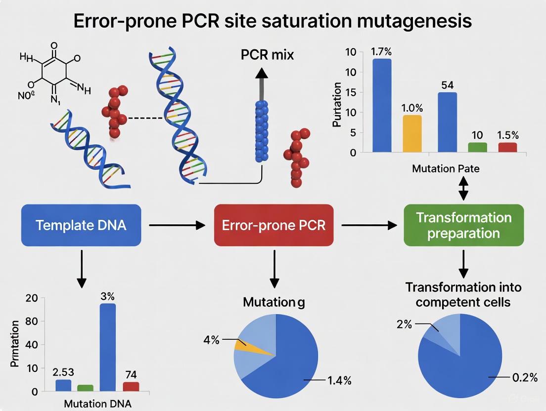
Error-prone PCR (epPCR) is a powerful directed evolution technique used to generate diverse genetic variants from a single gene template by introducing random mutations during PCR amplification [1]. By leveraging low-fidelity DNA polymerase under controlled conditions that reduce replication fidelity, researchers can create comprehensive mutant libraries for protein engineering, enzyme optimization, and functional genomics studies. This method represents a fundamental approach in the broader context of saturation mutagenesis research, enabling the exploration of sequence-function relationships without requiring prior structural knowledge.
The technique was originally developed by Leung et al. and has since become a workhorse method for combinatorial protein engineering [1]. Unlike site-saturation mutagenesis that targets specific residues, epPCR explores a wider mutational landscape, making it particularly valuable for optimizing enzyme properties such as thermostability, substrate specificity, and enantioselectivity when structural information is limited or when synergistic mutations across multiple residues are sought.
Principles and Mechanisms
Error-prone PCR introduces random mutations during DNA amplification through controlled manipulation of PCR conditions to reduce replication fidelity. The primary sources of variation stem from both polymerase misincorporation and DNA thermal damage [2].
Polymerase Errors occur when DNA polymerases incorporate incorrect nucleotides during strand elongation. The fidelity of DNA polymerases varies substantially between enzymes, with error rates ranging from approximately 1.1 errors per 10^6 base pairs for high-fidelity enzymes like KOD polymerase to significantly higher rates for non-proofreading enzymes [2]. These misincorporations are influenced by several factors:
- dNTP pool imbalances: Skewed deoxynucleotide triphosphate ratios increase misincorporation likelihood
- Magnesium concentration: Elevated Mg²⺠levels can reduce polymerase fidelity
- Template sequence context: Certain sequences are more prone to replication errors
- Extension times: Suboptimal extension parameters can promote misincorporation
Thermal Damage Errors represent a significant contributor to overall mutation rates, with three primary mechanisms:
- Depurination: Removal of purine bases (adenine or guanine) from the DNA backbone, with single-stranded DNA being particularly susceptible [2]
- Cytosine deamination: Conversion of cytosine to uracil, especially problematic at elevated temperatures [2]
- Oxidative damage: Oxidation of guanine to 8-oxoguanine, which can be mitigated by purging reactions with argon to reduce dissolved oxygen [2]
Thermal damage becomes increasingly significant with prolonged exposure to high temperatures, potentially reaching levels of 0.2-0.3% after one hour at 72°C (approximately 1 damaged base per 300-500 bases) [2].
Error Rate Quantification and Measurement
Advanced methods for quantifying epPCR error rates combine unique molecular identifier (UMI) tagging with high-throughput sequencing, enabling exceptional resolution in error detection [3]. This approach allows researchers to distinguish errors introduced during initial PCR from those occurring in subsequent amplification and sequencing steps, providing accurate per-cycle error rate measurements.
Table 1: Polymerase Error Rates and Preferences
| Polymerase | Error Rate (Substitutions/bp/cycle) | Dominant Substitution Types | Proofreading Activity |
|---|---|---|---|
| KOD Hot Start | ~1.1×10â»â¶ [2] | Not specified | Yes (3'→5' exonuclease) |
| Taq | ~1×10â»â´ [3] | A>G, T>C (20 cycles) | No |
| Phusion | ~4.6×10â»â· [3] | Not specified | Yes |
| Kapa HF | ~8.1×10â»â· [3] | C>T, G>A (20 cycles) | Yes |
| Tersus | ~1.3×10â»â¶ [3] | C>T, G>A (20 cycles) | Yes |
Different polymerases exhibit distinct substitution preferences, falling into two main categories: those predominantly generating C>T and G>A transitions, and those favoring A>G and T>C transitions [3]. This polymerase "fingerprint" significantly influences the resulting mutational spectrum and should be considered when designing epPCR experiments for specific applications.
Experimental Protocols
Standard Error-Prone PCR Protocol
Materials Required:
- Template DNA (high-purity plasmid prep, 0.1-1.0 ng/μL) [4]
- Mutagenic primers (if combining with targeted approaches)
- Low-fidelity DNA polymerase (e.g., GeneMorph II Random Mutagenesis Kit) [1]
- Modified nucleotide buffer (imbalanced dNTPs, elevated Mg²âº, Mn²âº)
- Standard PCR reagents and equipment
Procedure:
- Reaction Setup: Prepare 50μL reactions containing template DNA, 1× mutagenic buffer, 0.2mM each dNTP (or imbalanced ratios), 2-5mM MgCl₂, 0.3μM primers, and 1-2U/μL DNA polymerase [1].
Thermal Cycling:
- Initial denaturation: 94°C for 2 minutes
- 25-30 cycles of:
- Denaturation: 94°C for 15-30 seconds
- Annealing: 50-68°C for 30 seconds (optimize based on primer Tm)
- Extension: 72°C for 1-2 minutes/kb
- Final extension: 72°C for 5-10 minutes [1]
Product Analysis: Verify amplification by 1% agarose gel electrophoresis and purify using standard PCR purification kits.
Critical Parameters for Mutation Rate Control:
- Mg²⺠concentration: Increasing from 1.5mM to 5-7mM enhances error rates
- dNTP imbalances: Unequal dNTP concentrations (e.g., elevating dATP/dTTP while reducing dCTP/dGTP)
- Manganese addition: 0.1-0.5mM MnClâ‚‚ significantly increases misincorporation
- Template concentration: Higher template amounts reduce the number of replication cycles and resultant mutations
- Cycle number: Increasing cycles amplifies mutation accumulation
Improved Two-Stage PCR for Difficult Templates
For problematic templates such as plasmids containing P450-BM3 or Pseudomonas aeruginosa lipase A genes, an improved two-stage PCR method enhances success rates [5].
Workflow:
- First Stage (Megaprimer Generation): 5-10 cycles with both mutagenic primer and antiprimer (a non-mutagenic primer aiding DNA unwinding) at standard annealing temperatures
- Second Stage (Plasmid Amplification): 20 cycles with increased annealing temperature to eliminate oligonucleotide priming, allowing the megaprimer to drive amplification [5]
This approach is particularly valuable for saturation mutagenesis at single or multiple residues regardless of their location in the gene sequence and intrinsically avoids problems from palindromes, hairpins, or primer self-pairing [5].
Diagram 1: Two-stage PCR workflow
Library Construction Methods
Traditional Ligation-Dependent Cloning (LDCP):
- Primers incorporate restriction enzyme sites compatible with target plasmids
- PCR products and vectors are digested with appropriate restriction enzymes
- Ligation with DNA ligase recircularizes vectors [1]
- Limitations include significant loss of potential mutants, reducing library diversity
Circular Polymerase Extension Cloning (CPEC):
- A restriction enzyme- and ligase-free method offering superior efficiency
- High-fidelity DNA polymerase extends overlapping regions between insert and vector
- Single reaction forms circular molecules ready for transformation [1]
- Advantages: accelerated cloning, higher variant recovery, simplified procedure
Table 2: Cloning Method Comparison for Library Construction
| Parameter | LDCP (Traditional) | CPEC (Improved) |
|---|---|---|
| Efficiency | Limited efficacy, significant mutant loss | Higher variant recovery |
| Steps | Multiple: digestion, purification, ligation | Single PCR reaction |
| Time Requirement | Longer (overnight ligation possible) | Rapid (few hours) |
| Enzyme Dependence | Requires specific restriction enzymes | No restriction enzymes needed |
| Cost | Higher (multiple enzymes required) | Lower (fewer reagents) |
| Library Diversity | Reduced due to cloning bottlenecks | Better preservation of diversity |
Research Reagent Solutions
Table 3: Essential Reagents for Error-Prone PCR
| Reagent Category | Specific Examples | Function & Application Notes |
|---|---|---|
| Polymerases | GeneMorph II Kit, Taq, Mutazyme II | Low-fidelity enzymes for random mutagenesis; choice depends on desired error rate and mutational spectrum |
| Cloning Kits | NEB Q5 SDM Kit, CPEC method | Introduction of mutations into plasmids; CPEC offers advantages in efficiency and simplicity [1] [6] |
| Template Preparation | dam+ E. coli strains (e.g., DH5α) | Methylation-competent strains for subsequent DpnI digestion to remove template [4] |
| Error-Rate Modification | MnCl₂, unbalanced dNTPs, elevated Mg²⺠| Chemical mutagens to alter and control mutation frequency |
| Screening Tools | Restriction analysis, sequencing, functional assays | Identification and validation of desired mutants; high-throughput methods preferred for library screening |
Applications in Directed Evolution
Error-prone PCR serves as a foundational technology in directed evolution pipelines, enabling the improvement of enzyme properties through iterative rounds of mutation and selection. Key applications include:
- Thermostability enhancement through the B-FIT method targeting flexible regions
- Substrate scope expansion and enantioselectivity optimization via CASTing focusing on active site residues
- Organic solvent tolerance improvement for industrial biocatalysis
- pH profile modulation for applications under non-physiological conditions
The integration of epPCR with high-throughput screening methods creates a powerful platform for protein engineering, allowing researchers to explore vast sequence spaces and identify variants with desired properties that would be difficult to predict rationally.
Troubleshooting and Optimization
Common Challenges and Solutions:
- Low Mutation Frequency: Increase Mg²⺠concentration (5-7mM), add MnCl₂ (0.1-0.5mM), implement dNTP imbalances, or increase cycle number
- Poor Amplification: Optimize template quality and concentration, add DMSO (3-5%) for GC-rich templates, adjust annealing temperatures, or utilize the two-stage PCR protocol for difficult templates [5] [4]
- Limited Library Diversity: Implement CPEC cloning instead of traditional restriction-based methods, use higher template diversity, or combine with other mutagenesis methods [1]
- Template Carryover: Ensure complete DpnI digestion of methylated template DNA (from dam+ E. coli strains) [4]
Quantitative Error Monitoring: Employ high-throughput sequencing with unique molecular identifiers (UMIs) to accurately quantify error rates and profiles, enabling precise control over library quality and diversity [3].
Error-prone PCR remains an essential tool in the molecular biologist's toolkit, providing a robust method for generating diversity in directed evolution experiments. Through careful optimization of reaction conditions and integration with efficient cloning methodologies, researchers can create high-quality mutant libraries for advancing protein engineering and drug development initiatives.
The Rationale Behind Site Saturation Mutagenesis for Targeted Exploration
In the field of protein engineering and functional genomics, site saturation mutagenesis (SSM) stands as a powerful targeted approach that contrasts with non-targeted random mutagenesis methods. While error-prone PCR (epPCR) introduces mutations randomly throughout a gene, SSM provides a systematic methodology for investigating the function of specific amino acid positions by replacing them with all possible amino acid substitutions [7]. This application note delineates the rationale, advantages, and methodological frameworks for SSM, contextualized within broader directed evolution and functional analysis research, to guide researchers and drug development professionals in leveraging this technique for precise protein optimization and variant characterization.
SSM represents a sophisticated approach to systematic genetic exploration, transforming protein modification from educated guesswork into a comprehensive investigation of sequence-function relationships [7]. By methodically substituting every possible amino acid at specific positions, researchers can create "smarter libraries" that focus screening efforts on regions of interest, thereby significantly enhancing the efficiency of directed evolution campaigns [8]. This targeted strategy has proven instrumental in addressing diverse protein engineering challenges, from altering enzyme cofactor specificity to enhancing thermal stability.
SSM vs. Random Mutagenesis: A Comparative Rationale
Strategic Advantages of Targeted Exploration
The selection between SSM and random mutagenesis represents a fundamental strategic decision in protein engineering. While epPCR employs mutagenic buffers with elevated MgClâ‚‚ (7 mM), MnClâ‚‚, or unbalanced dNTP concentrations to introduce random mutations throughout a gene [9], SSM focuses investigative resources on predefined positions of interest. This focused approach offers several distinct advantages for hypothesis-driven protein engineering.
Table 1: Comparative Analysis of Site Saturation Mutagenesis vs. Random Mutagenesis
| Feature | Site Saturation Mutagenesis | Random Mutagenesis (epPCR) |
|---|---|---|
| Mutation Control | Targeted to specific residues | Random distribution across gene |
| Library Quality | Focused, "smarter" libraries [8] | Unbiased but with redundant coverage |
| Information Yield | Direct residue-function relationships | Global sequence-function landscape |
| Screening Efficiency | Higher hit rate per variant screened | Lower hit rate, requires high throughput [9] |
| Primary Applications | Protein engineering, critical residue identification, mechanism study [7] | Directed evolution when target regions unknown [10] |
| Technical Implementation | Two-stage PCR with mutagenic primers [5] | Modified PCR conditions with mutagenic agents [9] |
The precision of SSM enables researchers to address specific protein engineering challenges that are difficult to tackle with random approaches. For instance, SSM has been successfully employed to alter the coenzyme specificity of Candida methylica formate dehydrogenase (cmFDH) from NAD⺠to NADP⺠and to increase its thermostability by targeting specific positions in both the coenzyme binding and catalytic domains [8]. Similarly, large-scale SSM studies encompassing hundreds of human protein domains have systematically quantified the effects of over 500,000 missense variants, revealing that approximately 60% of pathogenic missense variants reduce protein stability [11].
Complementary Roles in Directed Evolution
Rather than mutually exclusive approaches, SSM and epPCR often play complementary roles in comprehensive protein engineering pipelines. epPCR serves as an exploratory tool when structural information is limited or when the target property involves distributed sequence determinants, while SSM enables focused optimization once key regions have been identified. The integration of both methods in successive rounds of directed evolution can accelerate the optimization process, with epPCR discovering beneficial regions and SSM intensively exploring those regions.
Figure 1: Decision framework for selecting mutagenesis strategies based on available structural information and research objectives. SSM requires prior knowledge of target regions, while epPCR offers broader exploration when such information is limited.
SSM Methodologies and Technical Implementation
Molecular Basis of SSM Techniques
SSM methodologies employ different molecular strategies to introduce targeted diversity, each with distinct advantages for specific experimental scenarios. The fundamental principle involves systematically replacing specific codons with degenerate codons (typically NNK or NNN, where N represents any nucleotide and K represents G or T) to encode all 20 amino acids at the targeted position.
Oligonucleotide-directed SSM utilizes mutagenic primers containing degenerate codons at the target positions. These primers are incorporated into the plasmid through whole-plasmid amplification approaches, such as the improved two-stage PCR method that functions effectively even with difficult-to-amplify templates [5]. In this method, the first PCR stage generates a megaprimer using both mutagenic and antiprimers (non-mutagenic primers that facilitate DNA uncoiling), while the second stage employs this megaprimer for plasmid amplification [5]. This method has been successfully applied to various enzymes including P450-BM3 from Bacillus megaterium, Pseudomonas aeruginosa and Candida antarctica lipases, and Aspergillus niger epoxide hydrolase [5].
Overlap extension PCR employs two separate PCR reactions that generate gene fragments with overlapping ends containing the desired mutations, followed by a second PCR reaction where these fragments serve as templates for full-length gene assembly [7]. Synthetic oligonucleotide approaches utilize pools of synthetic oligonucleotides encoding all possible variations at targeted positions, which are then cloned into expression vectors to create comprehensive variant libraries [7].
Advanced SSM Workflow for Large-Scale Functional Analysis
Recent advances in DNA synthesis and cloning technologies have enabled unprecedented scale in SSM applications. The "Human Domainome 1" study exemplifies this scale, employing microchip-based massive parallel synthesis (mMPS) to construct a library of 1,230,584 amino acid variants across 1,248 structurally diverse protein domains [11]. This approach systematically mutated every amino acid to all other 19 amino acids at every position in each domain, achieving 91% coverage of designed substitutions.
Table 2: Key Research Reagent Solutions for Site Saturation Mutagenesis
| Reagent/Category | Specific Examples | Function in SSM |
|---|---|---|
| Polymerase Systems | KOD Hot Start DNA polymerase [5] | High-fidelity amplification in two-stage PCR |
| Cloning Methods | Circular Polymerase Extension Cloning (CPEC) [1] | Efficient library construction without restriction enzymes |
| Degenerate Codons | NNK (encodes all 20 aa) | Creates diversity at targeted positions |
| Vector Systems | pETM11, pCDF1b [5] [1] | Protein expression for functional screening |
| Template Preparation | Plasmid isolation from desired host | Provides backbone for mutagenesis |
| Selection Assays | Abundance protein fragment complementation assay (aPCA) [11] | High-throughput functional screening |
The functional analysis of these comprehensive variant libraries employed an abundance protein fragment complementation assay (aPCA), where each protein domain was expressed as a fusion with a fragment of an essential enzyme, and cellular growth rate served as a proxy for protein abundance [11]. This innovative selection system enabled pooled cloning, transformation, and selection of hundreds of thousands of variants across diverse proteins in single experiments, ultimately yielding reproducible abundance measurements for 563,534 variants in 522 protein domains [11].
Figure 2: Advanced SSM experimental workflow integrating modern cloning and screening methodologies for comprehensive variant functional analysis.
Applications in Protein Engineering and Drug Development
Protein Optimization and Engineering
SSM has demonstrated remarkable success in addressing diverse protein engineering challenges. In enzyme engineering, SSM has been employed to alter cofactor specificity, enhance thermostability, improve substrate specificity, and increase resistance to organic solvents. The application of SSM to Candida methylica formate dehydrogenase (cmFDH) exemplifies this approach, where two rounds of SSM at positions 195, 196, and 197 in the coenzyme binding domain yielded double mutants D195S/Q197T and D195S/Y196L that dramatically altered coenzyme specificity from NAD⺠to NADPâº, increasing catalytic efficiency for NADP⺠by approximately 5×10â´-fold [8]. Simultaneously, SSM at position 1 in the catalytic domain identified the M1L mutant with improved thermostability, exhibiting 17% residual activity after incubation at 60°C compared to wild-type enzyme [8].
The precision of SSM makes it particularly valuable for engineering specific enzyme properties when structural information guides target selection. By focusing on residues within active sites, substrate-binding pockets, or known functional motifs, researchers can create focused libraries that yield significantly higher hit rates compared to random mutagenesis approaches. This strategy efficiently explores the sequence-function landscape around critical positions without the screening burden of comprehensively random libraries.
Functional Genomics and Variant Interpretation
Beyond protein engineering, SSM has emerged as a powerful tool for functional genomics and clinical variant interpretation. Large-scale SSM studies have enabled systematic quantification of variant effects across entire protein families, providing datasets for training and benchmarking computational variant effect predictors (VEPs) [11]. These comprehensive experimental datasets reveal fundamental principles of protein structure-function relationships, such as the observation that mutations in buried core regions are generally more detrimental than surface mutations, and that mutations to proline typically exert the strongest destabilizing effects, particularly in secondary structure elements [11].
Computational saturation mutagenesis approaches extend these experimental observations through in silico analysis of all possible missense variants in target proteins. For example, a comprehensive computational saturation mutagenesis study of adducin proteins (ADD1, ADD2, ADD3) employed multiple prediction tools (AlphaMissense, Rhapsody, PolyPhen-2, and PMut) to identify high-risk variants and characterize their potential structural and functional impacts [12]. This integrated computational approach identified glycine substitutions as particularly destabilizing due to effects on backbone flexibility, and clustered high-risk mutations in known regulatory regions including phosphorylation and calmodulin-binding sites [12].
The integration of experimental and computational SSM data provides powerful frameworks for clinical variant interpretation, distinguishing pathogenic mutations from benign polymorphisms, and elucidating molecular mechanisms underlying genetic diseases. These approaches are particularly valuable for rare variants where population data may be insufficient for statistical assessment of pathogenicity.
Site saturation mutagenesis represents a powerful methodology for targeted exploration of protein sequence-function relationships, offering precision and systematic analysis that complements broader random mutagenesis approaches. The technical evolution of SSM methodologies—from early oligonucleotide-directed methods to contemporary large-scale synthetic approaches—has enabled increasingly comprehensive functional characterization of protein variants. When strategically deployed within directed evolution campaigns or functional genomics studies, SSM provides efficient interrogation of specific positions or regions, yielding fundamental insights into protein structure-function relationships and accelerating the engineering of improved biocatalysts and therapeutic proteins. As DNA synthesis technologies continue to advance and computational prediction methods become increasingly sophisticated, the integration of experimental and in silico SSM approaches will further expand our ability to interpret variant effects and engineer proteins with novel functions.
Within the broader field of directed enzyme evolution, saturation mutagenesis stands as a powerful protein engineering strategy for probing and enhancing enzyme functions such as thermostability, substrate acceptance, and enantioselectivity [5]. Unlike random mutagenesis methods such as error-prone PCR (epPCR), which introduce mutations throughout the gene, saturation mutagenesis focuses on introducing a controlled set of mutations at specific, predefined amino acid positions [5] [13]. This approach enables the creation of high-quality variant libraries of a defined size, facilitating a more efficient exploration of the sequence-function landscape [14].
While several molecular biological methods exist for performing saturation mutagenesis, Overlap Extension PCR (OE-PCR) has proven to be a particularly versatile and efficient technique [15] [16]. This method is especially valuable for introducing degenerate bases at single or multiple codon locations, generating a precise series of amino acid substitutions in the encoded protein [14]. Furthermore, improved OE-PCR protocols have overcome many limitations of traditional methods, enabling simultaneous multiple-site large fragment insertion, deletion, and substitution, even for difficult-to-amplify templates [5] [16]. This application note details the principles, protocols, and key applications of OE-PCR for saturation mutagenesis, providing researchers with a robust framework for its implementation.
Key Principles and Comparative Advantages of OE-PCR
The Fundamental Workflow of Overlap Extension PCR
Overlap Extension PCR is a multi-stage technique that uses primers with complementary ends to seamlessly join DNA fragments. The core process can be broken down into several key stages, as illustrated in the workflow below.
Comparison of Saturation Mutagenesis Methods
The table below summarizes how OE-PCR compares to other common techniques used in saturation mutagenesis.
Table 1: Comparison of common saturation mutagenesis methods.
| Method | Key Principle | Primary Advantages | Common Limitations |
|---|---|---|---|
| Overlap Extension PCR (OE-PCR) | Uses primers with complementary ends to join DNA fragments and introduce mutations [14]. | Flexible; no restriction enzyme sites needed; suitable for multi-site mutagenesis and large fragments [16]. | Can require multiple PCR steps and optimization [15]. |
| QuikChange-Style | Uses complementary primers carrying the mutation in a site-directed mutagenesis protocol [5]. | Commercially available kits; straightforward for single-site mutations. | Limited to single sites; primer design constraints; fails with difficult templates [5]. |
| Error-Prone PCR (epPCR) | Uses low-fidelity PCR conditions to introduce random mutations throughout a gene [13]. | Simple; good for introducing random diversity across the entire gene. | Lacks precision; generates mostly neutral or deleterious mutations; biased mutation spectrum [17]. |
| CRISPR-Directed Evolution | Uses CRISPR-Cas systems for precise genome editing to introduce targeted diversity [13]. | Highly precise in vivo editing; can generate complex mutant libraries in genomic context. | Higher technical complexity; potential for off-target effects [13]. |
Improved versions of OE-PCR (IOEP) have been developed to address limitations like inefficient priming of large fragments. By adding primers that bind to the vector sequence during the final amplification stage, IOEP enables exponential amplification of the overlap extension product. This enhancement significantly increases the efficiency and success rate for cloning large and difficult-to-amplify fragments, with demonstrated success for constructs as large as 12 kb [16].
Detailed Experimental Protocol
This protocol describes an improved two-stage, two-primer OE-PCR method for efficient saturation mutagenesis, adapted from published studies [5] [16].
Research Reagent Solutions
The following table lists the essential materials required to execute this protocol successfully.
Table 2: Key reagents and materials for OE-PCR saturation mutagenesis.
| Reagent/Material | Specification/Function | Example Product (Source) |
|---|---|---|
| DNA Polymerase | High-fidelity, high-processivity enzyme for accurate amplification of large/gC-rich fragments. | Q5 DNA Polymerase [18] [15], PrimeSTAR GXL [16], KOD Hot Start [5] |
| Template DNA | Plasmid containing the wild-type gene of interest. | - |
| Oligonucleotides | Mutagenic primers and external primers for exponential amplification. | - |
| Restriction Enzyme | DpnI, which cleaves methylated DNA to digest the original template plasmid post-PCR. | DpnI (NEB) [5] [18] |
| Competent E. coli | High-efficiency cells for plasmid transformation after assembly. | DH5α [5] [16], Endura Electrocompetent [18] |
| Cloning Kit/Mix | Master mix for efficient assembly of PCR fragments. | NEBuilder HiFi DNA Assembly Master Mix [18] |
Step-by-Step Procedure
Stage 1: Primer and Template Preparation
- Primer Design: Design two mutagenic primers that are complementary to the same region on opposite strands. The primers should contain the desired degenerate codon (e.g., NNK, where N is A/T/G/C and K is G/T) at the target position. Ensure primers have a melting temperature (Tm) of at least 64°C and a minimum of 8 non-overlapping bases at the 3' end [19]. For improved OE-PCR (IOEP), also design two external primers that bind to the vector sequence flanking the insertion site [16].
- Primary PCR: Perform two separate primary PCRs using a high-fidelity polymerase. The goal is to generate two overlapping DNA fragments that collectively represent the entire plasmid, with the mutation at the overlap region.
- Reaction Setup:
- Template Plasmid DNA: 1-10 ng
- Forward Primer 1 & Reverse Mutagenic Primer (for Fragment A)
- Forward Mutagenic Primer & Reverse Primer 2 (for Fragment B)
- dNTPs: 200 µM each
- DNA Polymerase: per manufacturer's instructions (e.g., 0.625 U of PrimeSTAR GXL)
- Reaction Buffer: with Mg²⺠as required
- Cycling Parameters (Touchdown):
- Reaction Setup:
- Gel Purification: Verify the size and purity of the two PCR fragments by agarose gel electrophoresis. Excise the correct bands and purify the DNA using a gel extraction kit.
Stage 2: Overlap Extension and Exponential Amplification
- Overlap Extension Reaction:
- Combine the two purified fragments (e.g., 50-100 ng of each) in a single tube. For IOEP, also include the two external primers at this stage [16].
- Use a high-fidelity polymerase and the same cycling parameters as in the primary PCR, but without the initial denaturation step. The overlapping ends of the fragments will anneal and extend, forming a full-length circular plasmid.
- DpnI Digestion: To eliminate the original methylated template plasmid, treat the PCR product with DpnI (10 U per 50 µL reaction) for 1-2 hours at 37°C [5].
- Purification: Purify the DpnI-treated DNA using a standard PCR clean-up kit.
Stage 3: Cloning and Transformation
- Transformation: Transform 2-5 µL of the purified assembly product into competent E. coli DH5α cells using standard heat-shock or electroporation methods.
- Screening and Validation: Plate cells on selective media and incubate overnight. Screen resulting colonies by colony PCR or restriction digest. Validate the sequence of the mutated gene by Sanger sequencing of plasmid DNA from positive clones.
Critical Factors for Success
- Primer Design and Localization: Optimal primer design is critical. The direction and design of the "antiprimer" (a non-mutagenic primer used to complete complementary extension) are determining factors in successfully amplifying difficult templates [5]. Ensure sufficient overlap length (typically 15-25 bp) with high homology.
- Polymerase Selection: The choice of DNA polymerase significantly impacts success. Use a high-fidelity, high-processivity enzyme (e.g., Q5, KOD, PrimeSTAR GXL) to improve the quality and yield of full-length amplification, especially for large or complex fragments [15] [16].
- Handling Difficult Templates: For plasmids that are difficult to amplify (e.g., those with high GC-content or secondary structures), the improved two-stage PCR method, which uses the initial product as a megaprimer in a second PCR with adjusted annealing temperatures, has proven highly effective [5].
Applications in Directed Evolution
OE-PCR-based saturation mutagenesis is a cornerstone of modern directed evolution campaigns. Its primary applications include:
- Iterative Saturation Mutagenesis (ISM): This systematic strategy involves repeatedly performing saturation mutagenesis at different predefined sites (or "hotspots") in an enzyme. ISM is highly effective for optimizing properties like enantioselectivity and thermostability by exploring the combinatorial active-site saturation test (CAST) or B-FIT concepts [5].
- Deep Mutational Scanning: Saturation mutagenesis libraries can be used in massively parallel reporter assays (MPRAs) to functionally characterize thousands of single nucleotide variants in regulatory elements, aiding in the interpretation of disease-associated noncoding variants [17].
- Multi-Fragment Assembly: Improved OE-PCR enables the simultaneous insertion, deletion, or substitution of multiple large DNA fragments at different sites in a single vector, a powerful capability for complex metabolic engineering and synthetic biology projects [16].
Overlap Extension PCR provides a robust, flexible, and efficient platform for conducting saturation mutagenesis. Its ability to precisely randomize single or multiple amino acid positions, coupled with recent improvements that enhance its efficiency and expand its application to large DNA fragments, makes it an indispensable tool in the directed evolution workflow. By following the detailed protocol and considerations outlined in this application note, researchers can effectively leverage OE-PCR to engineer proteins with novel and enhanced functions, accelerating progress in biotechnology, drug development, and basic research.
Key Applications in Protein Engineering and Synthetic Biology
Error-prone PCR (epPCR) and site-saturation mutagenesis (SSM) represent two cornerstone methodologies in the field of protein engineering. These techniques facilitate the directed evolution of proteins by generating genetic diversity, enabling the development of enzymes and biosynthetic proteins with enhanced properties such as catalytic activity, stability, and substrate specificity. Within the context of a broader thesis on mutagenesis research, this application note details the key applications, methodologies, and reagent solutions that underpin their successful implementation in modern synthetic biology and drug development pipelines. The strategic application of these methods allows researchers to explore vast sequence-function landscapes efficiently [20].
Comparative Analysis of Mutagenesis Techniques
The selection of a mutagenesis strategy is critical to the success of a protein engineering campaign. Error-prone PCR introduces random mutations throughout a gene, making it ideal for exploring a wide mutational space when no prior structural knowledge is available. In contrast, Site-Saturation Mutagenesis allows for the focused randomization of specific codon locations, providing a more controlled and comprehensive exploration of key residues, often those implicated in catalytic activity or substrate binding [14] [20]. The following table summarizes their core characteristics and applications.
Table 1: Key Characteristics of epPCR and SSM
| Feature | Error-Prone PCR (epPCR) | Site-Saturation Mutagenesis (SSM) |
|---|---|---|
| Mutagenesis Scope | Random mutations across the entire gene sequence [20] | Focused mutagenesis at one or multiple pre-defined codon positions [14] [21] |
| Primary Application | Directed evolution without requiring structural data; improving general properties like stability [22] [20] | Investigating or optimizing specific active sites, binding pockets, or functional residues [14] [5] |
| Library Design | Uncontrolled; diversity depends on error-rate of polymerase [23] | Controlled and precise; uses degenerate codons (e.g., NNK) to access all possible amino acids at a site [14] [21] |
| Typical Throughput | Requires screening of large libraries (>10^5 variants) [22] | Library size is manageable and defined (theoretical maximum of 20 variants per codon) [14] |
| Integration with Automation | Well-suited for automated library construction and screening in biofoundries [24] | Highly amenable to automation for primer design, library construction, and high-throughput screening [25] [24] |
| Common Challenge | Biased mutation spectrum (preference for transitions) [17] | Requires prior knowledge (e.g., structural data) to select impactful positions for randomization [26] |
The quantitative performance of these methods is evidenced in numerous studies. For instance, in one saturation mutagenesis study of 20 disease-associated regulatory elements, researchers successfully measured the functional effects of over 30,000 single nucleotide substitutions and deletions, achieving near-complete coverage of all potential SNVs [17]. In a separate application, a combined directed evolution approach was used to co-evolve β-glucosidase for both enhanced activity and organic acid tolerance, leading to a 4.3-fold improvement in enzyme activity [26].
Experimental Protocols
Protocol 1: Site-Saturation Mutagenesis by Overlap Extension PCR
This protocol describes the creation of a high-quality variant library by introducing degenerate codons at specific positions via overlap extension PCR [14] [21].
Procedure:
- Primer Design: Design mutagenic primers containing degenerate codons (e.g., NNK or NNN, where K = G/T) at the target amino acid position(s). The primers must be complementary and have sufficient overlap for the extension reaction.
- First PCR Amplification: Perform two separate primary PCR reactions to generate overlapping gene fragments.
- Reaction A: Amplify the 5' fragment of the gene using a forward plasmid-specific primer and the reverse mutagenic primer.
- Reaction B: Amplify the 3' fragment using the forward mutagenic primer and a reverse plasmid-specific primer.
- Purification: Purify both PCR products from Step 2 to remove residual primers and polymerase.
- Overlap Extension PCR: Combine the purified fragments from Reaction A and B. In the first few cycles (without external primers), the overlapping ends of the fragments anneal and extend, forming full-length mutant genes. Then, add the external forward and reverse primers to amplify the full-length product.
- Digestion and Ligation: Digest the overlap extension PCR product and the target plasmid with the appropriate restriction enzymes. Purify the fragments and ligate the mutant gene insert into the plasmid backbone.
- Transformation and Library Creation: Transform the ligated product into a competent E. coli strain. Plate the cells on selective media to create the mutant library for subsequent screening [14].
Protocol 2: Error-Prone PCR for Random Mutagenesis
This protocol outlines the generation of a random mutant library using error-prone PCR, which is suitable for whole-gene diversification without a specific target site [22] [20].
Procedure:
- Reaction Setup: Set up the PCR reaction using a template plasmid containing the wild-type gene. To promote a high error rate, use a polymerase with low fidelity (e.g., Taq polymerase) and modify standard conditions:
- Increase the concentration of MgClâ‚‚ (e.g., 7 mM).
- Add a small, optimized concentration of MnClâ‚‚ (e.g., 0.5 mM).
- Use unbalanced dNTP concentrations (e.g., increase dATP and dTTP relative to dGTP and dCTP).
- Amplification: Run the PCR with a standard thermocycling program suitable for the gene of interest.
- Product Purification: Purify the error-prone PCR product to remove enzymes and unbalanced dNTPs.
- Downstream Cloning: The purified epPCR product can be cloned into an expression vector using various methods:
- Restriction/Ligation: If the product has terminal restriction sites.
- Homologous Recombination: In yeast or Bacillus subtilis systems, the product can be co-transformed with a linearized plasmid for in vivo assembly [22].
- Gibson Assembly: An isothermal, single-tube method based on overlapping homology.
- Library Transformation: Transform the assembled DNA into a suitable host organism to create the mutant library for high-throughput screening.
Workflow Visualization
The following diagram illustrates the logical sequence and key decision points in a directed evolution campaign utilizing epPCR and SSM.
Decision and Workflow for Directed Evolution
The Scientist's Toolkit: Research Reagent Solutions
Successful execution of mutagenesis experiments relies on a suite of specialized reagents and tools. The following table details essential materials and their functions.
Table 2: Key Research Reagents for Mutagenesis and Screening
| Reagent / Tool | Function / Application | Examples / Notes |
|---|---|---|
| Degenerate Oligonucleotides | Primers containing degenerate bases (NNK) for introducing all possible amino acid substitutions at a target codon in SSM [5] [21]. | Synthesized commercially; NNK reduces codon redundancy (32 codons for 20 amino acids). |
| Low-Fidelity Polymerase | Enzyme used in epPCR to introduce random mutations during DNA amplification [17] [20]. | Taq polymerase is commonly used under modified buffer conditions to increase error rates. |
| High-Fidelity Polymerase | Enzyme used in SSM protocols (e.g., Overlap Extension PCR) to minimize unwanted background mutations during amplification [5]. | Phusion or KOD Hot Start DNA polymerase are often preferred. |
| DpnI Restriction Enzyme | Digests the methylated parental DNA template post-PCR, enriching the final product for newly synthesized mutant DNA [5] [23]. | Critical for site-directed mutagenesis protocols to reduce background. |
| Specialized Vectors | Plasmid backbones optimized for cloning mutant libraries and expressing proteins in relevant hosts. | pET series for E. coli expression; integration plasmids for B. subtilis [17] [22]. |
| Competent Cells | High-efficiency bacterial or yeast cells for transforming mutant library DNA. | E. coli DH5α for plasmid propagation; specialized strains for protein expression. |
| Mass Photometry | A label-free technique for detecting molecular interactions and complex formation in solution, useful for screening binding events in libraries [21]. | Used to assess SpyTag-SpyCatcher binding in library screens. |
| Fluorescence-Activated Cell Sorting (FACS) | An ultra-high-throughput screening method for isolating variant-containing cells based on a fluorescent signal linked to the desired function [25]. | Enables screening of libraries with >100,000 variants in a few days. |
| Massively Parallel Reporter Assays (MPRAs) | Enables functional measurement of thousands of genetic variants (e.g., from saturation mutagenesis) simultaneously [17]. | Applied to saturation mutagenesis of 20 regulatory elements. |
| A-987306 | A 987306 is a potent, selective, and orally active histamine H4 receptor antagonist for research. It is For Research Use Only. Not for human consumption. | |
| iMAC2 | iMAC2, CAS:335166-00-2, MF:C19H22Br2Cl2FN3, MW:542.1 g/mol | Chemical Reagent |
Understanding Mutational Bias and Library Quality
In the field of protein engineering and functional genomics, error-prone PCR (epPCR) site saturation mutagenesis serves as a foundational technique for generating genetic diversity. This process is central to directed evolution experiments and deep mutational scanning (DMS) studies, which aim to elucidate genotype-phenotype relationships by systematically analyzing protein variants [27]. However, the practical application of these techniques is frequently compromised by mutational bias—systematic non-randomness in the types and locations of introduced mutations. Such biases can significantly skew library composition, reduce functional diversity, and ultimately lead to misleading biological conclusions or inefficient engineering campaigns.
The integrity of any downstream analysis or selection process is fundamentally dependent on the quality of the mutant library, which encompasses the evenness of variant distribution, the accurate representation of all intended mutations, and the minimization of non-functional sequences. A comprehensive understanding of the sources of mutational bias and the implementation of robust protocols to control library quality are therefore essential for researchers, scientists, and drug development professionals working in this domain. This document provides a detailed examination of these critical aspects, supported by structured data and actionable protocols.
Quantifying and Understanding Mutational Bias
Mutational bias refers to the non-stochastic deviations from theoretical mutation frequencies that occur during library construction. Recognizing and quantifying these biases is the first step toward mitigating their effects.
The following table summarizes the major sources of bias inherent to traditional error-prone PCR methods:
Table 1: Key Sources and Effects of Mutational Bias in Error-Prone PCR
| Source of Bias | Description | Impact on Library |
|---|---|---|
| Polymerase Specificity | Different DNA polymerases have distinct error signatures and preferences for specific nucleotide misincorporations [28]. | Skews the mutational spectrum (e.g., over-representation of transitions AG, TC over transversions) [29]. |
| Sequence Context | The local DNA sequence (e.g., high or low GC content) can influence the error rate at a given position [30]. | Uneven mutation distribution across the target gene, leading to "cold spots" and "hot spots". |
| PCR Conditions | Factors like MnClâ‚‚ concentration, unbalanced dNTP ratios, and increased MgClâ‚‚ are used to enhance error rates [23]. | Can exacerbate polymerase-specific biases and introduce additional sequence-specific artifacts if not carefully optimized. |
| Codon Degeneracy | Using NNN (where N is any base) randomization results in 32 codons encoding only 20 amino acids, with different stop codon frequencies [27]. | Non-uniform amino acid sampling; over-representation of some amino acids and multiple stop codons. |
The bias introduced by Taq polymerase, for instance, is particularly well-documented, with a much higher observed mutation rate at A/T bases compared to C/G bases [28] [27]. Furthermore, early saturation mutagenesis protocols that rely on doped or degenerate primers are susceptible to biases arising from DNA sequence, G/C content, and primer quality, which can distort the final library composition [30].
Impact of Mutational Bias on Library Quality and Experimental Outcomes
A biased library directly undermines the efficiency and success of a protein engineering or DMS campaign. An uneven distribution of variants means that the experimental screening effort may be wasted on characterizing an overabundance of certain mutations while missing others entirely. This sparse and non-uniform sampling of sequence space makes it difficult to identify rare, beneficial mutations or to accurately map the protein's fitness landscape [31]. Consequently, the conclusions drawn about which residues are critical for function, stability, or binding may be incomplete or statistically unreliable.
Strategies and Reagents for Reducing Mutational Bias
Several advanced methodological strategies have been developed to counteract mutational bias and construct higher-quality libraries.
Experimental Methods for Improved Library Construction
The table below compares several key protocols designed to generate more balanced mutant libraries.
Table 2: Comparison of Protocols for Reducing Mutational Bias
| Method | Core Principle | Key Advantage | Reference |
|---|---|---|---|
| Polymerase Blending | Using a combination of low-fidelity polymerases (e.g., Taq and Mutazyme) with complementary mutational spectra [28]. | Reduces the specific bias inherent to any single enzyme, creating a more uniform mutation distribution. | [28] |
| Megaprimer PCR | A two-stage, whole-plasmid PCR method that uses a mutagenic primer and a non-mutagenic "antiprimer" to generate a megaprimer [5]. | Overcomes difficulties with amplifying complex templates and avoids problems of primer self-pairing. | [5] |
| SLUPT (Synthesis of Libraries via dU-containing PCR Templates) | Utilizes a dU-containing single-stranded DNA template generated by PCR. Mutagenic primers are extended and ligated, followed by template degradation [32]. | High efficiency, very low background from the starting sequence, and excellent stoichiometric balance of nucleotides at varied positions. | [32] |
| One-Pot Saturation Mutagenesis | Employs strand-specific nicking enzymes to create ssDNA templates, followed by synthesis with degenerate primers and degradation of the wild-type strand [23]. | Allows customizable, multi-site saturation mutagenesis with high coverage and mutational efficiency in a single tube. | [23] |
| Semiconductor-Based Synthesis | Uses programmable semiconductor chips to synthesize thousands of predefined oligonucleotides in parallel [30]. | Enables complete user control over every variant in the library, eliminating synthesis-level bias and stop codons. | [30] |
These methods represent a significant evolution from purely random approaches. For example, the one-pot saturation mutagenesis method allows researchers to tile a region of interest with multiple primers, each containing three consecutive randomized bases (NNN) at a specific codon, enabling comprehensive and parallel mutagenesis [23]. Meanwhile, the semiconductor-based synthesis represents a shift towards fully rational library design, where the mutagenesis is "less random" and directly tailored to the researcher's specifications [30].
The Scientist's Toolkit: Essential Research Reagents
Successful library construction relies on a suite of specialized reagents. The following table details key solutions and their functions.
Table 3: Research Reagent Solutions for Saturation Mutagenesis
| Research Reagent | Function in Library Construction |
|---|---|
| Low-Fidelity Polymerase Blends | Engineered mixes of polymerases (e.g., from commercial kits) designed to reduce mutational bias during error-prone PCR [28] [27]. |
| Strand-Nicking Restriction Enzymes | Enzymes like Nt.BbvCI and Nb.BbvCI that nick specific DNA strands to create single-stranded templates for methods like one-pot mutagenesis [23]. |
| dU-containing dNTP Mixes | Nucleotide mixes used in PCR to create a template strand that can be selectively degraded by enzymes like Uracil DNA Glycosylase (UDG), as used in SLUPT and PFunkel methods [32] [23]. |
| Lambda Exonuclease | An enzyme that degrades one strand of double-stranded DNA, used in the SLUPT protocol to generate single-stranded DNA from a phosphorylated PCR product [32]. |
| Programmable Oligo Synthesis Platforms | Semiconductor-based systems that synthesize precisely defined oligonucleotide libraries, enabling the creation of bias-free, user-defined variant pools [30]. |
| M8-B | M8-B, MF:C22H25ClN2O3S, MW:433.0 g/mol |
| VU0364572 TFA | VU0364572 TFA, MF:C23H32F3N3O5, MW:487.5 g/mol |
A Detailed Protocol for One-Pot Saturation Mutagenesis
The following workflow and detailed protocol for one-pot saturation mutagenesis is adapted from Wrenbeck et al. and represents a robust method for generating high-quality, customizable libraries [23].
Diagram 1: One-Pot Saturation Mutagenesis Workflow
Step-by-Step Methodology
Part 1: Preparation of ssDNA Template
- Nick the Wild-type Plasmid: Set up a nicking reaction using the wild-type plasmid as a template. Use the restriction enzyme Nt.BbvCI (if the goal is to ultimately nick the sense strand first). Incubate at 37°C for 1 hour.
- Degrade the Nicked Strand: To the same tube, add Exonuclease III (ExoIII) and Exonuclease I (ExoI). ExoIII will processively degrade the nicked strand from the nick site, while ExoI will clean up any remaining single-stranded DNA. Incubate at 37°C for 1 hour, followed by enzyme inactivation at 70°C for 10-15 minutes. The product is a single-stranded DNA (ssDNA) template.
Part 2: Synthesize the First Mutant Strand
- Design Mutagenic Primers: Design a set of primers that tile across the region of interest. Each primer should contain a central
NNNtriplet to randomize the target codon, flanked by perfectly complementary wild-type sequence (~20-25 bp on each side). The primers must be the same sense as the degraded strand from Part 1. - Primer Annealing and Extension: In a new PCR tube, combine the ssDNA template from Part 1 with the pool of degenerate primers, high-fidelity Phusion polymerase, and dNTPs. Use a low primer-to-template ratio to ensure that, on average, only one primer binds to each template molecule. Run a limited number of PCR cycles (e.g., 10-15) to synthesize the complementary mutant strand. The product is a heteroduplex plasmid with one wild-type and one mutant strand.
- Purification: Purify the PCR product using a DNA clean-up kit to remove excess primers, enzymes, and dNTPs.
Part 3: Degrade the Wild-type Template Strand
- Nick the Wild-type Strand: Treat the purified heteroduplex plasmid from Part 2 with the other BbvCI variant (Nb.BbvCI), which will nick the strand opposite to the one nicked in Part 1 (the remaining wild-type strand). Incubate at 37°C for 1 hour.
- Degrade the Wild-type Strand: Add ExoIII and ExoI to degrade the nicked wild-type strand. Incubate at 37°C for 1 hour, followed by heat inactivation. This leaves the first synthesized mutant strand as the new template.
Part 4: Synthesize the Second Mutant Strand
- Second Strand Synthesis: To the reaction from Part 3, add a universal primer (complementary to the vector backbone) and high-fidelity Phusion polymerase. This step synthesizes the second strand, using the mutant strand as a template, resulting in a double-stranded mutant plasmid.
- Remove Template: Digest the product with DpnI to cleave any residual methylated wild-type starting plasmid that may have carried through.
- Library Completion: Transform the final DpnI-treated product into competent E. coli cells. After outgrowth, harvest the cells to obtain the plasmid library, which is now ready for quality control and functional screening.
Quality Assessment and Validation of Mutant Libraries
Rigorous quality control (QC) is non-negotiable for ensuring that the constructed library accurately represents the intended diversity and is free from major biases or errors.
Key Quality Control Metrics
- Sequencing Depth: The pre-selection library must be deeply sequenced using next-generation sequencing (NGS) to obtain a quantitative count of each variant. A minimum of 100-200 reads per variant is often required for reliable quantification [23]. Inadequate depth will fail to detect rare but potentially important variants.
- Assessment of Initial Bias: The NGS data from the pre-selection input library must be analyzed to determine the evenness of variant representation. This baseline is critical for distinguishing a mutation that is truly detrimental from one that was simply underrepresented from the start [31].
- Error Correction with UMIs: To mitigate the effects of PCR and sequencing errors, Unique Molecular Identifiers (UMIs) should be incorporated. UMIs are short, random DNA sequences attached to each original DNA molecule before amplification. Bioinformatic clustering of reads that share the same UMI allows for the computational correction of errors that occurred during library amplification and sequencing, resulting in a much cleaner and more accurate dataset [31].
Advanced Applications: Linking Method to Discovery
The transition from biased, low-quality libraries to controlled, high-fidelity libraries has enabled groundbreaking applications in basic and applied research. High-quality DMS studies, powered by advanced mutagenesis techniques, have allowed researchers to:
- Map antibody-antigen interfaces with single-residue resolution, identifying critical binding hotspots for therapeutic antibody optimization [31].
- Predict viral evolution by comprehensively identifying mutations in viral proteins (e.g., SARS-CoV-2 spike protein) that confer escape from neutralizing antibodies, thereby guiding vaccine design [27].
- Understand genetic interactions (epistasis) by revealing how the functional effect of one mutation depends on the presence of other mutations within the same gene or in different genes [27].
Diagram 2: From Controlled Synthesis to Discovery
The use of programmable semiconductor chips, for instance, exemplifies this progression. This technology allows for the synthesis of a pre-defined oligo pool where every variant is specified by the researcher, effectively merging large-scale DNA synthesis with rational design [30]. This approach directly addresses the core issue of bias, making the directed evolution process quicker, more efficient, and more reliable, as illustrated in the pathway above. This is particularly transformative for applications like therapeutic antibody engineering, where the goal is to find an optimal candidate in a vast sequence space.
From Theory to Bench: Practical Protocols for Library Construction and Screening
Site-saturation mutagenesis is a powerful directed evolution strategy for generating comprehensive variant gene libraries by introducing a precise series of amino acid substitutions at specific codon locations in a protein encoding sequence [14]. This technique uses degenerate oligonucleotide primers to systematically replace targeted codons, enabling researchers to explore structure-function relationships and improve protein properties such as thermostability, substrate specificity, and enzymatic activity without requiring prior structural knowledge [33]. When performed via overlap extension PCR, this method creates high-quality libraries that access amino acid substitutions unlikely to emerge through random mutagenesis techniques like error-prone PCR [14]. This protocol details the implementation of site-saturation mutagenesis within a broader research framework investigating error-prone PCR and saturation mutagenesis methodologies for protein engineering and drug development applications.
Principle of the Method
Site-saturation mutagenesis by overlap extension PCR utilizes degenerate codon representations (such as NNK, where N represents any nucleotide and K represents G or T) to randomize specific amino acid positions [5]. The NNK codon set encodes all 20 canonical amino acids while reducing redundancy from 64 to 32 codons and excluding two of the three stop codons [34]. The method employs two consecutive PCR stages: first, gene fragments containing mutated sequences are amplified using external primers and complementary internal primers bearing degenerate codons; second, these fragments undergo overlap extension where complementary ends anneal and are extended to form full-length mutated genes [14]. Compared to commercial site-directed mutagenesis kits that sometimes fail with difficult-to-amplify templates, this overlap extension approach demonstrates improved efficiency and reliability across various enzyme systems including P450-BM3, lipases, and epoxide hydrolases [5].
Table 1: Comparison of Mutagenesis Approaches
| Method | Key Features | Limitations | Best Applications |
|---|---|---|---|
| Site-Saturation Mutagenesis | Systematic codon replacement; focused diversity; high quality variants [14] | Requires screening; limited to targeted residues | Exploring specific active sites or regions [5] |
| Error-Prone PCR | Genome-wide random mutations; simple protocol [34] | Mutation bias; predominantly point mutations [34] | Broad exploration without structural data |
| Gene Site Saturation Mutagenesis (GSSM) | All possible single amino acid substitutions [33] | Resource-intensive screening | Comprehensive protein mapping |
Experimental Protocols
Primer Design and Calculation
Effective primer design is critical for successful site-saturation mutagenesis. Mutagenic primers should be 25-45 nucleotides long with the degenerate codon positioned near the center. Flanking sequences of 10-15 bases on each side ensure proper annealing. The NNK degeneracy is preferred over NNN as it reduces the codon set from 64 to 32 while maintaining coverage of all 20 amino acids and only one stop codon [34].
For multi-site saturation mutagenesis, primers must be designed to avoid complementarity that could form hairpins or primer-dimers. Melting temperatures (Tm) should be optimized for the specific PCR system, typically ranging between 60-72°C [5]. Table 2 provides example primers from actual studies.
Table 2: Exemplary Mutagenic Primers for Saturation Mutagenesis
| Target | Primer Name | Sequence (5' to 3') | Tm (°C) | Mutation Site |
|---|---|---|---|---|
| P450-BM3 | F87NNKF | GCAGGAGACGGGTTANNKACAAGCTGGACGCATG [5] | 64 | F87 |
| P450-BM3 | F87NNKR | CATGCGTCCAGCTTGTMNNTAACCCGTCTCCTGC [5] | 64 | F87 |
| Pseudomonas aeruginosa Lipase | M16-L17 NNK-PAL-F | CTGGCCCACGGCNNKNNKGGCTTCGACAAC [5] | 65 | M16-L17 |
Step-by-Step Procedure
Stage 1: Initial Fragment Amplification
Reaction Setup: Prepare two separate PCR reactions for each mutagenesis target:
- Reaction A: Template DNA (10-100 ng), Forward external primer (0.2-0.5 µM), Reverse mutagenic primer (0.2-0.5 µM)
- Reaction B: Template DNA (10-100 ng), Forward mutagenic primer (0.2-0.5 µM), Reverse external primer (0.2-0.5 µM)
PCR Conditions:
- Initial denaturation: 95°C for 2-5 minutes
- 25-30 cycles of:
- Denaturation: 95°C for 15-30 seconds
- Annealing: Tm of primers (55-68°C) for 30-60 seconds
- Extension: 72°C for 15-30 seconds per kb
- Final extension: 72°C for 5-10 minutes
Product Purification: Separate PCR products by agarose gel electrophoresis and extract using a gel purification kit. Quantify DNA concentration spectrophotometrically [14].
Stage 2: Overlap Extension PCR
Hybridization Reaction: Combine approximately 100-200 ng each of purified fragments A and B in a PCR tube without primers. Add PCR reagents except primers. Perform 5-10 cycles of:
- Denaturation: 95°C for 30 seconds
- Annealing: 45-55°C for 30-60 seconds
- Extension: 72°C for 30-60 seconds per kb
Full-Length Amplification: Add external primers (0.2-0.5 µM each) to the same tube. Perform 25-30 cycles using the same parameters as initial fragment amplification.
Product Analysis: Verify the full-length product by agarose gel electrophoresis against appropriate molecular weight standards [14] [5].
The following workflow diagram illustrates the complete experimental procedure:
Library Construction and Analysis
Cloning: Purify the overlap extension PCR product and clone into an appropriate expression vector using restriction enzyme digestion and ligation, or more efficient methods like Circular Polymerase Extension Cloning (CPEC) which can improve library coverage [1].
Transformation: Introduce the ligated DNA into competent Escherichia coli cells (such as DH5α or XL1-Blue) by electroporation or heat shock. Plate onto selective media and incubate overnight [5].
Library Quality Assessment:
- Sequence Verification: Sequence 10-20 random clones to determine mutation rate and diversity.
- Library Size: Ensure sufficient colonies to cover 95-99% of possible variants. The theoretical library size for a single position is 32 codon variants; for multiple positions, library size increases exponentially [34].
- Functional Screening: Express variants and screen for desired functional improvements using high-throughput assays.
The Scientist's Toolkit: Research Reagent Solutions
Table 3: Essential Reagents for Site-Saturation Mutagenesis
| Reagent/Category | Specific Examples | Function & Application Notes |
|---|---|---|
| Polymerases | KOD Hot Start DNA Polymerase [5], Phusion High-Fidelity DNA Polymerase [23] | High-fidelity amplification with proofreading activity for accurate library generation |
| Cloning Systems | T7 ligase [1], CPEC method [1] | Efficient ligation of PCR products into expression vectors; CPEC avoids restriction enzyme dependence |
| Vectors | pETM11 series [5], pCDF1b [1] | Protein expression vectors with appropriate selection markers and promoter systems |
| Competent Cells | E. coli DH5α [5], E. coli TOP10 [1] | High-efficiency transformation strains for library construction and propagation |
| Degenerate Codons | NNK (N=A/C/G/T, K=G/T) [34] | Encodes all 20 amino acids with only one stop codon; optimal for saturation mutagenesis |
| Selection Antibiotics | Ampicillin, Chloramphenicol [5] [35] | Selective pressure for plasmid maintenance during library construction |
| ML266 | ML266, MF:C24H22BrN3O4, MW:496.4 g/mol | Chemical Reagent |
| (S,R,S)-AHPC-Me hydrochloride | (S,R,S)-AHPC-Me hydrochloride, CAS:1948273-03-7, MF:C23H33ClN4O3S, MW:481.1 g/mol | Chemical Reagent |
Applications and Concluding Remarks
Site-saturation mutagenesis by overlap extension PCR provides a robust methodological framework for systematic protein engineering. This technique enables comprehensive exploration of sequence-function relationships at targeted positions, often revealing beneficial mutations inaccessible through random mutagenesis approaches [33]. When implemented within iterative saturation mutagenesis (ISM) strategies, where beneficial mutations from initial rounds are recombined and subjected to further randomization, this approach can efficiently navigate protein fitness landscapes [5].
The integration of site-saturation mutagenesis with high-throughput screening platforms and next-generation sequencing technologies creates powerful pipelines for directed evolution campaigns in both academic research and industrial drug development. As synthetic biology advances toward precision design, methodologies for constructing high-quality mutant libraries with comprehensive coverage and minimal bias remain essential for elucidating functional motifs in biomacromolecules and engineering novel functionalities [34].
Optimizing Error-Prone PCR to Control Mutation Rate and Reduce Bias
Error-prone PCR (epPCR) serves as a fundamental technique in directed evolution for generating genetic diversity from a single gene template. By introducing random mutations during PCR amplification, researchers can create comprehensive mutant libraries suitable for screening improved protein variants. The core principle involves utilizing low-fidelity DNA polymerase under conditions that promote misincorporation of nucleotides, thereby achieving mutation rates typically ranging from 1 to 20 base substitutions per gene [35]. Within the broader context of saturation mutagenesis research, epPCR provides a straightforward method for exploring sequence-function relationships without requiring prior structural knowledge, making it particularly valuable for initial diversification phases in protein engineering campaigns. However, the practical implementation of epPCR presents significant challenges in controlling mutation frequency and minimizing biochemical biases that can skew library representation and compromise screening effectiveness. This application note provides detailed methodologies and quantitative frameworks for optimizing epPCR parameters to achieve predictable mutation rates while mitigating common sources of bias.
Critical Parameters for Controlling Mutation Rates
Establishing Desired Mutation Frequencies
The mutation frequency in epPCR libraries profoundly impacts the probability of discovering improved variants. Libraries with very low mutation rates (m < 2) contain mostly single mutants, simplifying the identification of beneficial mutations but potentially missing synergistic effects. Conversely, highly mutated libraries (m > 8) enable exploration of multi-site interactions but dramatically reduce the fraction of functional clones [35]. Quantitative analysis demonstrates that the fraction of functional clones decreases exponentially with increasing mutation frequency up to approximately m = 8, though this trend may reverse in hypermutated libraries (m > 20) where functional clones occur at unexpectedly high frequencies [35].
Table 1: Relationship Between Mutation Frequency and Library Characteristics
| Average Mutations per Gene (m) | Functional Clones | Screening Considerations | Typical Applications |
|---|---|---|---|
| 1.7 - 2 | High percentage | Identifies single beneficial mutations | Initial rounds, stability optimization |
| 3 - 8 | Exponential decrease with m | Balanced diversity/function | Affinity maturation, substrate specificity |
| > 8 - 22.5 | Very low (≈0.17% at m=22.5) but functional clones present | Requires high-throughput screening | Exploring distant sequence space, multi-site synergies |
For most applications, maintaining mutation rates between 1-5 amino acid substitutions per protein provides an optimal balance between diversity and functionality. In a case study targeting a single-chain Fv antibody, libraries with m = 1.7, 3.8, and 22.5 all yielded clones with improved affinity after fluorescence-activated cell sorting (FACS), with the moderate error rate library (m = 3.8) providing the greatest affinity improvement [35].
Biochemical Optimization of Mutation Rate
Traditional epPCR protocols employ several biochemical manipulations to increase error rates, including: (1) increased concentration of Taq polymerase, (2) extended PCR extension time, (3) elevated concentration of MgClâ‚‚ (which stabilizes non-complementary base pairs), (4) increased concentration of dNTPs, and/or (5) addition of MnClâ‚‚ [23]. The use of Taq polymerase with an in-house dNTP mixture has been successfully implemented to achieve approximately 2% point mutation rates, with 3rd-to-5th-round PCR products typically selected for optimal diversity [36].
More recently, commercial random mutagenesis kits such as the GeneMorph II Random Mutagenesis kit have provided standardized platforms for controlling mutation frequency through proprietary enzyme blends and buffer formulations [1]. These systems offer more reproducible mutational spectra compared to traditional in-house formulations.
Table 2: DNA Polymerase Fidelity Measurements Under Standard Conditions
| Polymerase | Per-Base Error Rate (×10â»â¶) | Relative Fidelity | Dominant Substitution Types |
|---|---|---|---|
| Kapa HF | 5.9 | High | C>T, G>A |
| Taq-HS | 29.3 | Low | A>G, T>C |
| Encyclo | 10.6 | Medium | A>G, T>C |
| SD-HS | 21.6 | Low | A>T |
| Phusion | 0.9 | Very High | Not determined |
Error rate data adapted from quantitative measurements using unique molecular identifier tagging and high-throughput sequencing [3]. Polymerases cluster into distinct categories based on their substitution preferences, with some favoring transitions (C>T and G>A) while others predominantly introduce transversions.
Technical Approaches to Minimize PCR Bias
PCR amplification bias represents a significant challenge in epPCR library generation, potentially leading to uneven representation of sequence variants. The primary sources of bias include:
- Sequence-dependent amplification efficiency: Templates with high AT- or GC-content often amplify less efficiently, leading to underrepresentation in final libraries [37]. This effect becomes exponentially exaggerated over multiple cycles.
- Taq polymerase errors: Dominant sequence artifacts occur at rates approximately matching theoretical expectations (2-3.3×10â»âµ errors per nucleotide per duplication) [38]. These errors particularly impact diversity estimates in deep mutational scanning studies.
- Chimeric sequences and heteroduplex molecules: These artifacts can comprise up to 13% of sequences in standard 35-cycle amplifications, falsely inflating diversity measurements [38].
- Primer design limitations: Self-complementarity, hairpin formation, and palindromic sequences in primers can dramatically reduce amplification efficiency of specific variants [5].
Practical Strategies for Bias Reduction
Modification of standard amplification protocols can significantly reduce epPCR bias. Critical adjustments include:
- Cycle number optimization: Limiting amplification to 15-18 cycles followed by a reconditioning PCR step (3 additional cycles in a fresh reaction mixture) reduces heteroduplex formation and Taq error accumulation [38]. This modification decreased unique 16S rRNA sequences from 76% to 48% after chimera and error correction, indicating more accurate diversity representation.
- Polymerase selection: KAPA HiFi DNA polymerase demonstrates superior performance in amplifying regions with extreme GC-content, providing more uniform genomic coverage compared to traditional enzymes [37]. For AT-rich templates, additives like tetramethyleneammonium chloride (TMAC) increase melting temperature stability when used with compatible polymerases.
- Cloning method improvements: Traditional ligation-dependent cloning processes (LDCP) inevitably lose potential mutants during restriction digestion and ligation. Circular polymerase extension cloning (CPEC) provides an efficient restriction-free alternative, where high-fidelity DNA polymerase extends overlapping regions between insert and vector to form circular molecules [1]. In direct comparisons, CPEC yielded greater variant diversity from the same epPCR products.
- Unique molecular identifiers (UMIs): Incorporating random oligonucleotide tags before amplification enables bioinformatic correction of amplification biases during sequencing analysis [3]. Recent advances include homotrimeric nucleotide block UMIs that provide error-correcting capabilities through majority voting mechanisms, significantly improving molecular counting accuracy [39].
Integrated Experimental Protocols
Standardized Error-Prone PCR Protocol
Materials: Template DNA (10-100 ng), mutagenic primers, Taq DNA polymerase or specialized mutagenesis enzyme blend, 10× mutagenesis buffer (with Mg²âº), dNTP mix, MnClâ‚‚ (if required for error rate adjustment)
Procedure:
- Prepare reaction mixture with final concentrations of:
- 1× mutagenesis buffer
- 0.2 mM dATP and dGTP
- 1 mM dCTP and dTTP (nucleotide imbalance promotes misincorporation)
- 0.5 mM MnClâ‚‚ (optional for increased error rate)
- 5 U Taq polymerase
- 0.5 µM forward and reverse primers
- Template DNA (20-50 ng)
Perform thermal cycling:
- Initial denaturation: 94°C for 2 minutes
- 25-30 cycles of:
- Denaturation: 94°C for 15 seconds
- Annealing: 50-68°C (primer-specific) for 30 seconds
- Extension: 72°C for 1 minute per kb
- Final extension: 72°C for 5 minutes
Purify PCR products using silica membrane columns or magnetic beads.
Quantify mutation rate by sequencing 4-20 randomly selected clones (400-700 bp each) [35]. For libraries with m > 2, select clones from early to middle PCR rounds to maintain point mutation rates around 2% [36].
Enhanced Cloning via Circular Polymerase Extension Cloning (CPEC)
Materials: epPCR product, linearized vector with 15-20 bp overlaps with insert, high-fidelity DNA polymerase (e.g., TAKARA LA Taq), dNTPs, DpnI restriction enzyme
Procedure:
- Mix epPCR product and linearized vector in 1:3 molar ratio (insert:vector) in 1× PCR buffer with 0.2 mM dNTPs and 1 U high-fidelity polymerase.
Perform CPEC reaction:
- Initial denaturation: 94°C for 2 minutes
- 30 cycles of:
- Denaturation: 94°C for 15 seconds
- Annealing: 63°C for 30 seconds
- Extension: 68°C for 4 minutes (extension time adjusted based on total fragment size)
- Final extension: 72°C for 5 minutes
Digest template plasmid with DpnI (37°C for 1 hour) to eliminate methylated parental DNA.
Transform directly into competent E. coli cells via electroporation (2.5 kV/cm, 25 µF, 200 Ω) [1].
Plate transformed cells on selective media and harvest colonies for library analysis.
Research Reagent Solutions
Table 3: Essential Reagents for Error-Prone PCR Library Construction
| Reagent Category | Specific Examples | Function & Application Notes |
|---|---|---|
| Polymerases for epPCR | GeneMorph II Random Mutagenesis Kit, Taq DNA polymerase with adjusted buffer | Low-fidelity enzymes for introducing random mutations; commercial kits offer more reproducible mutation spectra |
| High-Fidelity Polymerases | KAPA HiFi, Phusion, TAKARA LA Taq | For bias-resistant amplification and CPEC cloning; KAPA HiFi provides superior GC-rich region coverage |
| Cloning Systems | CPEC method, Traditional restriction enzyme/Ligase | CPEC enables restriction-free cloning with higher variant recovery compared to ligation-dependent methods |
| Competent Cells | E. coli TOP10 electrocompetent, E. coli LMG194 | High-efficiency strains for library transformation; electrocompetent cells generally provide higher transformation efficiency |
| Specialized Additives | TMAC, MnClâ‚‚, unbalanced dNTPs | TMAC stabilizes AT-rich amplification; MnClâ‚‚ and nucleotide imbalance increase error rates in traditional epPCR |
Workflow Visualization
Effective optimization of error-prone PCR requires careful balancing of mutation frequency against library functionality while implementing robust strategies to minimize technical biases. The protocols and data frameworks presented herein provide researchers with evidence-based approaches for generating high-quality epPCR libraries suitable for comprehensive saturation mutagenesis studies. By integrating controlled biochemical mutagenesis with bias-resistant amplification and cloning methodologies, scientists can create diverse mutant libraries that maximize the probability of discovering beneficial protein variants for therapeutic and industrial applications. Future methodological developments will likely focus on increasingly sophisticated UMI designs and polymerase engineering to further enhance the precision and efficiency of random mutagenesis approaches.
Designing Degenerate Primers for Complete Amino Acid Coverage
In the field of protein engineering and directed evolution, site-saturation mutagenesis represents a powerful methodology for probing protein function and enhancing catalytic properties. This approach enables researchers to systematically replace specific amino acid residues within a protein sequence, facilitating the exploration of structure-activity relationships without relying on preconceived rational designs. Central to this technique is the strategic design of degenerate primers—synthetic oligonucleotides containing randomized codon regions that allow for the incorporation of all or most naturally occurring amino acids at targeted positions.
The strategic design of these primers directly dictates the quality and diversity of the resulting mutant library, impacting screening efficiency and the probability of identifying improved variants. Within the broader context of error-prone PCR research, saturation mutagenesis provides a targeted complement to random mutagenesis approaches, focusing diversity at residues predicted to be functionally important while reducing screening burdens through intelligent library design. This protocol details the principles and practical methodologies for designing degenerate primers that achieve comprehensive amino acid coverage, with specific applications in directed enzyme evolution and functional genomics studies.
Degenerate Codon Strategies and Amino Acid Coverage
The genetic code's degeneracy means that most amino acids are encoded by multiple codons. Degenerate primers utilize synthetic nucleotide mixtures at specific codon positions to create controlled, diverse variant libraries. The choice of degenerate codon strategy represents a critical balance between achieving complete amino acid coverage, minimizing redundancy, and avoiding unnecessary screening of identical amino acid variants. The most common degenerate codon systems are compared in Table 1.
Table 1: Comparison of Degenerate Codon Schemes for Saturation Mutagenesis
| Degenerate Codon | Number of Codons | Stop Codons | Amino Acids Covered | Key Advantages | Key Limitations |
|---|---|---|---|---|---|
| NNN | 64 | 3 (TAA, TAG, TGA) | All 20 | Theoretically complete coverage; all amino acids and stop codons | High redundancy (64-to-20); includes 3 stop codons; significant screening burden |
| NNK | 32 | 1 (TAG) | All 20 | All 20 amino acids encoded; reduced redundancy (32-to-20) | Includes one stop codon; slight amino acid bias |
| NNS | 32 | 1 (TAA) | All 20 | Similar to NNK; all 20 amino acids encoded | Includes one stop codon; slight amino acid bias |
| NDT | 12 | 0 | 12 (R, N, D, C, G, H, I, L, F, S, Y, V) | No stop codons; reduced redundancy | Only covers 12 amino acids; incomplete diversity |
| DBK | 18 | 0 | 18 (A, R, C, G, I, L, M, F, S, T, W, V) | No stop codons; broader coverage than NDT | Misses 2 amino acids (H, P); moderate redundancy |
The NNK codon (where N represents A/C/G/T and K represents G/T) represents the optimal compromise for most saturation mutagenesis applications, reducing the codon set from 64 to 32 while maintaining coverage of all 20 amino acids and only one stop codon [40] [41]. This strategy significantly decreases the screening effort required compared to NNN while preserving library completeness. Experimental validation of NNK-based libraries demonstrates observed amino acid frequencies closely matching theoretical expectations, confirming their reliability for creating high-quality mutant libraries [41].
Primer Design Parameters and Practical Considerations
Structural and Sequence Requirements
Successful primer design extends beyond codon degeneracy to encompass several critical structural parameters:
Flanking Sequences: Each arm flanking the degenerate codon should typically be 15-20 nucleotides in length, possessing a minimum of six G/C bases to ensure stable annealing during PCR [40]. These regions must perfectly match the template sequence to prevent mispriming.
Melting Temperature (Tm): The non-degenerate portions of the primer should exhibit a Tm of approximately 70-95°C for the QuikChange protocol, with ideal G/C content maintained between 45-55% [40]. The degenerate central region will inherently have a lower Tm but is buffered by the high-Tm flanking sequences.
Secondary Structure: Primers must be designed to avoid self-complementary palindromic sequences, particularly on the 3' and 5' ends, which promote primer-dimer formation. Highly stable hairpin loops should also be avoided through careful sequence analysis [40].
Design Workflow and Validation
The following diagram outlines the systematic workflow for designing and validating degenerate primers:
Notably, for saturation mutagenesis, desalted primers without specialized HPLC or gel purification have been successfully employed with a success rate exceeding 95% in high-throughput applications, significantly reducing both cost and turnaround time [40].
Experimental Protocol: Saturation Mutagenesis Using the QuikChange Method
This protocol adapts the Stratagene QuikChange Site-Directed Mutagenesis Kit for saturation mutagenesis applications, enabling reliable construction of single-site saturation libraries [40].
Reagent Setup
- Template DNA: 20 ng of methylated plasmid DNA (most standard E. coli strains yield methylated DNA)
- Primers: 6 pmol of each complementary degenerate primer (2 μM concentration in deionized H2O)
- PCR Master Mix: 1X Pfu reaction buffer, 200 μM of each dNTP, 1 unit of PfuTurbo DNA polymerase
- Digestion Enzyme: 5 units of DpnI restriction enzyme
- Transformation: 50 μL of chemically competent E. coli cells (e.g., TOP10)
PCR Amplification and Digestion
Reaction Assembly: Combine template DNA, primers, and PCR master mix in a 25 μL total reaction volume.
Thermal Cycling:
- Initial Denaturation: 95°C for 2 minutes
- 16 Cycles of:
- Denaturation: 95°C for 30 seconds
- Annealing: 55°C for 1 minute
- Extension: 68°C for 10 minutes (adjust for larger plasmids)
- Final Extension: 68°C for 10 minutes
Parental Template Digestion: Cool reactions on ice, then add 5 units of DpnI. Incubate at 37°C for 1 hour to cleave methylated and hemimethylated parental DNA molecules while leaving newly synthesized mutant DNA intact.
Transformation and Library Validation
Transformation: Transform 5 μL of DpnI-treated reaction into 50 μL of chemically competent TOP10 E. coli cells using standard heat-shock protocol (30 seconds at 42°C).
Recovery and Plating: Add 250 μL SOC media, incubate with shaking at 37°C for 1 hour, and plate 100-150 μL onto LB agar plates with appropriate antibiotic.
Validation: Typically, 100-500 colonies are obtained per reaction. Successful randomization is confirmed by sequencing the plasmid library pool, which should reveal approximately equal quantities of all four bases at each position of the targeted codon [40].
Advanced Applications and Integration with High-Throughput Methods
The saturation mutagenesis framework described serves as foundation for sophisticated protein engineering workflows. The integration of degenerate primer-based library construction with high-throughput screening platforms enables comprehensive functional analysis, an approach central to deep mutational scanning (DMS) [31].
In DMS, saturation mutagenesis libraries are subjected to functional challenges, with variant frequencies before and after selection quantified via next-generation sequencing (NGS). This generates fitness scores for thousands of variants in a single experiment, mapping the protein's fitness landscape [31]. Recent advances have applied these principles at remarkable scale, with one study reporting the functional analysis of over 500,000 missense variants across more than 500 human protein domains, revealing that approximately 60% of pathogenic missense variants reduce protein stability [11].
The workflow below illustrates how degenerate primer-based saturation mutagenesis integrates into a comprehensive DMS pipeline:
For specialized applications, alternative strategies like chip-based oligonucleotide synthesis enable mutagenesis of entire protein domains, achieving coverage exceeding 90% of designed amino acid substitutions [11]. However, degenerate primer-based methods remain the most accessible and cost-effective approach for targeting specific protein regions.
Research Reagent Solutions
Table 2: Essential Reagents for Degenerate Primer-Based Saturation Mutagenesis
| Reagent/Resource | Specification/Function | Application Notes |
|---|---|---|
| Degenerate Primers | Desalted, 30-40 nt, 2 μM working concentration | NNK codons for complete amino acid coverage; avoid specialized purification [40] |
| High-Fidelity DNA Polymerase | PfuTurbo or similar high-fidelity enzyme | Maintains sequence accuracy during amplification [40] |
| Template Plasmid | Methylated, 20 ng/reaction | Standard preparation from dam+ E. coli strains [40] |
| DpnI Restriction Enzyme | 5 units/reaction, 37°C digestion | Selective degradation of methylated parental template [40] |
| Competent E. coli Cells | Chemically competent (e.g., TOP10) | 50 μL cells/transformation; avoid electroporation to prevent bias [40] |
| NGS Validation | >500x coverage, plasmid library prep | Quantifies randomization efficiency and library quality [41] |
Fluorescence-activated cell sorting (FACS) has emerged as a powerful methodology for high-throughput screening in protein engineering and functional genomics. This technology enables researchers to rapidly analyze and isolate rare variants from immense libraries generated through techniques such as error-prone PCR and site saturation mutagenesis. By measuring fluorescence signals corresponding to specific protein functions—such as binding affinity, expression level, or enzymatic activity—FACS can process millions of individual cells within minutes, dramatically accelerating the identification of improved variants [35] [42]. Within the context of error-prone PCR and saturation mutagenesis research, FACS provides an essential tool for navigating vast sequence spaces and recovering functional clones that would be impractical to identify through conventional screening methods.
The integration of FACS into directed evolution pipelines has proven particularly valuable when screening libraries with high mutation frequencies. Studies have demonstrated that even heavily mutated libraries (averaging >20 mutations per gene) contain recoverable functional clones at frequencies exceeding theoretical expectations, suggesting that FACS enables researchers to exploit non-additive genetic interactions (epistasis) that can lead to dramatic functional improvements [35] [43]. This application note details experimental protocols and methodologies for implementing FACS-based screening to isolate enhanced protein variants from randomized libraries.
FACS Applications in Mutagenesis Research
Error-Prone PCR Library Screening
Error-prone PCR generates genetic diversity through polymerase infidelity, creating libraries with mutation frequencies ranging from subtle (1-2 mutations/gene) to extensive (>20 mutations/gene). FACS enables quantitative analysis and isolation of functional clones across this mutation spectrum. Research on single-chain Fv (scFv) antibodies demonstrated that while the fraction of functional clones generally decreases exponentially with increasing mutation frequency, hypermutated libraries (m = 22.5 mutations/gene) contained significantly more active clones than predicted, with approximately 0.17% of the library (∼10,000 clones) retaining hapten binding activity [35]. Critically, these functional clones included variants with substantially improved affinity, indicating that FACS can effectively mine heavily mutated sequence space for gain-of-function mutations, many of which map to residues distant from the binding site [35].
Table 1: Functional Clone Distribution in Error-Prone PCR Libraries
| Average Mutation Rate (m) | Functional Clones | Affinity Improvement | Library Characteristics |
|---|---|---|---|
| 1.7 (Low) | Higher percentage | Moderate improvement | Traditional stepwise evolution |
| 3.8 (Moderate) | Intermediate percentage | Greatest improvement | Balanced diversity/function |
| 22.5 (High) | 0.17% of library | Significant improvement | Access to synergistic mutations |
Saturation Mutagenesis and Deep Mutational Scanning
Saturation mutagenesis systematically targets specific residues or regions to explore all possible amino acid substitutions, generating comprehensive variant libraries for deep mutational scanning (DMS). The SMuRF (Saturation Mutagenesis-Reinforced Functional Assays) framework exemplifies the integration of saturation mutagenesis with FACS-based functional screening [44]. This approach has been successfully applied to disease-related genes such as FKRP and LARGE1, enabling functional characterization of all possible coding single-nucleotide variants and resolving variants of uncertain significance [44].
In SMuRF implementations, researchers employ a "block-by-block" strategy where target genes are divided into non-overlapping regions (e.g., 6 blocks for FKRP, 10 for LARGE1). Each block undergoes separate saturation mutagenesis and FACS screening, enabling comprehensive coverage without requiring barcode sequencing [44]. This methodology significantly reduces costs and technical barriers compared to conventional DMS, making functional variant mapping accessible to standard research laboratories.
Table 2: Saturation Mutagenesis Applications with FACS Screening
| Application | Target Genes | Functional Assay | Key Outcomes |
|---|---|---|---|
| Dystroglycanopathy variant interpretation | FKRP, LARGE1 | α-DG glycosylation (IIH6C4 antibody) | Functional scores for all coding SNVs; VUS resolution |
| Antibody affinity maturation | scFv antibodies | Antigen binding (fluorescent conjugates) | Isolation of high-affinity clones with distant mutations |
| Enzyme engineering | Various enzymes | Surface display activity sensors | Improved catalytic efficiency & stability |
Experimental Protocols
FACS-Based Screening of scFv Antibody Libraries
This protocol describes the screening of error-prone PCR-generated scFv libraries displayed on E. coli, adapted from methodology that successfully isolated higher-affinity antibody variants [35].
Library Construction and Mutagenesis
- Template Preparation: Use a plasmid containing the wild-type scFv gene (e.g., pSD192 for digoxigenin-binding scFv) as template for error-prone PCR [35].
- Error-Prone PCR: Set up 30-cycle PCR reactions under mutagenic conditions:
- 1.5 mM MgClâ‚‚ (final concentration)
- 0.5 mM MnClâ‚‚
- Nucleotide bias: 0.35 mM dATP, 0.40 mM dCTP, 0.20 mM dGTP, 1.35 mM dTTP
- Thermostable DNA polymerase (e.g., Taq polymerase)
- Cycle parameters: 3 min at 94°C; 30 cycles of (1 min at 94°C, 2 min at 50°C, 3 min at 72°C); final extension 5 min at 72°C [35] [43].
- Vector Ligation: Digest PCR products and vector (e.g., pB30DN) with appropriate restriction enzymes (EcoRI or EcoRI/SphI). Purify fragments and ligate (10 μg insert with 15 μg vector) in 400 μL total volume for 24 hours at 16°C [35].
- Transformation and Library Validation: Electroporate ligation products into expression host (e.g., E. coli LMG194). Plate serial dilutions to determine library size. Sequence 4-20 random clones to estimate actual mutation frequency [35].
Cell Surface Display and Staining
- Culture and Induction: Grow transformed libraries overnight at 37°C. Subculture 1:100 and grow at 25°C to OD600 = 0.4-0.6. Induce expression with 0.2% arabinose for 6 hours at 25°C [35].
- Fluorescent Labeling: Label cells with fluorescently conjugated antigen (e.g., digoxigenin-BODIPY-FL). Use concentration approximating Kd of wild-type scFv for initial sorts; decrease concentration in subsequent sorts to increase stringency [35] [42].
- Staining Procedure: Wash cells twice with staining buffer (1% FBS, 2.5 mM EDTA in PBS). Resuspend in staining buffer containing fluorescent antigen. Incubate 15 minutes at room temperature, protected from light. Wash twice to remove unbound label [35] [45].
FACS Analysis and Sorting
- Instrument Setup: Calibrate FACS sorter (e.g., BD FACSAria, Cytek Aurora) using non-induced cells and wild-type display cells as negative and positive controls, respectively [35] [45].
- Gating Strategy:
- Gate population based on forward and side scatter to exclude debris and aggregates.
- Set fluorescence gates to collect top 0.1-1% of fluorescent cells for initial sort; increase stringency in subsequent sorts.
- Sorting Parameters: Sort at 1000-3000 events/second to maintain viability and efficiency. Collect sorted cells into rich media for immediate outgrowth [35] [46].
- Iterative Enrichment: Repeat sorting for 3-5 rounds with increasing stringency (decreased antigen concentration or increased competitor). Between sorts, regrow collected cells and reinduce for next sort [35].
Validation and Characterization
- Clone Analysis: After final sort, plate individual clones and screen for antigen binding. Sequence variants to identify mutations.
- Affinity Measurement: Quantitatively measure affinity of improved clones using flow cytometric titration or surface plasmon resonance.
SMuRF Protocol for Saturation Mutagenesis
This protocol implements the SMuRF framework for comprehensive saturation mutagenesis with FACS-based functional screening [44].
Platform Preparation
- Cell Line Engineering:
- Generate knockout of endogenous gene of interest (GOI) in HAP1 cells (GOI-KO) using CRISPR/Cas9.
- Validate knockout by functional assay and sequencing.
- Transduce with Lenti-DAG1 to overexpress α-dystroglycan, enhancing assay sensitivity [44].
- Vector Construction:
- Clone GOI into lentiviral vector with weak UbC promoter for controlled expression (e.g., Lenti-UbC-FKRP-EF1α-BSD).
- Confirm expression level matches physiological relevance to avoid artifactural suppression of variant effects [44].
Saturation Mutagenesis (PALS-C Method)
- Oligo Pool Design: Design 64-nt reverse PCR primers containing all possible codon substitutions for targeted regions. Divide GOI into non-overlapping blocks (e.g., 6 for FKRP) [44].
- Variant Introduction:
- Perform Programmed Allelic series with Common procedures (PALS-C) using wild-type plasmid as template.
- Step 1: Single-tube reaction with entire oligo pool.
- Subsequent steps: Process each block separately in single-tube reactions [44].
- Lentiviral Production: Package variant libraries into lentiviral particles using standard packaging systems.
Functional Screening
- Cell Transduction: Transduce GOI-KO Lenti-DAG1 HAP1 cells with lentiviral variant libraries at low MOI to ensure single-copy integration.
- IIH6C4 Staining:
- Harvest cells 48-72 hours post-transduction.
- Wash with PBS and stain with IIH6C4 antibody (specific for glycosylated α-DG) in FACS buffer.
- Incubate 30 minutes on ice, wash, and incubate with fluorescent secondary antibody if necessary [44].
- FACS Analysis and Sorting:
- Analyze IIH6C4 fluorescence by FACS.
- Gate cells based on glycosylation level: sort populations with restored glycosylation (high fluorescence) and deficient glycosylation (low fluorescence).
- Collect at least 1,000 cells per population for genomic DNA extraction [44].
Sequencing and Functional Scoring
- Variant Recovery: Extract genomic DNA from sorted populations. Amplify integrated variants by PCR.
- Next-Generation Sequencing: Sequence amplicons by Illumina NGS. Analyze sequence enrichment between functional and non-functional populations.
- Functional Score Calculation: Calculate functional score for each variant based on normalized enrichment in functional versus non-functional populations [44].
The Scientist's Toolkit: Research Reagent Solutions
Table 3: Essential Reagents for FACS-Based Variant Screening
| Reagent/Category | Specific Examples | Function & Application Notes |
|---|---|---|
| Display System | E. coli Lpp-OmpA′ fusion [35]; Yeast surface display [42]; Mammalian cell display [42] | Presents recombinant proteins on cell surface for FACS detection. Choice depends on required post-translational modifications. |
| Mutagenesis Reagents | Taq polymerase with biased dNTPs [35] [43]; Mutagenic bacterial strains [35]; Saturation oligo pools [44] | Introduces random or targeted mutations during library construction. Error rate controlled by Mn²⺠concentration and nucleotide bias. |
| Fluorescent Probes | BODIPY-FL-EDA conjugates [35]; IIH6C4 antibody [44]; SYTO9/PI viability stains [47] | Labels cells based on target binding, expression, or viability. Concentration should approximate Kd for effective affinity-based sorting. |
| Cell Culture & Selection | Arabinose induction systems [35]; Blasticidin selection [44]; SOC recovery media [35] | Maintains selective pressure and enables controlled expression of displayed proteins during library amplification. |
| Sorting Instruments | BD FACSAria; Cytek Aurora; Sony SH800 [45] [47] | High-speed cell sorters capable of processing >10,000 events/second. Nozzle size (70-100 μm) optimized for eukaryotic/prokaryotic cells. |
| Pomalidomide-PEG4-C-COOH | Pomalidomide-PEG4-C-COOH, MF:C23H29N3O10, MW:507.5 g/mol | Chemical Reagent |
| Thalidomide-O-amido-C8-NH2 | Thalidomide-O-amido-C8-NH2, CAS:1950635-15-0, MF:C23H30N4O6, MW:458.5 g/mol | Chemical Reagent |
Technical Considerations and Optimization
Mutation Frequency Optimization
The optimal mutation frequency for random mutagenesis libraries represents a balance between diversity generation and functional retention. Quantitative studies indicate that moderate mutation rates (m = 3-8 mutations/gene) often yield the greatest affinity improvements, though higher mutation rates (m > 20) can access synergistic mutations unreachable through stepwise mutagenesis [35] [43]. When designing error-prone PCR experiments, note that actual mutation distributions often deviate from Poisson expectations due to PCR efficiency factors, affecting functional clone frequencies [43].
FACS Parameter Optimization
- Stringency Control: Increase sorting stringency progressively through successive rounds by:
- Throughput Considerations: For library sizes >10⸠variants, use pre-enrichment steps (e.g., magnetic bead sorting) before FACS to reduce processing volume [42].
- Expression Normalization: For display-based systems, use dual-color staining to normalize for expression differences (one channel for expression level, another for function) [42].
Advanced FACS Applications
Recent technological advances have expanded FACS applications in high-throughput screening:
- Secretory Pathway Analysis: Molecular sensors on mother yeast cells (MOMS) enable FACS-based detection of extracellular metabolites at 100 nM sensitivity, allowing screening of 10â· single cells at 3,000 cells/second [46].
- Antimicrobial Resistance Testing: Quantitative FACS can determine antibiotic susceptibility of fastidious bacteria within 90 minutes by monitoring growth inhibition through nucleic acid intercalation (SYTO9/PI staining) [47].
- CAR-T Cell Kinetics: FACS provides superior correlation with cytokine levels compared to qPCR in cellular kinetic studies, making it preferred for pharmacodynamic analyses [45].
Within the broader framework of error-prone PCR and site saturation mutagenesis research, the engineering of regulatory genetic elements represents a shift from random exploration to targeted design. Promoters and Ribosome Binding Sites (RBS) are pivotal control points for gene expression, directly influencing transcriptional and translational efficiency [48]. Traditional methods for optimizing these elements often relied on labor-intensive, iterative single mutations. The integration of site-saturation mutagenesis—a technique that systematically replaces specific codons to generate all possible amino acid substitutions at a given position—with high-throughput screening technologies now enables the comprehensive exploration of sequence-function relationships in these regions [49] [34]. This approach allows researchers to generate vast genetic diversity in a targeted manner, creating libraries of promoter and RBS variants that can be screened for desirable properties such as tailored expression levels, inducibility, or host compatibility [48].
Key Methodologies and Workflows
The engineering of promoters and RBSs relies on robust methodologies for library generation and screening. The following workflow encapsulates the core process from library design to variant isolation.
Library Design and Construction
The foundation of a successful engineering project lies in the construction of a high-quality mutant library.
- Target Region Selection: For bacterial promoters, semi-rational design typically focuses on the -35 and -10 boxes, as single-nucleotide changes in these core regions can dramatically alter promoter strength and transcription factor binding affinity [48]. In RBS engineering, the key target is the Shine-Dalgarno sequence and its spacing to the start codon, which directly modulates translation initiation rates [48].
- Oligonucleotide Design: Saturation is achieved using degenerate primers containing NNK codons (where N is any nucleotide and K is G or T). This scheme encodes all 20 canonical amino acids while reducing codon redundancy and stop codon frequency compared to a fully degenerate NNN mixture [48] [34].
- Library Construction via Overlap Extension PCR: This efficient two-step PCR method utilizes degenerate primers to introduce massive numbers of mutations while leveraging relatively inexpensive oligonucleotides [48].
- Fragment Generation: In the first PCR, the target gene or promoter region is amplified using degenerate primers and flanking primers, creating mutated fragments with overlapping ends.
- Fragment Assembly: In a second PCR, these fragments are mixed and assembled into full-length variants without the need for additional primers, through their complementary overhangs [48].
This method efficiently generates libraries with diversities ranging from 10â´ to 10â· variants, making it suitable for high-throughput functional screening [48].
High-Throughput Screening with FACS
Following library construction, Fluorescence-Activated Cell Sorting (FACS) enables rapid isolation of optimized variants.
- Reporter System: The promoter or RBS library is typically cloned upstream of a fluorescent reporter gene (e.g., GFP). Expression levels of the fluorescent protein directly correlate with the functional strength of the engineered element [48].
- Sorting Process: Cells are subjected to multiple rounds of positive and negative sorting based on fluorescence intensity. This iterative process rapidly converges the library from hundreds of thousands of variants to a few with the desired phenotype, such as very high or very low expression [48].
- Timeline: The entire process from library construction and transformation to sequence verification typically requires 6-9 days, with FACS screening taking an additional 3-5 days for trained personnel [48].
Table 1: Comparison of Key Mutagenesis Methods for Library Generation
| Method | Key Principle | Advantages | Typical Library Diversity | Best Suited For |
|---|---|---|---|---|
| Error-Prone PCR (epPCR) [49] | Low-fidelity PCR introduces random mutations. | Simple; requires no prior structural information. | Varies widely | Broad, untargeted exploration of sequence space. |
| Site-Saturation Mutagenesis [48] [34] | Degenerate primers target specific residues for randomization. | Focuses diversity on key regions; semi-rational. | 10^4 - 10^7 variants | Engineering specific domains, promoters, or RBSs. |
| CRISPR-HDR [50] | CRISPR-Cas9-induced breaks repaired with mutagenic donor templates. | Enables chromosomal diversification at native loci. | Highly scalable with sgRNA libraries | Functional genomics in native regulatory contexts. |
Application Notes & Experimental Protocols
Protocol: Engineering an Inducible Promoter via Saturation Mutagenesis and FACS
This protocol details the steps to engineer a bacterial inducible promoter by randomizing its transcription factor binding sites.
1. Objectives:
- To randomize specific nucleotides within the operator region of a repressor-controlled promoter.
- To isolate variants with improved dynamic range (high induced expression and low basal expression) using FACS.
2. Materials:
- Plasmid Template: Contains the parent inducible promoter fused to a GFP reporter gene.
- Oligonucleotides: Degenerate primers targeting the operator site(s) with an NNK codon scheme, and flanking amplification primers.
- PCR Reagents: High-fidelity DNA polymerase (e.g., KAPA HiFi HotStart or Platinum SuperFi II), dNTPs, buffer [34].
- Host Strain: An E. coli or other microbial strain lacking the cognate repressor protein to prevent selection pressure during cloning.
- FACS Instrument.
3. Procedure: Day 1: Library Construction 1. Perform Overlap Extension PCR: - Primary PCR: Amplify the promoter-reporter cassette using the degenerate primers and flanking primers. Use a high-fidelity polymerase to minimize unwanted secondary mutations. - Purify the PCR product. - Assembly PCR: Use the purified product as the sole template for a second PCR to assemble the full-length, mutated promoter-reporter constructs. 2. Digest and Purify the assembled DNA and the destination vector backbone with appropriate restriction enzymes. 3. Ligate the mutated insert and the vector backbone. 4. Transform the ligation product into the host strain. Plate a small aliquot to estimate library size and culture the rest for plasmid extraction.
Day 2-3: Library Preparation for FACS 1. Isolate the library plasmid pool from the cultured cells. 2. Transform the plasmid library into the final screening strain that contains the repressor protein and any other necessary genetic background.
Day 4-6: FACS Screening 1. First Sort (Negative Selection for Low Basal Expression): - Grow two cultures: one uninduced and one induced. - Analyze the uninduced culture by FACS and collect the bottom 5-10% of cells with the lowest fluorescence (tightest repression). 2. Second Sort (Positive Selection for High Induced Expression): - Induce the collected population from the first sort. - Analyze by FACS and collect the top 5-10% of cells with the highest fluorescence (strongest induction). 3. Repeat the negative and positive selection cycle 1-2 more times to enrich for the best performers. 4. Plate the final sorted population to obtain single colonies.
Day 7-9: Validation 1. Pick 50-100 single colonies and culture them in deep-well plates. 2. Measure fluorescence in both induced and uninduced states to calculate dynamic range. 3. Sequence the promoter region of the top-performing clones to identify the beneficial mutations.
Critical Data Interpretation
- Handling Redundancy: Be aware that the NNK codon library will contain multiple codons for the same amino acid. Functional analysis should therefore focus on the amino acid sequence rather than the nucleotide sequence.
- Context Dependence: The effect of a mutation can be influenced by neighboring sequences. Combinatorial effects in multi-site mutants are common, which is why screening the full library is essential instead of characterizing single-point mutations individually.
Table 2: Key Reagents and Solutions for Promoter/RBS Engineering
| Research Reagent / Tool | Function / Application | Example Products / Notes |
|---|---|---|
| High-Fidelity DNA Polymerase | Amplifies gene fragments with minimal error rates during library construction. | KAPA HiFi HotStart, Platinum SuperFi II, Hot-Start Pfu [34]. |
| Degenerate Oligonucleotide Pools | Source of genetic diversity for saturation mutagenesis. | Synthesized with NNK codons; available via high-throughput chip-based synthesis [34]. |
| Fluorescent Reporter Proteins | Serves as a quantitative proxy for promoter strength or RBS efficiency. | GFP, YFP, RFP, etc. |
| Fluorescence-Activated Cell Sorter (FACS) | Enables high-throughput screening and isolation of variant cells based on fluorescence. | Requires a suitably engineered fluorescent reporter system [48]. |
Troubleshooting and Optimization
- Low Library Diversity: This can result from inefficient PCR amplification or transformation. Titrate the amount of template DNA in the primary PCR and use electrocompetent cells for transformation to ensure high efficiency.
- Poor Enrichment During FACS: If clear population shifts are not observed between sorts, re-evaluate the induction conditions and the functionality of the repressor/activator system. Ensure the FACS gating strategy is correctly set.
- High Unmutated Background: Optimize the efficiency of the overlap extension PCR and subsequent restriction digestion. Using a dU-containing single-stranded DNA template (SLUPT method) can dramatically reduce background from the starting template [32].
The strategic application of site saturation mutagenesis and high-throughput screening to promoter and RBS engineering provides a powerful pathway to optimize gene expression for synthetic biology and metabolic engineering. By moving beyond random mutagenesis to targeted, data-driven design, researchers can efficiently solve complex challenges in transcriptional and translational control, accelerating the development of advanced microbial cell factories and diagnostic tools.
Library Construction in Challenging Hosts like Bacillus subtilis
Library Construction in Challenging Hosts like Bacillus subtilis represents a critical methodology in directed evolution, particularly for enzymes whose substrates cannot traverse the cell membrane. While Escherichia coli has traditionally served as the primary host for library generation due to its high transformation efficiency and rapid growth, its cytoplasmic expression system presents significant limitations for screening enzymes with impermeable substrates [51] [22]. Bacillus subtilis emerges as an attractive alternative host due to its generally recognized as safe (GRAS) status, excellent protein secretion capability, and well-established fermentation processes [51] [22]. However, researchers face considerable challenges in generating mutant libraries in B. subtilis, including limited library size, plasmid instability, and heterozygosity issues [51] [22].
This application note details a robust protocol for constructing large random mutant libraries in B. subtilis via chromosomal integration of error-prone PCR (epPCR) products. This method effectively circumvents plasmid-related instability and achieves library sizes exceeding 5 × 10^5 mutants per microgram of DNA—sufficient for most directed evolution campaigns—within a single day [51]. The protocol is presented within the broader context of thesis research on error-prone PCR site saturation mutagenesis, providing drug development professionals with a standardized workflow for optimizing enzyme activity and expression in this industrially relevant host.
The following diagram illustrates the comprehensive workflow for library construction in B. subtilis, from error-prone PCR through to high-throughput screening of mutant libraries.
Key Methodologies and Data Comparison
Researchers employ multiple strategies for library generation and strain improvement in B. subtilis. The table below summarizes three prominent approaches, highlighting their applications, advantages, and limitations.
Table 1: Comparison of Library Construction and Strain Improvement Methods in Bacillus subtilis
| Method | Application | Key Advantage | Library Size/Output | Time Requirement | Technical Limitations |
|---|---|---|---|---|---|
| Chromosomal epPCR Integration [51] [22] | Directed evolution of enzyme activity and secretion | Solves plasmid instability and heterozygosity; fast implementation | (5.31 \times 10^5) mutants/µg DNA | 1 day | Limited by transformation efficiency |
| ARTP Mutagenesis & Protoplast Fusion [52] | Whole-cell mutagenesis for metabolic engineering | Broader genomic diversity without need for genetic information | MK-7 titer increased from 75 mg/L to 196 mg/L | Days to weeks | Requires screening of random mutations |
| T7 RNAP-Guided Base Editing (BS-MutaT7) [53] | Targeted in vivo continuous evolution | High processivity over 5 kb region; accelerated evolution | Mutation rates up to (5.8 \times 10^{-5}) per base per generation | Continuous | Requires specialized genetic construction |
The selection of an appropriate method depends on the research objectives. Chromosomal epPCR integration is ideal for focused evolution of specific enzymes, while ARTP mutagenesis offers a non-targeted approach for overall strain improvement. The emerging BS-MutaT7 system enables continuous evolution of large genomic regions without manual intervention [53].
Detailed Experimental Protocol
Strain and Growth Conditions
- Bacterial Strain: Utilize B. subtilis SCK6, which features an artificially inducible master regulator ComK for enhanced competence [51] [22]. For increased transformation efficiency, employ the derived SCK6A strain, which expresses the homologous recombination-promoting protein NgAgo under a xylose-inducible promoter [51].
- Growth Media:
- YN Medium (0.7% yeast extract, 1.8% nutrient broth): For preparing supercompetent cells [51] [22].
- LB Medium (1% tryptone, 0.5% yeast extract, 0.5% NaCl): For routine cultivation and selection of transformants [51].
- 2× Super-Rich (2× SR) Medium (3% tryptone, 5% yeast extract, 0.6% K₂HPO₄, pH 7.2): For fermentation and enzyme expression studies [51].
- Antibiotics: Use appropriate antibiotics for selection based on the resistance marker in your insertion construct. Typical concentrations include: Zeocin (20 mg/L), Erythromycin (5 mg/L), Chloramphenicol (5 mg/L) [51].
Step-by-Step Library Construction Protocol
Step 1: Error-Prone PCR (epPCR)
Perform epPCR on the target gene using standard mutagenesis conditions. Adjust Mn²⺠concentration and nucleotide bias to achieve a mutation frequency of 1-2 amino acid substitutions per gene, as optimal mutation rates balance diversity with protein functionality [54].
Step 2: Assembly of Insertion Construct
Generate the insertion construct via a PCR-based multimerization method that fuses three key components:
- Flanking Regions (LF/RF): Amplify 500-1000 bp homologous sequences from the target chromosomal integration site (e.g., amyE locus) [51].
- Antibiotic Resistance Marker (AbR): Amplify a selectable marker (e.g., Zeocin resistance gene) [51].
- epPCR Product: The mutagenized gene of interest.
Use overlap extension PCR to assemble these fragments in the order: LF-AbR-epPCR product-RF. This linear construct will facilitate chromosomal integration via homologous recombination at the target locus [51].
Step 3: Preparation of Supercompetent B. subtilis SCK6 Cells
- Inoculate B. subtilis SCK6 or SCK6A into 4 mL YN medium with appropriate antibiotics.
- Incubate overnight (~12 h) at 37°C with shaking at 220 rpm.
- Dilute the culture to OD₆₀₀ = 1.0 in fresh YN medium supplemented with 1.5% (w/v) xylose (for SCK6A to induce NgAgo expression).
- Incubate for 2 h at 37°C with shaking. These cells are now supercompetent for transformation [51] [22].
Step 4: Transformation and Library Generation
- Mix 100-500 ng of the insertion construct with different volumes of supercompetent cells (100-500 µL) in a 1.5 mL microcentrifuge tube.
- Incubate at 37°C with shaking at 220 rpm for 90 minutes to allow for DNA uptake and homologous recombination.
- Plate the transformation mixture on selective LB agar plates containing the appropriate antibiotic.
- Incubate plates at 37°C until colonies appear (typically 24-48 h) [51] [22].
Step 5: Library Analysis and Validation
- Determine Library Size: Count the colonies to calculate the total number of transformants. The protocol typically yields approximately (5.31 \times 10^5) mutants per µg of insertion construct [51].
- Verify Mutation Diversity: Sequence a random subset of colonies (20-50) to confirm the presence and distribution of mutations in the target gene.
- Assess Library Quality: Ensure that the majority of colonies contain integrated constructs by replica plating or colony PCR.
High-Throughput Screening
For screening the mutant library for improved enzyme activity:
- Replica Plating: Transfer colonies from the selective plates to screening plates containing the target substrate (e.g., 50 mg/L chlorpyrifos for Methyl Parathion Hydrolase activity) [51].
- Activity Assay: Incubate plates at 37°C for 12 h and identify mutants with enhanced activity based on the size of transparent halos (hydrolysis zones) around colonies [51].
- Validation: Select mutants with larger halos than the control strain for further verification in liquid culture assays.
The Scientist's Toolkit
Table 2: Essential Research Reagents for Library Construction in B. subtilis
| Reagent/Strain | Function/Application | Key Features |
|---|---|---|
| B. subtilis SCK6 Strain [51] [22] | Host for library construction | Artificially inducible ComK for high transformation efficiency ((10^5) transformants/µg for integration plasmids) |
| NgAgo (enhanced variant) [51] | Promotes homologous recombination | Increases transformation efficiency when co-expressed in SCK6A strain |
| epPCR Reagents [54] | Generation of mutant gene library | Utilizes Mn²⺠and biased nucleotide ratios to introduce random mutations |
| Homologous Flanking Regions [51] | Chromosomal integration | 500-1000 bp sequences homologous to target locus (e.g., amyE) for efficient recombination |
| Antibiotic Resistance Markers [51] | Selection of successful transformants | Zeocin, erythromycin, or chloramphenicol resistance genes for robust selection |
| YN Medium with Xylose [51] [22] | Preparation of supercompetent cells | Optimized for inducing competence in SCK6/SCK6A strains |
| Uzansertib phosphate | PIM Inhibitor 1 Phosphate|RUO|PIM1 Kinase Research | PIM Inhibitor 1 Phosphate is a potent, cell-permeable PIM1 kinase inhibitor for cancer research mechanisms. This product is For Research Use Only. Not for human or veterinary use. |
| 2-Hydroxy-3,4,5,6-tetramethoxychalcone | 2-Hydroxy-3,4,5,6-tetramethoxychalcone, MF:C19H20O6, MW:344.4 g/mol | Chemical Reagent |
Technical Considerations
Optimization Strategies
- Increasing Transformation Efficiency: Three key parameters significantly enhance transformation efficiency: (1) co-expression of homologous recombination-promoting proteins like NgAgo; (2) increasing the number of competent cells; and (3) extending the length of homologous flanking regions to 500-1000 bp [51].
- Addressing ComK-Induced Toxicity: While ComK overexpression enhances competence, it can inhibit DNA replication and cell division, ultimately leading to cell death [55]. To mitigate this, researchers can employ suppressor mutations that upregulate stress response pathways (SigB or Spx), though this may reduce ComK levels and competence [55].
- Alternative Mutagenesis Methods: For whole-cell mutagenesis without target gene information, Atmospheric and Room Temperature Plasma (ARTP) mutagenesis offers a efficient alternative with a broad detection spectrum and high positive mutation rate [52].
Advanced Applications
- Metabolic Engineering: Combine random mutagenesis with metabolic engineering to enhance production of valuable compounds like menaquinone-7 (MK-7). After ARTP mutagenesis, overexpress mutated key enzymes (MenD, MenA, Dxs, Dxr) in the MK-7 biosynthetic pathway to significantly increase titers [52].
- Continuous Evolution Systems: Implement the BS-MutaT7 system for targeted in vivo hypermutation. This approach uses fusions of base deaminases with T7 RNA polymerase to enable continuous evolution of specific genomic regions over 5 kb with mutation rates 37,000-fold higher than the genomic background [53].
Chromosomal integration of epPCR products in B. subtilis represents a powerful methodology for constructing mutant libraries in this challenging host. This protocol addresses fundamental limitations of plasmid-based systems, including instability and heterozygosity, while achieving library sizes sufficient for most directed evolution campaigns. The method's rapid implementation—completed within a single day—significantly accelerates research timelines compared to traditional approaches that first construct libraries in E. coli before transferring to B. subtilis.
When applied within a thesis framework focused on error-prone PCR site saturation mutagenesis, this protocol enables comprehensive investigation of enzyme structure-function relationships and optimization of biocatalytic properties. The integration of this method with emerging techniques like ARTP mutagenesis and continuous evolution systems provides drug development professionals with a versatile toolkit for engineering B. subtilis as a robust host for pharmaceutical enzyme production and metabolic engineering applications.
Solving Common Challenges: A Troubleshooting Guide for Robust Mutagenesis
Overcoming Difficult-to-Amplify Templates with Improved PCR Protocols
In the field of directed evolution and site saturation mutagenesis, the polymerase chain reaction (PCR) serves as a fundamental tool for creating diverse genetic libraries. However, non-homogeneous amplification due to sequence-specific efficiencies presents a significant obstacle, particularly in multi-template PCR reactions where parallel amplification of diverse DNA molecules is required. This imbalance in amplification efficiency often results in skewed abundance data, compromising the accuracy and sensitivity of subsequent analyses and creating biased mutant libraries that do not adequately represent the intended sequence diversity [56].
The exponential nature of PCR means that even slight differences in amplification efficiency between templates can lead to drastic representation disparities. For instance, a template with an amplification efficiency just 5% below the average will be underrepresented by a factor of approximately two after only 12 PCR cycles—a common cycle number in PCR-based library preparation [56]. This problem is particularly pronounced in error-prone PCR site saturation mutagenesis research, where accurate representation of all variants is crucial for identifying improved enzyme properties, including thermostability, substrate specificity, and enantioselectivity [5] [33].
Understanding the Root Causes of Amplification Difficulties
Sequence-Specific Factors Affecting PCR Efficiency
Recent research has challenged long-standing PCR design assumptions by identifying specific molecular mechanisms that contribute to poor amplification efficiency. Through deep learning interpretation frameworks, scientists have discovered that specific motifs adjacent to adapter priming sites are closely associated with inefficient amplification. Contrary to conventional wisdom, GC content alone does not fully explain amplification disparities, as demonstrated by controlled experiments with GC-balanced pools that still exhibited significant efficiency variations [56].
The primary mechanism causing low amplification efficiency appears to be adapter-mediated self-priming, where sequences form secondary structures that interfere with proper primer binding and extension. This phenomenon is particularly problematic in mutagenesis experiments where consistent amplification across all variants is essential for library quality [56].
Technical Challenges in Mutagenesis PCR
Traditional site saturation mutagenesis methods often encounter difficulties with difficult-to-amplify templates, especially when targeting complex genomic regions or utilizing plasmids with challenging secondary structures. These challenges can include:
- Formation of stable secondary structures in GC-rich regions that resist denaturation
- Primer-dimer formation and non-specific amplification that compete with target amplification
- Polymerase stalling at complex DNA structures
- Non-homogeneous amplification across variant libraries [5] [57]
These technical challenges are especially prevalent in whole-plasmid amplification approaches used in protocols such as QuikChange, where amplification of complex templates like those containing P450-BM3 genes from Bacillus megaterium often fails without specialized optimization [5].
Optimized PCR Strategies for Challenging Templates
Polymerase Selection and Reaction Composition
The choice of DNA polymerase significantly impacts the success of amplifying difficult templates, particularly in mutagenesis applications. Proofreading polymerases with high processivity and fidelity are essential for maintaining sequence accuracy during library generation.
Table 1: Polymerase Selection Guide for Difficult Templates
| Polymerase Type | Best Applications | Key Features | Recommended Additives |
|---|---|---|---|
| Q5 High-Fidelity | GC-rich templates (up to 80% GC), long amplicons | >280x fidelity of Taq, ideal for long/difficult amplicons | Q5 High GC Enhancer |
| OneTaq Hot Start | Routine and GC-rich PCR | 2x fidelity of Taq, supplied with GC buffer | OneTaq High GC Enhancer |
| KOD Hot Start | Saturation mutagenesis, whole-plasmid amplification | High processivity, minimal sequence bias | DMSO, Betaine |
| Phusion | XXL templates (>10 kb), complex secondary structures | High fidelity, efficient long-range amplification | Varies by template |
For GC-rich templates (defined as sequences with ≥60% GC content), specialized polymerase formulations with GC enhancers can dramatically improve results. These enhancers contain additives that help inhibit secondary structure formation and increase primer stringency [57]. When using standalone polymerases (as opposed to master mixes), researchers gain flexibility to optimize Mg2+ concentration and additive ratios, which is crucial for challenging amplification scenarios [57].
Advanced PCR Protocol Optimization
Two-Stage Megaprimer PCR for Whole-Plasmid Amplification
For difficult-to-amplify templates in saturation mutagenesis, an improved two-primer, two-stage PCR method has demonstrated superior performance compared to traditional methods. This protocol is particularly valuable for random mutagenesis experiments where template complexity causes amplification failure in conventional approaches [5].
Experimental Protocol: Two-Stage Megaprimer PCR
- First Stage - Megaprimer Generation
- Assemble reaction with: 10-100 ng plasmid template, 0.2-0.5 μM mutagenic primer, 0.2-0.5 μM antiprimer, 1X polymerase buffer, 200 μM dNTPs, 1.5-2.0 mM MgCl₂, and 1 U/μL proofreading polymerase
- Thermal cycling: Initial denaturation at 95°C for 2 min; 5-10 cycles of: 95°C for 30 sec, 45-55°C for 30 sec, 68-72°C for 1-2 min/kb
- Critical: The antiprimer (a non-mutagenic primer) helps complete complementary extension and assists in opening and uncoiling DNA
- Second Stage - Plasmid Amplification
- Without purification, increase annealing temperature to 65-72°C
- Perform 20-25 cycles with extended elongation times (1-2 min/kb)
- Digest template with DpnI to eliminate methylated parental DNA
- Transform into competent cells for library generation [5]
This method's efficiency stems from its ability to handle templates resistant to amplification by conventional protocols, with megaprimer size and antiprimer design being determining factors for success [5].
Parameter Optimization for GC-Rich and Complex Templates
Table 2: Comprehensive PCR Optimization Parameters for Difficult Templates
| Parameter | Standard Range | Optimized for Difficult Templates | Mechanistic Rationale |
|---|---|---|---|
| Mg²⺠Concentration | 1.5-2.0 mM | 1.0-4.0 mM (0.5 mM increments) | Facilitates primer binding and polymerase activity; reduces electrostatic repulsion |
| Annealing Temperature | 5°C below Tm | Gradient: 45-72°C | Increased stringency reduces non-specific binding in early cycles |
| Additives | None | DMSO (1-10%), Betaine (0.5-2M), Glycerol (1-10%) | Reduces secondary structure formation; increases primer specificity |
| Extension Time | 1 min/kb | 2-4 min/kb | Allows polymerase to resolve through complex secondary structures |
| Cycle Number | 25-30 | 35-40 | Increases yield for low-efficiency amplifications |
| Polymerase Amount | Standard protocol | 1.5-2X concentration | Overcomes inhibition from secondary structures |
For particularly challenging GC-rich regions, a thermal gradient approach with incremental increases in annealing temperature during the first few cycles can significantly improve specificity. This "touch-up" PCR protocol starts at lower annealing temperatures (45-50°C) for several cycles, then increases by 2-3°C increments every 5 cycles until reaching the optimal annealing temperature [58]. Additionally, hot-start PCR methods prevent non-specific amplification by keeping the polymerase inactive until the first high-temperature denaturation step, significantly improving yield and specificity in complex mutagenesis reactions [59].
The Scientist's Toolkit: Essential Reagents and Materials
Table 3: Research Reagent Solutions for Difficult PCR Templates
| Reagent Category | Specific Products | Function & Application |
|---|---|---|
| Specialized Polymerases | Q5 High-Fidelity DNA Polymerase, OneTaq DNA Polymerase, Phusion | High fidelity amplification; specialized buffers for GC-rich templates |
| GC Enhancers | OneTaq GC Enhancer, Q5 High GC Enhancer | Proprietary additive mixes that reduce secondary structure formation |
| Proofreading Enzymes | Pfu DNA Polymerase, Tli DNA Polymerase | 3′→5′ exonuclease activity for error correction in long amplicons |
| Hot-Start Systems | GoTaq G2 Hot Start, antibody-based inactivation | Prevents non-specific priming during reaction setup |
| Additive Reagents | DMSO, Betaine, Formamide, 7-deaza-2'-deoxyguanosine | Reduces secondary structures; increases primer stringency |
| Direct Amplification Kits | Q5 Blood Direct 2X Master Mix | Amplification directly from blood samples; resistant to inhibitors |
Predictive Modeling for Amplification Efficiency
Recent advances in deep learning approaches have enabled the prediction of sequence-specific amplification efficiencies based solely on sequence information. One-dimensional convolutional neural networks (1D-CNNs) trained on synthetic DNA pools can now predict amplification efficiencies with high performance (AUROC: 0.88), allowing researchers to identify and potentially redesign sequences with poor amplification characteristics before library synthesis [56].
The CluMo (Motif Discovery via Attribution and Clustering) framework enables researchers to identify specific sequence motifs associated with poor amplification efficiency, providing mechanistic insights into PCR failure. This approach has demonstrated a fourfold reduction in the required sequencing depth to recover 99% of amplicon sequences—a significant advantage in mutagenesis library screening applications [56].
Quality Assessment and Validation Methods
Efficiency Quantification and Reproducibility
Verifying amplification efficiency across mutant libraries requires orthogonal validation methods. Researchers should employ:
- qPCR Efficiency Quantification: Using dilution curves to experimentally quantify amplification efficiencies of representative sequences
- Cross-Platform Validation: Comparing results across different PCR systems and polymerases
- Pool Diversity Testing: Verifying that amplification efficiencies remain consistent across different pool compositions [56]
Experimental data demonstrates that sequences identified as having low amplification efficiency show reproducible under-representation, being "effectively drowned out completely by cycle number 60" in serial amplification experiments. This reproducibility confirms that poor amplification is an intrinsic property of specific sequences rather than a stochastic artifact [56].
Comparison of Detection Technologies
For quantitative assessment of mutagenesis library distributions, digital PCR (dPCR) offers advantages over traditional quantitative real-time PCR (qPCR) for certain applications. dPCR demonstrates superior sensitivity and precision, particularly for detecting low-abundance targets within complex mixtures—a critical factor when assessing representation in mutagenesis libraries [60].
Table 4: qPCR vs. dPCR for Mutagenesis Library Analysis
| Parameter | Quantitative Real-Time PCR (qPCR) | Digital PCR (dPCR) |
|---|---|---|
| Sensitivity | Good for medium/high abundance targets | Superior for low abundance targets |
| Precision | Moderate (intermediate variability) | High (low intra-assay variability) |
| Quantification Method | Relative to standard curve | Absolute counting of molecules |
| Multiplexing Capability | Limited by spectral overlap | Improved for multiple targets |
| Inhibitor Tolerance | Moderate | High (due to partitioning) |
| Best Application | Routine efficiency measurement | Low-abundance variant detection |
Implementing optimized PCR protocols for difficult-to-amplify templates in site saturation mutagenesis requires a systematic approach. Researchers should:
- Pre-screen sequences using predictive models where available to identify potential amplification challenges
- Select appropriate polymerase systems based on template characteristics (GC content, secondary structure potential)
- Implement two-stage or touch-down protocols for particularly challenging templates
- Validate amplification efficiency across the mutant library using orthogonal methods
- Utilize homogeneous library design principles to minimize representation bias
By addressing the fundamental mechanisms causing non-homogeneous amplification—particularly adapter-mediated self-priming—researchers can significantly improve the quality and representation of mutagenesis libraries. The protocols and optimization strategies outlined here enable more effective exploration of sequence space in directed evolution experiments, ultimately accelerating the development of novel enzymes with improved properties for research, industrial, and therapeutic applications.
In the field of directed evolution and protein engineering, error-prone PCR and site saturation mutagenesis constitute powerful techniques for probing protein function and generating novel enzyme variants. The success of these sophisticated methodologies hinges on a foundational step: robust primer design. For researchers and drug development professionals, flawed primers can sabotage months of experimental work, leading to inconclusive results, wasted resources, and failed reactions. This application note details the primary pitfalls in mutagenic primer design—specifically the formation of hairpins, primer-dimers, and other secondary structures—and provides validated protocols to avoid them, ensuring the generation of high-quality mutant libraries.
The challenges are particularly pronounced in site saturation mutagenesis, where primers must incorporate degenerate bases (e.g., NNK codons) to randomize target amino acid positions, often while dealing with "difficult-to-amplify" templates such as GC-rich genes or large plasmids [5] [61]. By integrating thermodynamic principles with practical experimental workflows, this guide provides a comprehensive framework for designing, troubleshooting, and executing successful saturation mutagenesis experiments.
Primer Design Fundamentals and Quantitative Parameters
Core Design Parameters for Mutagenic Primers
The design of primers for saturation mutagenesis must satisfy more stringent criteria than standard PCR primers, as they must reliably incorporate mutations while faithfully amplifying the template. The following parameters are critical for success [62] [63]:
- Length: For standard mutagenesis, primers between 18–30 nucleotides are common. For methods like In-Fusion cloning, the 3' end should have 18–25 nucleotides complementary to the template, with an additional 15-nucleotide 5' overlap for recircularization [64].
- GC Content: Aim for 40–60% GC content. This ensures sufficient binding stability without promoting non-specific annealing. A "GC clamp"—one or two G or C bases at the 3' end—enhances binding, but avoid more than three G/C in the last five bases [62].
- Melting Temperature ((Tm)): Primers should have a (Tm) between 50–65°C, with a "sweet spot" of 60–64°C for many high-fidelity polymerases. The (T_m) values for a primer pair should not differ by more than 2°C to ensure synchronous binding during the annealing step [62].
- Mutation Placement: The desired mutation(s) should be located in the center of the primer for traditional methods [63]. For inverse PCR methods, mutations are placed within the homologous 5' overhang [64].
Quantitative Stability Thresholds for Secondary Structures
The thermodynamic stability of secondary structures is quantified by the change in Gibbs free energy (ΔG). More negative ΔG values indicate more stable, and therefore more problematic, structures [65]. The table below summarizes key thresholds to evaluate during in silico design.
Table 1: Thermodynamic Parameters for Evaluating Primer Secondary Structures
| Structure Type | Description | Stability Threshold (ΔG) | Impact on Reaction |
|---|---|---|---|
| Hairpin Loop | Intramolecular folding, especially in long primers (>40 bp) | ΔG > -9 kcal/mol is tolerable [65] | Sequesters primer, prevents binding; if 3' end is involved, can cause self-amplification [65]. |
| Self-Dimer | Two copies of the same primer anneal | ΔG > -9 kcal/mol is ideal [62] | Depletes primer concentration, generates short amplicon artifacts. |
| Cross-Dimer | Forward and reverse primers anneal to each other | ΔG > -9 kcal/mol is ideal [62] | Depletes both primers, generates primer-dimer artifacts, reduces yield. |
Troubleshooting and Experimental Protocols
A Two-Step PCR Protocol for Difficult Templates
Standard QuikChange-style mutagenesis can fail with complex templates. The following two-step megaprimer PCR protocol, adapted from Sanchis et al. and subsequent improvements, has proven highly effective for difficult-to-amplify genes like cytochrome P450-BM3 [5] [61].
Diagram: Two-Step Megaprimer PCR Workflow
Step 1: Megaprimer Generation
Materials:
- Template DNA: 10–50 ng of plasmid DNA containing the target gene (e.g., pRSFDuet-1-P450-BM3) [61].
- Primers: One mutagenic primer (forward or reverse) with degenerate NNK codons and one non-mutagenic ("silent" or "antiprimer") primer [5] [61].
- Polymerase: KOD Hot Start DNA Polymerase (or other high-fidelity polymerase) [5] [61].
- PCR Reagents: dNTPs, appropriate buffer.
Method:
- Prepare a 50 µL PCR reaction mix [61]:
- 5 µL of 10x KOD hot start polymerase buffer
- 3 µL of 25 mM MgSO₄
- 5 µL of 2 mM dNTPs
- 1.5 µL of 10 µM forward mutagenic primer
- 1.5 µL of 10 µM reverse non-mutagenic primer
- 1 µL of template DNA (50 ng)
- 1 µL of KOD Hot Start DNA Polymerase
- Nuclease-free water to 50 µL.
- Run the following PCR cycles [5]:
- Initial Denaturation: 95°C for 2 minutes.
- Amplification (28 cycles):
- Denaturation: 95°C for 20 seconds.
- Annealing: 45–55°C for 10 seconds. Use a gradient to optimize.
- Extension: 70°C for 30 seconds per kb of the fragment length.
- Final Extension: 70°C for 2 minutes.
- Hold: 4°C.
- Purification: Analyze the PCR product on an agarose gel. Excise and purify the short DNA fragment (the "megaprimer") using a commercial PCR purification kit [61]. Quantify the concentration.
Step 2: Whole Plasmid Amplification
Method:
- Prepare a 50 µL PCR reaction mix using the purified megaprimer [61]:
- The entire purified megaprimer (typically 50–100 ng)
- 10–50 ng of the original plasmid template
- Other PCR components as in Step 1.
- Run the following PCR cycles [5]:
- Initial Denaturation: 95°C for 2 minutes.
- Amplification (20–24 cycles):
- Denaturation: 95°C for 20 seconds.
- Annealing: 65–72°C for 30 seconds. Increased temperature eliminates priming by short oligonucleotides.
- Extension: 70°C for 4–6 minutes (depending on plasmid size).
- Final Extension: 70°C for 5 minutes.
- Hold: 4°C.
- Digestion: Treat the PCR product with DpnI restriction enzyme (10 U/µL) for 2–6 hours at 37°C to digest the methylated parental template DNA [66] [61].
- Transformation: Transform 2–5 µL of the DpnI-treated DNA into competent E. coli cells (e.g., DH5α or BL21(DE3)) using a standard heat-shock protocol. Plate cells on LB agar with the appropriate antibiotic and incubate overnight at 37°C [61].
Troubleshooting Common Scenarios
Even with careful design, experiments can fail. The table below outlines common problems and their solutions.
Table 2: Troubleshooting Guide for Failed Mutagenesis Experiments
| Problem | Potential Causes | Corrective Actions |
|---|---|---|
| No Colonies After Transformation | Inefficient PCR amplification, toxic sequences, flawed primer design, or incompetent cells [66] [63]. | - Check PCR product on a gel. - Desalt DNA before transformation [66]. - Use positive control DNA to verify cell competence. - Screen for toxic protein sequences [66]. |
| Low Mutagenesis Efficiency (High % of Parental Sequence) | Incomplete DpnI digestion or low-quality megaprimer [66] [61]. | - Ensure DpnI enzyme is active and digestion time is sufficient. - Gel-purify the megaprimer from Step 1 to remove residual primers and non-specific products [61]. - Increase the number of cycles in the second PCR step. |
| Non-Specific Amplification / Multiple Bands | Primers with low specificity, annealing temperature too low, or too much template DNA [66] [67]. | - Increase annealing temperature in a gradient PCR [67] [62]. - Use primer design software (e.g., Primer-BLAST) to check specificity [62]. - Reduce the amount of template DNA to 10–20 ng [66]. |
| Primer-Dimer Formation | High self-complementarity between primers, especially at the 3' ends [67] [62]. | - Redesign primers to avoid 3' complementarity. - Use thermodynamic tools (e.g., OligoAnalyzer) to screen designs; discard primers with dimer ΔG < -9 kcal/mol [62] [65]. - Increase annealing temperature. |
The Scientist's Toolkit: Essential Research Reagents
Successful implementation of these protocols requires high-quality reagents selected for their specific roles in overcoming the challenges of saturation mutagenesis.
Table 3: Essential Reagents for High-Efficiency Saturation Mutagenesis
| Reagent / Tool | Function / Rationale | Examples / Specifications |
|---|---|---|
| High-Fidelity Polymerase | Amplifies template with low error rates, essential for avoiding secondary mutations outside the target site. Crucial for GC-rich templates. | KOD Hot Start [5] [61], PrimeSTAR Max [64], Phusion, Q5 [67]. |
| Cloning Kit (Seamless) | For methods based on inverse PCR; enables efficient recircularization of the linear, mutated plasmid without traditional ligation. | In-Fusion Cloning kits [64]. |
| Competent Cells | High-efficiency cells are required for robust library generation, especially with large plasmids. | E. coli DH5α (cloning), BL21(DE3) (expression). Homemade or commercial >10⸠CFU/µg [5] [61]. |
| Primer Design Software | Automates and validates primer design against key parameters (Tm, GC%, secondary structures, specificity). | NCBI Primer-BLAST [62], Primer3 [62], TeselaGen Design Module [63], Takara Bio's online tool [64]. |
| Thermodynamic Analysis Tool | Quantifies the stability (ΔG) of potential hairpins and dimers, allowing for objective screening of candidate primers. | IDT OligoAnalyzer Tool [66] [65], mFold [65]. |
| PCR Cleanup/Gel Extraction Kit | Critical for purifying the megaprimer from the first PCR step, removing salts, primers, and enzymes that inhibit the second PCR. | QIAquick PCR Purification Kit, Zymo Research kits [61]. |
Meticulous primer design is the cornerstone of successful site saturation mutagenesis. By adhering to the fundamental parameters of length, (T_m), and GC content, rigorously screening for destabilizing secondary structures using thermodynamic principles, and employing robust experimental protocols like the two-step megaprimer PCR, researchers can overcome common pitfalls. The integration of these strategies, supported by the recommended toolkit of reagents and software, will significantly enhance the quality and diversity of mutant libraries, thereby accelerating directed evolution campaigns and drug development pipelines.
Strategies to Maximize Transformation Efficiency and Library Size
In site saturation and error-prone PCR mutagenesis research, the success of directed evolution campaigns is fundamentally constrained by two technical bottlenecks: the diversity of the mutant library created and the transformation efficiency with which this library can be introduced into a host organism for functional screening [68] [69]. While mutagenesis techniques can generate theoretical sequence spaces exceeding 10^20 variants, practical library sizes in expression systems like yeast surface display are typically limited to 10^7 to 10^9 unique variants—a tiny fraction of the possible diversity [69]. This application note details integrated strategies to maximize both transformation efficiency and functional library size within the context of a broader thesis on error-prone PCR and site saturation mutagenesis research, providing actionable protocols for researchers and drug development professionals.
Core Principles and Strategic Considerations
The Transformation Efficiency Imperative
Transformation efficiency, measured in colony-forming units per microgram of DNA (CFU/µg), directly determines how much of a mutagenesis library can be functionally screened. Electroporation typically achieves efficiencies of 10^10 to 3×10^10 CFU/µg, significantly outperforming chemical transformation (10^6 to 5×10^9 CFU/µg) [70]. For large libraries (>10^7 variants), electroporation is therefore essential, as it allows for adequate coverage of sequence space [71] [70].
The optimal mutation rate in error-prone PCR libraries represents a critical balance. While low mutation rates preserve function, they yield fewer unique functional clones. Conversely, very high mutation rates produce mostly unique sequences but few that retain function [68] [43]. An optimal rate exists that maximizes the number of unique, functional variants, enabling access to beneficial mutations that may require synergistic interactions [68].
Addressing Library Size Limitations in Eukaryotic Systems
Yeast surface display provides eukaryotic folding machinery and post-translational modifications but faces inherent library size constraints of 10^7 to 10^9 variants, representing a 100 to 1000-fold reduction compared to phage display systems [69]. This limitation stems from the biological process of transforming yeast, which relies on permeabilized cell walls rather than highly efficient viral infection mechanisms [69]. Overcoming this constraint requires integrated optimization across library construction, transformation, and screening stages.
Table 1: Transformation Efficiency Requirements for Different Cloning Applications
| Application | Recommended Transformation Efficiency (CFU/µg) | Preferred Transformation Method |
|---|---|---|
| Routine cloning & subcloning | ~1 × 10^6 | Chemical transformation (heat shock) |
| Difficult cloning (blunt-end, large inserts) | ~1 × 10^8 to 1 × 10^9 | Chemical transformation or electroporation |
| Genomic/cDNA library construction | >1 × 10^10 | Electroporation |
| Large plasmid transformation (>30 kb) | >1 × 10^10 | Electroporation |
Experimental Protocols
High-Efficiency Yeast Electroporation for Library Transformation
This optimized protocol achieves transformation efficiencies up to 10^8 CFU/µg, enabling sufficient coverage of diversified genomic libraries with only 0.1 µg of DNA per reaction [71].
Materials:
- Saccharomyces cerevisiae strain EBY100 (ATCC MYA-4941)
- pYD1 vector or similar yeast display vector (Addgene #73447)
- Electroporation system (e.g., Bio-Rad Gene Pulser Xcell) with 2 mm gap cuvettes
- Sorbitol, lithium acetate, dithiothreitol (DTT), calcium chloride
- Synthetic-defined media lacking tryptophan (SD/-trp) for selection
- Size-selection magnetic beads (e.g., Takara Bio)
Method:
- Plasmid Preparation: Isolate plasmid DNA using a commercial mini-prep kit. Perform additional cleanup with size-selection magnetic beads to achieve optimal purity (260/280 ratio ≈1.8, 260/230 ratio ≈2.0) [71].
Preparation of Electrocompetent Cells:
- Grow EBY100 yeast cells in appropriate media to mid-logarithmic phase (OD600 0.6-0.8).
- Harvest cells by centrifugation and resuspend in conditioning buffer containing 100 mM lithium acetate, 10 mM DTT, and 1 M sorbitol.
- Incubate for 30 minutes at 30°C with gentle mixing.
- Wash cells twice with cold 1 M sorbitol and once with cold 10% glycerol.
- Resuspend in a small volume of cold 1 M sorbitol to concentrate 100-fold from original culture [71].
Electroporation:
- Mix 100 µL of electrocompetent cells with 100 ng of purified library DNA in a pre-chilled electroporation cuvette.
- Apply electrical pulse (typical parameters: 1.5 kV, 25 µF, 200 Ω for S. cerevisiae).
- Immediately add 1 mL of recovery medium (e.g., YPD with 1 M sorbitol) and incubate at 30°C for 1 hour [71].
Selection and Analysis:
- Plate transformed cells on SD/-trp plates to select for transformants.
- Incubate at 30°C for 2-3 days until colonies appear.
- Calculate transformation efficiency using the formula:
Validation: Include controls to validate the protocol [71]:
- Yeast without plasmid on YPD agar: Expected growth
- Yeast without plasmid on SD/-trp agar: Expected no growth
- pYD1 plasmid alone on SD/-trp agar: Expected growth
Site-Saturation Mutagenesis by Overlap Extension PCR
This protocol introduces degenerate base combinations at specific codon locations to generate high-quality variant gene libraries of a defined size [14].
Materials:
- High-fidelity DNA polymerase with 3'→5' exonuclease activity (e.g., Pfu, Phusion)
- Degenerate oligonucleotide primers containing NNK codons (N = A/T/G/C, K = G/T)
- DpnI restriction enzyme
- DH5α or other dam+ E. coli strain for template preparation
Method:
- Primer Design: Design complementary primers containing the desired degenerate codons, flanked by 11-15 bases of complementary sequence on either side. For multi-site mutagenesis, ensure primers have non-overlapping 3' ends and complementary 5' ends to prevent primer dimerization and enable use of PCR products as subsequent templates [72].
Overlap Extension PCR:
- Perform first-round PCR in two separate reactions generating overlapping fragments.
- Use 0.1-1.0 ng/µL of methylated plasmid template from a dam+ E. coli strain.
- For GC-rich templates, add 3% DMSO to reduce secondary structures.
- PCR conditions: Initial denaturation 95°C for 2 min; 30 cycles of 95°C for 30 sec, 45-68°C for 30 sec (based on primer Tm), 72°C for 1 min/kb; final extension 72°C for 5-10 min [4].
Template Removal and Transformation:
- Combine PCR products and treat with DpnI (37°C for 1 hour) to digest methylated parental template.
- Transform into competent E. coli cells without ligation, as host repair enzymes will seal nicks in the plasmid [4].
Screening and Validation:
- Screen colonies by restriction fragment length polymorphism (RFLP) if introducing/ablating restriction sites.
- Sequence confirmed clones to verify desired mutations and absence of secondary mutations [4].
Error-Prone PCR for Random Mutagenesis
This protocol utilizes unbalanced dNTP concentrations and biased metal ion conditions to increase polymerase error rates [73].
Materials:
- Error-prone PCR kit (e.g., JBS Error-Prone Kit) or individual components
- Taq polymerase (non-proofreading)
- Unbalanced dNTP mixture (e.g., elevated dCTP/dTTP)
- Mn²âº-containing error-prone solution
Method:
- Reaction Setup:
- Assemble 50 µL reaction containing: 5 µL 10× reaction buffer, 2 µL unbalanced dNTP mix, 20-100 pmol primers, 2-50 ng template DNA, 2-5 units Taq polymerase.
- Add 5 µL 10× error-prone solution (containing Mn²âº) last to prevent precipitation [73].
Thermocycling:
- 30 cycles of: 94°C for 30 sec, 45-68°C for 30 sec (primer-specific), 72°C for 1 min/kb [73].
- Higher Mg²⺠concentrations (up to 7 mM) and Mn²⺠substitution further increase error rates.
Library Construction:
- Purify PCR products and clone into appropriate expression vector.
- Transform using high-efficiency electroporation to maximize library coverage [73].
Table 2: Comparison of Mutagenesis Methods
| Method | Mutagenesis Rate | Key Features | Best Applications |
|---|---|---|---|
| Site-saturation mutagenesis [14] | Targeted to specific codons | Complete randomization at specific positions; high quality, defined libraries | Mapping functional residues; focused evolution of active sites |
| Error-prone PCR [73] | 0.6-2.0% per gene | Introduces random mutations throughout gene; simple protocol | General protein evolution; exploring unknown sequence space |
| DNA shuffling [73] | ~0.7% per gene | Recombines mutations from related genes; mimics sexual evolution | Recombining beneficial mutations from different homologs |
The Scientist's Toolkit: Essential Research Reagents
Table 3: Key Research Reagent Solutions for Mutagenesis and Transformation
| Reagent/Category | Specific Examples | Function/Application |
|---|---|---|
| High-Fidelity Polymerases | Phusion, Pfu, Vent | Amplification for site-directed mutagenesis; produces blunt ends for efficient circularization [4] |
| Error-Prone PCR Systems | JBS Error-Prone Kit | Enhanced mutational rate via unbalanced dNTPs and Mn²⺠[73] |
| Specialized Cloning Strains | DH5α, Mach1 T1R | dam+ for DpnI digestion; endA1 for improved plasmid quality; phage resistance [4] [70] |
| Yeast Surface Display System | EBY100 strain + pYD1 vector | Aga1p-Aga2p display system; GAL1 inducible promoter; TRP1 selection [71] |
| Electroporation Systems | Bio-Rad Gene Pulser Xcell | High-efficiency transformation for library construction [71] |
| Library Quality Control Tools | Next-generation sequencing, Flow cytometry | Assessing library diversity, expression levels, and display efficiency [69] |
Advanced Integration Strategies
Sequential Enrichment for Maximizing Functional Diversity
When building comprehensive libraries exceeding practical transformation limits, implement sequential enrichment:
- Construct multiple smaller sub-libraries targeting different protein regions or mutation types
- Screen each sub-library independently under appropriate selection pressures
- Combine beneficial mutations from different sub-libraries through DNA shuffling or overlap extension PCR
- Screen the recombined library for additive or synergistic effects [69]
This approach provides comprehensive coverage of sequence space while working within transformation efficiency constraints.
Smart Library Design Principles
Maximize functional diversity within size-constrained libraries through computational design:
- Use structural information to focus diversification on regions most likely to yield functional improvements
- Employ sequence analysis to identify conserved and variable regions in protein families
- Apply machine learning algorithms to predict mutation effects and optimize library composition [69]
- For promoter engineering, target -35/-10 regions, operator sequences, or ribosomal binding sites based on desired expression characteristics [74]
Workflow Visualization
Diagram 1: Integrated workflow for maximizing transformation efficiency and library size in mutagenesis studies. This workflow encompasses strategic mutagenesis method selection, quality-controlled library construction, high-efficiency transformation, and functional screening. Critical optimization points include smart library design to maximize functional diversity within practical constraints and electroporation to achieve transformation efficiencies >10^8 CFU/µg necessary for adequate library coverage.
Maximizing transformation efficiency and library size requires integrated optimization across the entire directed evolution workflow. Strategic selection of mutagenesis methods, implementation of high-efficiency electroporation protocols, application of smart library design principles, and utilization of appropriate host strains and vectors collectively enable researchers to overcome the inherent limitations in library diversity. For drug development professionals and researchers engaged in error-prone PCR and site saturation mutagenesis, these protocols provide a foundation for constructing and screening comprehensive variant libraries that maximize the probability of identifying improved proteins for therapeutic and industrial applications.
Addressing the Limitations of Error-Prone PCR and Traditional QuikChange
In the field of protein engineering and directed evolution, error-prone PCR (epPCR) and site-saturation mutagenesis are foundational techniques for creating genetic diversity. However, researchers often face significant limitations with these methods, including restricted mutagenesis spectrum, low efficiency on large plasmids, and poor library quality. Traditional approaches like the QuikChange protocol can fail with difficult-to-amplify templates and are often limited to introducing single mutations [5]. This application note details improved methodologies that overcome these constraints, enabling more efficient and comprehensive mutagenesis for advanced research and drug development applications.
Quantitative Comparison of Mutagenesis Methods
The table below summarizes the key limitations of conventional methods and corresponding improvements offered by advanced protocols:
Table 1: Comparative Analysis of Mutagenesis Methods and Their Limitations
| Method | Key Limitations | Impact on Research | Reported Improvement |
|---|---|---|---|
| Traditional QuikChange | Fails with difficult-to-amplify templates; limited to single residues; primer design restrictions [5]. | Restricted application scope; inefficient for multi-site mutagenesis. | Two-stage PCR: Successful application to P450-BM3, Pseudomonas aeruginosa lipase, and other recalcitrant targets [5]. |
| Standard Error-Prone PCR (epPCR) | Favors certain mutation types; difficult to control rate; low throughput; high cloning loss with ligation-dependent cloning [23] [1]. | Biased mutant libraries; significant reduction in library breadth and diversity. | CPEC cloning: Increased variant recovery; accelerated process; elimination of restriction enzyme dependencies [1]. |
| Low Mutation Rate Libraries | Limited exploration of sequence space; stepwise improvement requires multiple iterations [35]. | May miss beneficial combinations of mutations (epistatic effects). | Hypermutated Libraries (m=22.5): Functional clones at unexpectedly high frequency; isolation of high-affinity scFv antibodies [35]. |
Table 2: Performance Metrics of Advanced Mutagenesis and Cloning Techniques
| Technique | Key Parameter | Performance Outcome | Experimental Context |
|---|---|---|---|
| Two-Stage PCR Mutagenesis [5] | Application spectrum | Successfully randomized sites in P450-BM3, Candida antarctica lipase, Aspergillus niger epoxide hydrolase. | Overcame amplification failures encountered with traditional protocols. |
| CPEC vs. LDCP [1] | Cloning efficiency | CPEC yielded a greater number of functional DsRed2 gene variants compared to traditional cut-and-paste ligation. | Direct comparison using the same epPCR products for library generation. |
| High Error-Rate Libraries [35] | Functional clone frequency | At m=22.5, ~0.17% of clones were functional, yielding high-affinity binders. | Flow cytometric analysis and sorting of scFv antibody libraries displayed on E. coli. |
Improved Experimental Protocols
Two-Stage Whole-Plasmid Saturation Mutagenesis
This protocol addresses the failure of QuikChange with difficult-to-amplify templates by employing a megaprimer-based approach [5].
- Step 1: Primer Design. Design a mutagenic primer and an antiprimer (a non-mutagenic primer to aid extension and plasmid opening). For multi-residue saturation, primers containing degenerate codons (NNK) are used.
- Step 2: First-Stage PCR (Megaprimer Generation). Set up the PCR reaction with plasmid template, mutagenic primer, antiprimer, and a high-fidelity DNA polymerase (e.g., KOD Hot Start). Use a low annealing temperature for a limited number of cycles (e.g., 5-10 cycles) to generate the megaprimer.
- Step 3: Second-Stage PCR (Plasmid Amplification). Increase the annealing temperature to eliminate non-specific priming. Perform ~20 cycles to amplify the entire plasmid using the megaprimer.
- Step 4: Template Digestion and Transformation. Treat the PCR product with DpnI to digest the methylated parental template. Purify the product and transform into competent E. coli cells.
One-Pot Saturation Mutagenesis for Deep Mutational Scanning
This method generates comprehensive mutant libraries from a single pot reaction, ideal for deep mutational scanning [23].
- Step 1: Prepare ssDNA Template. Nick the wild-type plasmid backbone with Nt.BbvCI or Nb.BbvCI restriction enzyme. Degrade the nicked strand using Exonuclease III and Exonuclease I to create a single-stranded template.
- Step 2: Synthesize First Mutant Strand. Use a mixture of degenerate primers (with NNN at target codons) tiling across the region of interest and Phusion polymerase to synthesize the complementary mutant strand. Use a low primer-to-template ratio to ensure one primer anneals per template. Column purify the product.
- Step 3: Degrade Wild-Type Template Strand. Nick the remaining wild-type strand with the opposite BbvCI variant not used in Step 1. Degrade this strand with ExoIII and ExoI.
- Step 4: Synthesize Second Mutant Strand. Synthesize the second mutant strand using a universal primer. Digest the product with DpnI to remove any residual starting template. Transform, harvest, and sequence the final library.
Circular Polymerase Extension Cloning (CPEC) for epPCR Libraries
CPEC eliminates the inefficiencies of ligation-dependent cloning, maximizing the diversity of epPCR libraries [1].
- Step 1: Generate Mutant Insert via epPCR. Perform error-prone PCR on your target gene using a mutagenic kit (e.g., GeneMorph II Random Mutagenesis Kit) with primers containing overlaps to the linearized vector.
- Step 2: Linearize Vector Backbone. Amplify the vector backbone using high-fidelity PCR or digest with restriction enzymes, ensuring ends have homology to the insert.
- Step 3: CPEC Reaction. Mix the purified mutant insert and linearized vector in a 1:1 molar ratio. Use a high-fidelity DNA polymerase (e.g., Phanta Max Super-Fidelity DNA Polymerase) for the extension reaction. The protocol: 94°C for 2 min (initial denaturation); 30 cycles of 94°C for 15 s, 63°C for 30 s, 68°C for 4 min; final extension at 72°C for 5 min.
- Step 4: Transform. Directly transform the CPEC reaction product into competent E. coli cells without purification.
The Scientist's Toolkit: Essential Research Reagents
Table 3: Key Reagent Solutions for Advanced Mutagenesis workflows
| Reagent / Material | Function | Application Notes |
|---|---|---|
| KOD Hot Start DNA Polymerase | High-fidelity amplification in two-stage PCR [5]. | Essential for difficult-to-amplify templates due to high processivity and fidelity. |
| Phanta Max Super-Fidelity DNA Polymerase | High-efficiency amplification of large DNA fragments [75]. | Used in SMLP method for fragments up to 20 kb; suitable for CPEC. |
| Nt.BbvCI & Nb.BbvCI | Nicking enzymes for ssDNA template generation [23]. | Critical for one-pot saturation mutagenesis; ensure compatible site in plasmid. |
| Exonuclease III (ExoIII) | Degrades nicked double-stranded DNA [23]. | Used in conjunction with nicking enzymes to create single-stranded templates. |
| Exonuclease I (ExoI) | Degrades single-stranded DNA [23]. | Removes residual primers and single-stranded DNA after nicking. |
| DNPI Restriction Enzyme | Digests methylated parental DNA template [5] [23]. | Crucial step in most PCR-based mutagenesis to reduce background. |
| DeepChek Software | Analysis of NGS data for variant calling [76]. | Compatible with multiple sequencing platforms for detecting majority and minority mutations. |
Concluding Remarks and Implementation
The methodologies detailed herein provide robust solutions to longstanding challenges in molecular mutagenesis. The two-stage PCR method enables saturation mutagenesis of previously intractable templates. One-pot saturation mutagenesis simplifies the generation of complex, high-quality libraries for deep mutational scanning. Furthermore, replacing traditional ligation-dependent cloning with CPEC for epPCR products significantly enhances library diversity and recovery. By integrating these protocols, researchers can accelerate protein engineering campaigns, improve the exploration of sequence-function relationships, and more effectively develop novel enzymes and therapeutics. When implementing these methods, careful attention to primer design, template quality, and the use of high-fidelity polymerases is paramount for success.
In the field of directed evolution and protein engineering, site-saturation mutagenesis is a fundamental technique for probing enzyme function and enhancing catalytic properties. However, many traditional methods, such as the widely used QuikChange protocol, often fail when dealing with difficult-to-amplify templates, including plasmids containing genes for P450-BM3 or Pseudomonas aeruginosa lipase [5]. The megaprimer approach has emerged as a powerful and efficient alternative, enabling researchers to overcome these limitations through a simple two-primer, two-stage polymerase chain reaction (PCR) method [5].
The core principle of the megaprimer method involves the initial generation of a large mutagenic DNA fragment (the megaprimer), which is then used in a second PCR to amplify the entire plasmid, thereby incorporating the desired mutation [5] [77]. This technique is particularly valuable in the context of error-prone PCR site saturation mutagenesis research, as it facilitates the creation of high-quality libraries with reduced screening effort—a critical advantage given that screening typically represents the bottleneck in directed evolution experiments [5].
Key Methodological Variations and Applications
Several advanced implementations of the megaprimer approach have been developed to address specific research needs. The table below summarizes the principle and primary application of three key variants.
Table 1: Key Variations of the Megaprimer Approach
| Method Name | Principle | Primary Application |
|---|---|---|
| Two-Stage Megaprimer PCR [5] | A single two-stage PCR using a mutagenic primer and an antiprimer (a non-mutagenic primer aiding DNA uncoiling). The first stage generates the megaprimer; the second uses it for whole-plasmid amplification. | Saturation mutagenesis at one or more residues in difficult-to-amplify templates (e.g., P450-BM3, lipases). |
| MEGAWHOP [78] | A two-step process where a megaprimer is synthesized and purified in the first step, then used as a primer in a second "whole plasmid" PCR. | Efficient introduction of single or multiple mutations; a reliable alternative when QuikChange fails. |
| PTO-QuickStep [79] | Streamlined protocol using phosphorothioate (PTO) oligonucleotides. A single conventional PCR generates the megaprimer, and 3’ overhangs are exposed via alkaline iodine cleavage. | Fast, efficient cloning and random mutagenesis library creation without the need for pre-cloning the gene into an expression vector. |
These methods offer distinct advantages. The Two-Stage PCR intrinsically avoids problems arising from palindromes, hairpins, or self-pairing in oligonucleotides that plague methods based on overlapping primers [5]. MEGAWHOP shines for the introduction of multiple mutations within a single fragment [78]. PTO-QuickStep simplifies the workflow by replacing two parallel asymmetric PCRs with a single conventional PCR, reducing preparation time and removing unwanted by-products [79].
Table 2: Quantitative Performance of Megaprimer Methods
| Method | Efficiency/Complexity | Key Experimental Findings |
|---|---|---|
| Two-Stage Megaprimer PCR [5] | Successfully applied to multiple enzymes (P450-BM3, C. antarctica lipase, A. niger epoxide hydrolase). | Optimal performance determined by megaprimer size and antiprimer direction/design. |
| Single-Tube Megaprimer PCR [77] | Average mutagenesis efficiency of 82% (across seven distinct mutated proteins). | No intermediate purification required; uses flanking primers with different melting temperatures (Tm). |
| MegAnneal [80] | Library size of ~107 cfu/µg DNA/transformation. | Restriction enzyme-free; uses randomly mutated single-stranded megaprimers and uracil-containing template to minimize wild-type background. |
Essential Reagents and Materials
A successful megaprimer experiment requires careful selection of reagents. The following table catalogs the key components.
Table 3: Research Reagent Solutions for Megaprimer Mutagenesis
| Reagent/Kit | Function/Role | Specific Example |
|---|---|---|
| High-Fidelity DNA Polymerase | Critical for accurate amplification during megaprimer synthesis and whole-plasmid PCR. | PrimeSTAR GXL DNA Polymerase (for robust amplification of large plasmids up to ~10 kb) [81]. |
| Template Plasmid | The DNA vector containing the wild-type gene to be mutated. Prepared from standard miniprep. | Plasmids such as pETM11-P450-BM3 (8474 bp) have been successfully used [5]. |
| DpnI Restriction Enzyme | Digests the methylated parental template plasmid post-PCR, enriching for the newly synthesized mutated plasmid in the transformation. | Added directly to the PCR product for 5-15 minutes before transformation [78]. |
| Competent E. coli Cells | For propagation of the mutated plasmid after PCR and DpnI digestion. | Standard cloning strains like E. coli DH5α [5] or XL10-Gold [81] are commonly used. |
| Phosphorothioate (PTO) Oligos | Modified oligonucleotides used in PTO-QuickStep; the PTO bond is cleaved by iodine to expose 3' overhangs. | Oligos with two PTO modifications create a "fail-safe" mechanism for efficient megaprimer generation [79]. |
Detailed Experimental Protocol: MEGAWHOP
The following workflow and corresponding protocol detail the MEGAWHOP method, a widely used and effective implementation of the megaprimer approach [78].
Diagram 1: MEGAWHOP Workflow
Protocol: MEGAWHOP (Megaprimer PCR of Whole Plasmid)
Guidelines: Optimize PCR conditions if needed. For larger inserts (>1 kb), increase the amount of megaprimer and extend elongation times. Always include a negative control (no megaprimer) to assess background [78].
I. MegaPrimer Synthesis
- Primer Design: Design a primer containing the desired mutation as for the QuikChange protocol. For the first PCR, use the T7forward primer and the mutagenic reverse primer if the mutation is closer to the beginning of the gene, or the T7reverse and the mutagenic forward primer if the mutation is closer to the terminal part of the gene [78].
- PCR Setup: Assemble the reaction with the following components [78]:
- 1 µL Template DNA (from Miniprep)
- 0.25 µL T7prom primer (50 µM)
- 0.25 µL Mutagenic reverse Primer (50 µM)
- 23.5 µL Water
- 25 µL Platinum Taq Polymerase MM (2X)
- PCR Cycling [78]:
- Denature: 94 °C for 2:00 minutes
- 30 cycles of:
- Denature: 94 °C for 0:30 minutes
- Annealing: 62 °C for 2:30 minutes
- Elongation: 72 °C for 0:20 minutes (adjust for polymerase and fragment size)
- Final Elongation: 72 °C for 5:00 minutes
- Confirmation and Purification: Confirm the successful creation of a megaprimer (band of 100-400 bp) via agarose gel electrophoresis. Purify the PCR product using a kit (e.g., GeneJET PCR Purification Kit), eluting in 20 µL of elution buffer instead of the standard 50 µL to concentrate the product [78].
II. Mutagenic PCR and Cloning
- Mutagenic PCR Setup: Use the purified megaprimer product as a primer. Run a control PCR without the megaprimer. Assemble the reaction as follows [78]:
- 1 µL Template DNA (from Miniprep)
- 2.5 µL MegaPrimer (purified product)
- 9 µL Water
- 12.5 µL High-Fidelity DNA Polymerase MM (e.g., Phusion, 2X)
- PCR Cycling [78]:
- Denature: 98 °C for 2:00 minutes
- 30 cycles of:
- Denature: 98 °C for 0:30 minutes
- Annealing: 62 °C for 2:30 minutes
- Elongation: 72 °C for 4:00 minutes (adjust for polymerase and plasmid size)
- Final Elongation: 72 °C for 5:00 minutes
- Template Digestion and Transformation: Add 1 µL of DpnI (FastDigest) directly to the PCR tube and incubate for 5-15 minutes to digest the methylated parental template. Transform the digested product into competent E. coli cells (e.g., DH5α). Expect significantly more colonies on the sample plates compared to the control plate [78].
The megaprimer approach represents a robust and versatile solution for site-directed and saturation mutagenesis, particularly when confronting templates that are recalcitrant to amplification by other methods. Its flexibility, as demonstrated by variants like the two-stage PCR, MEGAWHOP, and PTO-QuickStep, allows researchers to tailor the technique to their specific project needs, whether for single amino acid probing or the construction of complex mutant libraries. By integrating this method into directed evolution workflows, scientists can effectively overcome technical barriers, thereby accelerating the pace of protein engineering and drug development research.
Beyond the Basics: Comparative Analysis and Validation of Mutagenesis Strategies
In the field of protein engineering, directed evolution has emerged as a powerful forward-engineering process that harnesses Darwinian principles within a laboratory setting to tailor proteins for specific applications [82]. The 2018 Nobel Prize in Chemistry awarded to Frances H. Arnold for her pioneering work in this area underscores its transformative impact on modern biotechnology and industrial biocatalysis [82]. Two foundational techniques in the directed evolution toolkit are error-prone PCR (epPCR) and site saturation mutagenesis (SSM), which represent distinct philosophical approaches to creating genetic diversity.
Error-prone PCR employs random mutagenesis to introduce changes throughout a gene sequence, while site saturation mutagenesis adopts a more targeted, semi-rational approach by systematically randomizing specific amino acid positions [83] [82] [7]. Understanding the strengths, weaknesses, and optimal applications of each method is crucial for researchers aiming to engineer proteins with enhanced stability, novel catalytic activity, or altered substrate specificity. This application note provides a direct comparison of these techniques, supported by experimental protocols and quantitative data to inform strategic methodological choices in research and development.
Fundamental Principles and Comparative Analysis
Error-Prone PCR: Controlled Randomness
Error-prone PCR is a modified version of traditional PCR designed to intentionally reduce replication fidelity during DNA amplification [84]. This technique uses "sloppy" polymerization conditions to introduce random mutations across the entire gene of interest. The mechanism relies on several key adjustments to standard PCR conditions: using error-prone polymerases that lack proofreading activity, creating imbalanced deoxynucleotide triphosphate (dNTP) concentrations, and adding manganese ions (Mn²âº) to destabilize the polymerase's accuracy [82] [84]. The mutation rate can be tuned by adjusting Mn²⺠concentration, typically targeting 1-5 base mutations per kilobase, resulting in an average of one or two amino acid substitutions per protein variant [82].
A significant limitation of epPCR is its non-random bias. DNA polymerases intrinsically favor transition mutations (purine-to-purine or pyrimidine-to-pyrimidine) over transversion mutations (purine-to-pyrimidine or vice versa) [82]. Combined with the degeneracy of the genetic code, this bias means epPCR can only access approximately 5-6 of the 19 possible alternative amino acids at any given position, constraining the explorable sequence space [82].
Site Saturation Mutagenesis: Focused Exploration
Site saturation mutagenesis represents a more targeted approach that systematically randomizes one or more specific codons to create libraries containing all possible amino acid substitutions at chosen positions [83]. This technique is particularly valuable when structural or functional information guides residue selection, such as active site residues in enzymes or suspected functional domains [83] [85]. SSM transforms protein modification from educated guesswork into a comprehensive investigation of sequence-function relationships at defined locations [7].
The power of SSM lies in its ability to explore combinatorial mutations that would be statistically improbable to obtain through random mutagenesis. While epPCR primarily produces single base changes, SSM can simultaneously mutate two or more bases within the same codon, enabling access to amino acid substitutions that require multiple nucleotide changes [83]. This capability is particularly valuable for exploring non-intuitive mutations that would be unlikely to occur naturally or through random mutagenesis approaches.
Direct Comparison of Technical Parameters
Table 1: Comprehensive Comparison of Error-Prone PCR and Site Saturation Mutagenesis
| Parameter | Error-Prone PCR | Site Saturation Mutagenesis |
|---|---|---|
| Mutagenesis Approach | Random, throughout gene | Targeted, specific residues |
| Library Size | Large (10â´-10â· variants) | Smaller, more focused (32 variants for single codon) |
| Amino Acid Coverage | Limited (~5-6 of 19 possible substitutions per position) | Comprehensive (all 20 amino acids) |
| Structural Information Required | None | Beneficial but not always essential |
| Mutation Bias | Yes (transition favored over transversion) | Minimal with proper degenerate codon design |
| Best Applications | Exploring global sequence space, improving stability, directed evolution without structural data | Active site engineering, elucidating residue function, optimizing specific regions |
| Screening Throughput Demand | High (large libraries) | Moderate (smaller, smarter libraries) |
| Key Advantage | Simplicity, no prior structural knowledge needed | Comprehensive exploration of targeted positions |
| Primary Limitation | Non-random mutation spectrum, limited amino acid access | Requires identification of target sites |
Table 2: Quantitative Comparison of Experimental Outcomes from Representative Studies
| Study | Method | Rounds of Evolution | Improvement Factor | Key Findings |
|---|---|---|---|---|
| β-Galactosidase Evolution [85] | DNA Shuffling | 7 | 10x kcat/KM | 39-fold decrease in native activity; 2.7-fold preference retained for native substrate |
| β-Galactosidase Evolution [85] | Site Saturation Mutagenesis | 1 | 180x kcat/KM | 700,000-fold inversion of specificity; significantly more active and specific variants |
| DsRed2 Library Construction [1] | epPCR + CPEC | 1 | N/A | Higher cloning efficiency than restriction enzyme-based methods |
Experimental Protocols and Workflows
Error-Prone PCR Protocol
Principle: Error-prone PCR introduces random mutations during amplification by reducing the fidelity of DNA polymerization through modified reaction conditions and specialized enzyme blends [82] [84].
Reagents and Equipment:
- Template DNA (10-100 ng)
- Error-prone polymerase (e.g., Mutazyme, GeneMorph II Random Mutagenesis Kit)
- Forward and reverse primers specific to target gene
- Imbalanced dNTP mixture (e.g., higher dATP concentration)
- MgClâ‚‚ (elevated concentration, 5-7 mM)
- MnClâ‚‚ (0.1-0.5 mM)
- Standard PCR reagents (buffer, nuclease-free water)
- Thermocycler
- Agarose gel electrophoresis equipment
- DNA purification kit
Procedure:
- Reaction Setup: Combine in a PCR tube: 10-100 ng template DNA, 1× error-prone PCR buffer, 0.2 mM each dNTP (or imbalanced according to kit specifications), 0.5 μM forward primer, 0.5 μM reverse primer, 5-7 mM MgCl₂, 0.1-0.5 mM MnCl₂, and 1-2 units error-prone DNA polymerase. Adjust total volume to 50 μL with nuclease-free water.
Thermal Cycling: Program thermocycler with the following parameters:
- Initial denaturation: 94°C for 2 minutes
- 25-30 cycles of:
- Denaturation: 94°C for 30 seconds
- Annealing: 50-60°C for 30 seconds (optimize based on primer Tm)
- Extension: 72°C for 1 minute per kb of amplicon
- Final extension: 72°C for 5-10 minutes
- Hold at 4°C
Product Analysis and Purification: Verify amplification success by analyzing 5 μL of product on an agarose gel. Purify the remaining PCR product using a DNA purification kit according to manufacturer's instructions. Elute in nuclease-free water or appropriate buffer for downstream applications.
Cloning and Library Construction: Clone the mutated PCR products into an expression vector using efficient cloning methods such as Circular Polymerase Extension Cloning (CPEC), which has demonstrated superior efficiency compared to traditional restriction enzyme-based methods [1]. Transform into competent Escherichia coli cells and plate on selective media to create the variant library.
Site Saturation Mutagenesis Protocol
Principle: Site saturation mutagenesis systematically replaces specific amino acid codons with degenerate codons (NNK or NNN, where N=A/G/C/T, K=G/T) to create all possible amino acid substitutions at targeted positions [83] [7].
Reagents and Equipment:
- Plasmid DNA containing wild-type gene (50-100 ng)
- High-fidelity DNA polymerase with proofreading activity
- Mutagenic primers containing degenerate codons
- DpnI restriction enzyme
- T4 polynucleotide kinase
- T4 DNA ligase
- Competent E. coli cells
- Agar plates with appropriate antibiotic
- Thermocycler
- DNA purification kit
Procedure:
- Primer Design: Design mutagenic primers that flanκ the target codon(s) and incorporate degenerate NNK sequences (where N=A/G/C/T, K=G/T). The NNK codon provides 32 possible codons covering all 20 amino acids with only one stop codon. Ensure primers have sufficient overlapping sequence (typically 15-20 bases) on both sides of the mutagenic site for proper annealing.
PCR Amplification: Set up PCR reaction containing: 50-100 ng plasmid template, 1× high-fidelity PCR buffer, 0.2 mM each dNTP, 0.5 μM each forward and reverse mutagenic primer, and 1-2 units high-fidelity DNA polymerase. Use the following thermocycling conditions:
- Initial denaturation: 95°C for 2 minutes
- 18-25 cycles of:
- Denaturation: 95°C for 30 seconds
- Annealing: 55-65°C for 1 minute (optimize based on primer Tm)
- Extension: 72°C for 2 minutes per kb of plasmid
- Final extension: 72°C for 5-10 minutes
Template Removal and Product Purification: Digest parental (methylated) template DNA by adding 1 μL of DpnI restriction enzyme directly to the PCR reaction and incubating at 37°C for 1-2 hours. Purify the digested product using a DNA purification kit.
Ligation and Transformation: Ligate the nicked circular DNA products using T4 DNA ligase (optional for some methods). Transform 1-5 μL of the ligation product into competent E. coli cells. Plate transformed cells on selective agar plates and incubate overnight at 37°C.
Library Validation: Isolate plasmid DNA from multiple colonies and sequence to verify mutation distribution and library quality before proceeding to functional screening.
Research Reagent Solutions and Essential Materials
Table 3: Essential Research Reagents for Mutagenesis Experiments
| Reagent/Material | Function | Example Products | Application Notes |
|---|---|---|---|
| Error-Prone Polymerase | Low-fidelity amplification | Mutazyme, GeneMorph II Random Mutagenesis Kit | Tune mutation rate with Mn²⺠concentration |
| High-Fidelity Polymerase | Accurate amplification for SSM | KOD Hot Start, Q5, Phusion | Essential for SSM to avoid unwanted secondary mutations |
| Degenerate Oligonucleotides | Introducing targeted diversity | Custom NNK-codon primers | NNK covers all 20 amino acids with one stop codon |
| Cloning Kit | Library construction | CPEC, Gibson Assembly, Restriction enzyme-based | CPEC shows higher efficiency for epPCR libraries [1] |
| Competent Cells | Library transformation | E. coli DH5α, XL1-Blue, BL21(DE3) | High efficiency (>10ⷠcfu/μg) crucial for library diversity |
| dNTP Solutions | Nucleotide substrates | Various commercial suppliers | Use imbalanced concentrations for epPCR |
| DpnI Enzyme | Template removal | New England Biolabs, Thermo Scientific | Digests methylated parental DNA in SSM |
| Selection Antibiotics | Selective pressure | Ampicillin, Kanamycin, Chloramphenicol | Concentration depends on vector and host system |
Strategic Implementation and Application Guidelines
Method Selection Framework
Choosing between error-prone PCR and site saturation mutagenesis requires careful consideration of research goals, available structural information, and screening capacity:
Select Error-Prone PCR when:
- No prior structural or functional information is available
- Exploring global sequence space for improved stability or expression
- Targeting multiple undefined regions simultaneously
- Seeking unexpected solutions through completely random exploration
Select Site Saturation Mutagenesis when:
- Structural data identifies specific residues of interest
- Studying active site mechanics or binding pocket optimization
- Previous epPCR rounds have identified "hotspot" regions
- Comprehensive analysis of specific position functionality is needed
Case Study: β-Galactosidase Evolution
A direct comparison of these methods in evolving β-galactosidase into a β-fucosidase demonstrated the power of targeted approaches. While traditional DNA shuffling required seven rounds of evolution to achieve a 10-fold improvement in kcat/KM for the novel substrate, a single round of site saturation mutagenesis at three active site residues produced variants with a 180-fold improvement and a dramatic 700,000-fold inversion of substrate specificity [85]. This case highlights how SSM can yield superior results more efficiently when appropriate target residues can be identified.
Integrated Approaches
The most effective protein engineering strategies often combine both techniques sequentially: initial epPCR screens identify beneficial regions or hotspots, followed by SSM to comprehensively explore those specific positions [82]. This hybrid approach leverages the exploratory power of random mutagenesis with the focused efficiency of saturation techniques, potentially accelerating the engineering of desired protein properties.
Error-prone PCR and site saturation mutagenesis represent complementary approaches in the directed evolution toolkit. Error-prone PCR offers a straightforward method for global exploration of sequence space without requiring structural information, while site saturation mutagenesis provides targeted, comprehensive analysis of specific residues. The choice between these methods should be guided by available structural information, screening capacity, and specific research objectives. As demonstrated in comparative studies, SSM can deliver dramatically improved outcomes more efficiently when applicable target sites can be identified. However, both techniques continue to evolve with improvements in cloning efficiency, library construction, and screening methodologies, further enhancing their utility for protein engineering and drug development applications.
The Rise of Synthetic Site Saturation Variant Libraries (SSVLs)
Site saturation mutagenesis (SSM) constitutes a powerful method in the directed evolution of proteins, enabling researchers to systematically explore a protein's sequence space and investigate the relationship between sequence, structure, and function. Traditional approaches to SSM, particularly those relying on error-prone PCR (epPCR), have been fundamental to protein engineering but suffer from significant technical limitations including amplification biases, incomplete access to mutational space, and codon bias. The emergence of synthetic Site Saturation Variant Libraries (SSVLs) represents a paradigm shift in the field, offering researchers unprecedented control, precision, and completeness in variant library generation. This technological advancement is particularly relevant within the broader context of error-prone PCR site saturation mutagenesis research, as it addresses many of the methodological constraints that have historically limited the efficiency and effectiveness of directed evolution campaigns.
Synthetic SSVLs leverage recent breakthroughs in massively parallel oligonucleotide synthesis to systematically replace specific amino acid positions with all possible amino acid substitutions in a single, optimized library. This approach has demonstrated remarkable efficiency, generating >99% of desired variants with high uniformity of representation—a significant improvement over traditional methods. For researchers and drug development professionals, this transition from stochastic mutagenesis to precision library design enables more comprehensive exploration of protein function, more reliable identification of functional variants, and ultimately, accelerated development of novel enzymes, therapeutics, and biosensors.
Comparative Analysis: Traditional vs. Synthetic Approaches
Technical Limitations of Error-Prone PCR
Error-prone PCR has served as a workhorse technique in directed evolution for decades, introducing random mutations through reduced-fidelity polymerase reactions. While this method has yielded successes, quantitative analysis reveals fundamental constraints. Research demonstrates that in epPCR libraries with moderate mutation frequencies (average of 1.7-8 base substitutions per gene), the fraction of functional clones decreases exponentially (r² = 0.99) as mutation frequency increases [35]. Surprisingly, even highly mutated libraries (m = 22.5 substitutions per gene) can maintain functional clones at higher-than-expected frequencies, though the overall proportion remains small [35].
The methodological limitations of epPCR extend beyond mutation frequency concerns. Traditional epPCR suffers from intrinsic sequence biases, particularly a preference for transitions (purine-to-purine or pyrimidine-to-pyrimidine changes) over transversions, and a specific preference for T/A transversions [29]. This results in non-uniform coverage of the mutational landscape and incomplete sampling of amino acid substitutions. Furthermore, epPCR offers no control over codon usage, potentially introducing undesirable sequence motifs or premature stop codons that reduce library quality and efficiency.
The SSVL Advantage
Synthetic SSVLs address these limitations through precision DNA synthesis rather than enzymatic amplification. The technical comparison between these approaches reveals significant advantages for synthetic libraries:
Table 1: Comparative Analysis of Mutagenesis Methods
| Parameter | Error-Prone PCR | Degenerate (NNK/NNS) | Synthetic SSVLs |
|---|---|---|---|
| Eliminates sequence bias | No | No | Yes |
| Number of codons available | Unknown | 32 | All 64 |
| Prevents undesirable motifs | No | No | Yes |
| Allows codon optimization | No | No | Yes |
| Avoids stop codons | No | Yes | Yes |
| Variant representation uniformity | Low | Moderate | High |
| Library quality verification | Limited | Limited | NGS-verified |
This comprehensive comparison, derived from commercial SSVL providers [86], highlights the technical superiority of synthetic approaches. The availability of all 64 codons provides researchers with complete control over amino acid substitutions and codon optimization for specific expression systems. The elimination of sequence bias ensures more uniform sampling of sequence space, while NGS verification of library quality provides confidence in library composition before commencing resource-intensive screening campaigns.
SSVL Application Notes: Implementation and Workflows
Key Research Applications
GPCR Engineering and Characterization
Synthetic SSVLs have demonstrated particular utility in G-protein coupled receptor (GPCR) engineering, where they outperform epPCR by providing greater variant representation and simplifying downstream validation. In application notes benchmarking SSVLs against epPCR libraries using glucose activation assays in yeast, SSVLs produced superior variant representation while providing access to complete variant diversity [87]. This comprehensive coverage is critical for understanding the sequence-function relationships in these pharmacologically important membrane proteins.
Oncogenic Mutation Characterization
SSVL technology has enabled systematic characterization of oncogenic mutations, particularly in challenging targets like KRAS. Large-scale saturation mutagenesis screens using synthetic libraries allow researchers to characterize and catalog mutations in this critical oncogene, addressing the significant challenge of tumor evolution in drug development [86]. The precision and completeness of SSVLs make them ideally suited for building comprehensive mutation databases that inform both basic cancer biology and therapeutic development.
Disease-Associated Genetic Variant Interpretation
The application of saturation mutagenesis to functional interpretation of disease-related genetic variants represents another emerging application. SMuRF (Saturation Mutagenesis-Reinforced Functional) assays employ SSVL-like approaches to generate functional scores for small-sized variants in disease-related genes [88]. This protocol enables high-throughput, cost-effective interpretation of unresolved variants across a broad array of disease genes, addressing a critical bottleneck in genomic medicine.
Experimental Design Considerations
Library Design Strategies
Effective implementation of SSVL technology requires careful library design planning. Researchers must determine whether to screen positions individually (one position per well in a 96-well plate) or pooled (all positions in a single tube) based on their screening throughput and objectives [86]. The number of amino acids to screen at each position (1-20) must be balanced against library size and screening capacity. Modern library design tools provide interactive interfaces to streamline this process, offering real-time optimization feedback and automated statement of work generation [86].
Region Selection Criteria
For successful SSVL implementation, region selection should prioritize structurally or functionally important sites based on available structural data, evolutionary conservation, or previous mutational studies. In enzyme engineering, CASTing (Combinatorial Active-site Saturation Testing) and B-FIT (B-Factor Iterative Test) approaches systematically target residues around the active site or those with high B-factors (indicating flexibility) [5]. For non-coding regions, selection should focus on disease-associated regulatory elements with prior evidence of functional impact, such as promoters of TERT, LDLR, and enhancers of SORT1, BCL11A [29].
SSVL Protocols: Technical Implementation
Synthetic Library Construction Workflow
The construction of synthetic SSVLs follows a standardized workflow that ensures high-quality library generation:
Diagram 1: SSVL construction workflow.
Library Design and Oligonucleotide Synthesis
The process begins with target region identification and library specification using dedicated design tools. Researchers upload their target sequence and specify positions for randomization and desired amino acid diversity. The design tools provide instant feedback on potential design issues, enabling rapid optimization [86]. Following design finalization, massively parallel oligonucleotide synthesis occurs using proprietary silicon-based DNA synthesis platforms that enable base-by-base precision at unprecedented scales [86].
Quality Control and Normalization
Following synthesis, libraries undergo rigorous quality control through next-generation sequencing (NGS) to verify that all desired variants are present in correct ratios [86]. This NGS verification confirms uniform variant representation—a critical differentiator from traditional methods. Libraries are then normalized by mass to ensure equal representation of each variant position, eliminating biases that commonly plague epPCR libraries [86]. The final product delivers >99% of desired variants with minimal unwanted sequences or stop codons.
Functional Screening and Validation
Following library construction, the critical process of functional screening commences:
Diagram 2: Functional screening workflow.
Delivery and Screening
SSVL libraries are delivered in formats compatible with high-throughput screening—typically individual positions in 96-well plates or pooled libraries in single tubes [86]. Library delivery to appropriate host systems varies by application, with nucleofection commonly used for mammalian cell line establishment [88]. Following delivery, functional screening employs assays tailored to the target protein, ranging from fluorescence-activated cell sorting (FACS) for surface-displayed proteins [35] to reporter gene assays for transcriptional regulators [29].
Analysis and Validation
Hit identification from screening campaigns relies on next-generation sequencing of enriched populations or individual clones. For regulatory element SSVLs, functional scores are generated by comparing variant enrichment between selected and unselected populations [29]. Validated hits undergo secondary validation in appropriate biological contexts to confirm functional improvements before advancing to further engineering or development.
Essential Research Tools and Reagents
Successful implementation of SSVL technology requires specific research tools and reagents:
Table 2: Essential Research Reagents for SSVL Implementation
| Reagent/Resource | Function | Application Notes |
|---|---|---|
| Twist SSVL Platforms | Pre-designed variant libraries | Provides >99% desired variants; NGS-verified quality; customizable codon usage [86] |
| Library Design Tools | Automated library design and optimization | Intuitive interfaces with real-time error checking; automated SOW generation [86] |
| High-Fidelity Polymerases | Amplification of synthetic constructs | KOD Hot Start DNA polymerase recommended for difficult templates [5] |
| Restriction Enzymes (DpnI) | Template digestion | Selective digestion of methylated template DNA post-amplification [5] |
| NGS Platforms | Library quality assessment and hit identification | Verification of library composition and uniformity; analysis of variant enrichment [86] [29] |
| Specialized Vectors | Library cloning and expression | Modified pGL4.11/pGL4.23 for regulatory elements; system-specific expression vectors [29] |
| FACS Instrumentation | High-throughput screening | Isolation of functional variants based on binding or activity [35] |
Synthetic Site Saturation Variant Libraries represent a significant methodological advancement over traditional error-prone PCR approaches, offering researchers unprecedented control, precision, and completeness in protein engineering campaigns. The quantifiable benefits of SSVLs—including >99% variant coverage, elimination of sequence biases, and NGS-verified quality—translate to more efficient directed evolution pipelines and more reliable functional characterization.
As the field advances, emerging applications in regulatory element characterization [29], disease variant interpretation [88], and comprehensive protein characterization [86] demonstrate the expanding utility of SSVL technology. The integration of increasingly sophisticated library design algorithms with expanding DNA synthesis capabilities promises to further accelerate this trajectory, potentially enabling whole-protein scanning mutagenesis at unprecedented scales.
For researchers and drug development professionals, the adoption of SSVL methodology addresses critical bottlenecks in functional genomics and protein engineering. By providing comprehensive, bias-free access to mutational space, these powerful tools are transforming our ability to decipher sequence-function relationships and engineer novel biological functions—ultimately accelerating the development of new therapeutics, enzymes, and biosensors.
Introducing Sequence Saturation Mutagenesis (SeSaM) for Reduced Bias
In the field of directed evolution and functional genomics, site saturation mutagenesis is a fundamental technique for probing gene function and engineering novel protein properties. Traditional error-prone PCR (epPCR) methods have been widely adopted for this purpose but suffer from consistent limitations that restrict the diversity and quality of mutant libraries. These limitations include a strong polymerase-induced bias that favors transitions over transversions, a predominance of single nucleotide substitutions, and a non-random distribution of mutations across the gene sequence [89] [90]. The Sequence Saturation Mutagenesis (SeSaM) method was developed specifically to overcome these constraints, providing a chemo-enzymatic random mutagenesis approach that generates more comprehensive and less biased sequence diversity [90] [91]. This protocol details the implementation of SeSaM, a method that minimizes polymerase bias and enables the creation of mutant libraries enriched with transversions and consecutive nucleotide exchanges, thereby expanding the accessible sequence space for protein engineering and functional variant characterization.
Principle and Advantages of the SeSaM Method
Core Technological Concept
The SeSaM method operates through a four-step, PCR-based process that decouples mutation incorporation from polymerase-driven amplification, thus bypassing the inherent nucleotide substitution preferences of DNA polymerases [90] [91]. The fundamental innovation involves the use of universal bases or degenerate nucleotides to randomly introduce mutations at every position in the gene sequence, unlike epPCR which relies on polymerase misincorporation during amplification [90]. This technique systematically generates a collection of DNA fragments of varying lengths, introduces universal or degenerate bases at fragment ends, and then converts these bases to standard nucleotides, creating a library with a high frequency of transversions and consecutive mutations [89] [90].
Comparative Advantages over Traditional Methods
The SeSaM method offers several distinct advantages that make it particularly valuable for directed evolution campaigns and functional studies:
- Reduced Mutational Bias: By avoiding polymerase-dependent misincorporation, SeSaM generates a mutational spectrum that is complementary to epPCR, with significantly higher transversion frequencies (
16.22–22.58%for G→T and6.38–9.69%for G→C compared to standard epPCR) [89]. - Consecutive Nucleotide Exchanges: SeSaM increases the occurrence of consecutive nucleotide exchanges by
10âµâ€“10â¶-fold compared to epPCR, with up to30%of sequenced mutants containing consecutive mutations [89] [90]. This enables simultaneous randomization of entire codons, dramatically expanding accessible amino acid diversity. - Comprehensive Sequence Coverage: The method applies mutations uniformly across the entire gene sequence rather than at polymerase-favored positions, ensuring more thorough exploration of sequence-function relationships [90].
- Tunable Mutation Spectra: Through the use of different degenerate nucleotides and optimized protocols (SeSaM-Tv+, SeSaM-Tv-II, SeSaM-P/R), researchers can adjust mutational biases to suit specific experimental needs [90] [91].
SeSaM Experimental Workflow
The following diagram illustrates the comprehensive four-step SeSaM protocol:
Step-by-Step Protocol Description
Step I: Generation of Single-Stranded DNA Fragments
The process begins with PCR amplification of the target gene using a biotinylated forward primer and standard reverse primer in the presence of both standard nucleotides and α-phosphothioate nucleotides [91]. The phosphothioate nucleotides are randomly incorporated throughout the gene sequence. The resulting PCR products are then treated with iodine under alkaline conditions, which specifically cleaves the phosphothioate bonds, generating a pool of single-stranded DNA fragments of varying lengths. Biotinylated fragments are isolated using streptavidin-coated magnetic beads, and non-biotinylated strands are removed using DNA melting solution (0.1 M NaOH) [91]. This step creates the foundation for random mutagenesis by producing fragments that terminate at every possible position within the gene.
The single-stranded DNA fragments from Step I are elongated using terminal deoxynucleotidyl transferase (TdT), which adds one or more universal or degenerate bases to the 3'-ends [90] [91]. Universal bases such as deoxyinosine (dITP) can pair with all four standard nucleotides, while degenerate bases (e.g., dPTP, dKTP) pair with specific subsets of nucleotides, allowing control over mutational bias [89] [91]. In the SeSaM-Tv+ protocol, this step is optimized to enrich for transversions. The elongation reaction uses an oligonucleotide with three distinct parts: a "mutational part" containing universal/degenerate bases, an "adhesive part" to assist annealing in subsequent steps, and a "redundant part" connected via a phosphothioate bond for removal after ligation [91].
Step III: Synthesis of Full-Length Mutant Genes
A single-stranded template is synthesized using a reverse primer, and the elongated fragments from Step II are annealed to this template due to complementarity in the adhesive region [91]. The fragments are then extended to full-length using DNA polymerase, with the single-stranded template serving as the scaffold. Reverse primers in a subsequent PCR reaction anneal to the newly synthesized full-length strands, generating double-stranded genes that contain nucleotide analogs in one strand and standard nucleotides in the other [91]. Methylated and hemimethylated parental templates are removed by DpnI digestion, similar to the QuikChange site-directed mutagenesis method but with only one non-mutagenic primer [91].
Step IV: Replacement of Universal/Degenerate Bases
In the final step, the nucleotide analog-containing strands are used as templates in a PCR reaction that replaces universal or degenerate bases with standard nucleotides [90] [91]. This replacement randomly introduces point mutations at positions where universal/degenerate bases were incorporated. The resulting mutant library is then cloned into an appropriate expression vector, transformed into a host organism (typically E. coli), and screened for desired functionalities. Sequencing of random clones validates the mutation profile, showing the characteristic bias toward transversions and consecutive mutations [91].
Key Research Reagents and Solutions
Table 1: Essential Reagents for SeSaM Protocol Implementation
| Reagent Category | Specific Examples | Function in Protocol |
|---|---|---|
| Specialized Nucleotides | α-phosphothioate dNTPs (dATPαS, dGTPαS, dTTPαS, dCTPαS) | Creates cleavage sites for generating random-length DNA fragments [91] |
| Universal/Degenerate Bases | Deoxyinosine (dITP), dPTP, dKTP, dITP | Introduces random mutations during replacement with standard nucleotides [89] [91] |
| Enzymes | Terminal deoxynucleotidyl transferase (TdT), ThermoPhage RNA Ligase II, DNA polymerase | Fragment elongation, ligation, and amplification [91] |
| Cleavage Reagents | Iodine (in ethanol) | Specifically cleaves phosphothioate bonds [91] |
| Purification Systems | Streptavidin-coated magnetic beads, Biotinylated primers | Isolation of specific DNA fragments [91] |
Quantitative Comparison of Mutagenesis Methods
Table 2: Performance Comparison Between SeSaM and Error-Prone PCR Methods
| Parameter | SeSaM-Tv+ Method | Traditional Error-Prone PCR |
|---|---|---|
| Transversion Frequency | Approximately half the frequency of SeSaM [89] | |
| Consecutive Mutations | Extremely rare (increased by |
|
| Mutation Distribution | Uniform across gene sequence [90] | Polymerase-specific hot spots [90] |
| Amino Acid Diversity | Broad, including non-conservative substitutions [90] | Limited, predominantly conservative changes [90] |
| Key Innovation | Universal/degenerate base incorporation [90] | Polymerase misincorporation [90] |
Applications in Directed Evolution and Functional Genomics
The SeSaM technology has been successfully applied in numerous directed evolution campaigns across various enzyme classes, demonstrating its practical utility for protein engineering:
- Enzyme Thermostability: Evolution of phytase with increased thermostability for industrial applications [90].
- Solvent Tolerance: Engineering of proteases with enhanced detergent tolerance and cellulases for improved resistance to ionic liquids [90].
- Catalytic Efficiency: Optimization of monooxygenases for improved catalytic efficiency using alternative electron donors [90].
- Analytical Applications: Directed evolution of glucose oxidase for enhanced performance in analytical applications [90].
- Functional Characterization: SMuRF (Saturation Mutagenesis-Reinforced Functional) assays for interpreting disease-related genetic variants, enabling high-throughput functional scoring of variants in disease genes [88].
These applications highlight SeSaM's versatility in addressing diverse protein engineering challenges, particularly where traditional epPCR methods have failed to generate sufficient diversity or specific types of mutations needed for functional improvements.
Advanced SeSaM Methodologies
SeSaM-Tv+ and Subsequent Improvements
The original SeSaM method has been refined through several iterations to enhance its capabilities. The SeSaM-Tv+ protocol specifically enriches for transversions using a optimized combination of degenerate bases (dPTP, dKTP, dITP) and carefully selected DNA polymerases [89]. Further advancements led to SeSaM-Tv-II, which employs a chimeric polymerase in Step III to increase transversion frequency and consecutive mutation rates [90]. The SeSaM-P/R method introduced alternative degenerate nucleotides (dRTP and dPTP) for more efficient substitution of thymine and cytosine bases, achieving consecutive mutation rates of up to 30% with 2-4 consecutive mutations [90]. These methodological improvements have progressively expanded the sequence space accessible through random mutagenesis, providing protein engineers with powerful tools for navigating fitness landscapes.
Computational Saturation Mutagenesis
Recent advances have complemented experimental SeSaM with computational approaches. In silico saturation mutagenesis enables researchers to predict the structural and functional impacts of all possible amino acid substitutions before embarking on laboratory experiments [92]. This computational framework utilizes multiple prediction tools (AlphaMissense, Rhapsody, PolyPhen-2, PMut) to assess pathogenicity and stability effects, helping prioritize targets for experimental validation [92]. The integration of computational and experimental saturation mutagenesis represents a powerful combined approach for efficient protein optimization and functional variant characterization.
Sequence Saturation Mutagenesis represents a significant advancement over traditional error-prone PCR methods by systematically addressing their inherent biases and limitations. Through its unique four-step process involving phosphothioate nucleotide incorporation, universal/degenerate base elongation, and template-directed reconstruction, SeSaM generates more diverse mutant libraries with enhanced transversion frequencies and consecutive mutations. This protocol provides researchers with a robust toolkit for implementing SeSaM in directed evolution projects and functional genomics studies, enabling more comprehensive exploration of sequence-function relationships across diverse biological contexts.
In error-prone PCR (epPCR) site saturation mutagenesis research, the success of directed evolution campaigns hinges on the quality and diversity of the mutant libraries generated. Without proper validation, researchers risk screening libraries with insufficient diversity, harboring biases that can lead to wasted resources and failed experiments. The generation of a mutant library in alternative hosts like Bacillus subtilis often faces challenges of "small library size, plasmid instability, and heterozygosity" [22]. This application note establishes robust, implementable protocols for validating library diversity, ensuring that your mutagenesis experiments provide meaningful, high-quality results for drug development and protein engineering.
Core Techniques for Assessing Sequence Diversity
Accurately measuring the sequence diversity of PCR-amplified DNA requires standards and methods calibrated for this specific purpose. Two principal techniques, one based on biochemical analysis and the other on sequencing, provide complementary validation data.
AmpliCot: DNA Hybridization Kinetics
The AmpliCot technique exploits the principles of DNA hybridization kinetics to estimate sequence diversity. This method is highly suitable for initial, rapid assessments of library complexity. The underlying principle is that the rate at which single-stranded DNA molecules in a pool find and anneal to their complements is directly proportional to the diversity of sequences present; more diverse libraries will anneal more slowly. The reaction is typically monitored using a double-strand DNA-binding dye, and the resulting data provides an estimate of the number of unique sequences. This method is particularly valuable for its relative speed and lower cost compared to deep sequencing [93].
Next-Generation Sequencing (NGS) Analysis
Direct sequencing via NGS platforms, such as Illumina, provides the most definitive assessment of library diversity. It allows for the direct enumeration of unique sequences and the identification of any biases in nucleotide distribution or mutation frequency. For a comprehensive analysis, the following QC parameters should be evaluated in the resulting FASTQ files [94]:
- Per-Base Sequence Quality: Assessed by tools like FastQC. Quality tends to be lower in the first few cycles and at the end of reads.
- Per-Base Sequence Content: In a random library, an equal representation of A, C, T, and G bases is expected, resulting in parallel lines on a sequence composition plot. Bias from random hexamer priming is common at the start of reads.
- GC Content: The observed GC distribution of the library should match the theoretical expectation based on the gene of interest and the organism, helping to identify potential contamination [94].
Table 1: Comparison of Key Diversity Validation Techniques
| Technique | Principle | Key Output | Advantages | Limitations |
|---|---|---|---|---|
| AmpliCot Analysis | DNA hybridization kinetics | Estimate of unique sequence count | Cost-effective; rapid; no specialized equipment beyond a real-time PCR machine | Does not provide individual sequence information |
| NGS (Illumina) | Direct high-throughput sequencing | Exact sequence variants and their frequencies | Gold standard; provides exhaustive data on diversity and bias | Higher cost and computational burden for data analysis |
Experimental Protocols
Protocol: Validation Using AmpliCot Analysis
This protocol is adapted from methods used to validate a modular library of known sequence diversity [93].
1. Principle: Denatured PCR amplicons from the mutant library are allowed to reanneal. The rate of hybridization is measured fluorescently and used to calculate the effective sequence diversity.
2. Reagents:
- Purified epPCR amplicon library
- Double-stranded DNA (dsDNA) binding dye (e.g., SYBR Green I)
- Appropriate buffer (e.g., 10 mM Tris-HCl, pH 8.0, 1 mM EDTA, 100 mM NaCl)
3. Procedure:
- Step 1: Dilute the purified epPCR amplicon to a standardized concentration (e.g., 50 ng/μL) in the required buffer.
- Step 2: Add the dsDNA-binding dye to the solution according to the manufacturer's instructions.
- Step 3: Denature the DNA completely by heating the mixture to 95°C for 10 minutes in a real-time PCR machine.
- Step 4: Rapidly cool the reaction to the desired annealing temperature (e.g., 60-65°C, depending on the library's Tm).
- Step 5: Continuously monitor the fluorescence of the dsDNA-binding dye over a period of 1-4 hours.
- Step 6: Analyze the resulting cot curve. The time taken for half of the DNA to reanneal (Cot1/2) is proportional to the sequence complexity of the library. Compare this value to standards of known diversity for quantification [93].
Protocol: Validation by Next-Generation Sequencing
This protocol outlines the steps from library preparation to primary bioinformatic QC [94].
1. Principle: The mutant library is prepared for sequencing, and the resulting data is processed to directly count unique sequence variants and assess quality.
2. Reagents:
- Purified epPCR amplicon library
- Library preparation kit (e.g., Illumina)
- QC reagents (e.g., for Fragment Analyzer or Bioanalyzer)
3. Procedure:
- Step 1: Library Preparation. Fragment the amplicon (if necessary), repair ends, add Illumina-compatible adapters via ligation, and index the library via a limited-cycle PCR.
- Step 2: Library QC (Pre-Sequencing). Perform fragment analysis to verify the size distribution of the final library. The ideal distribution is typically a tight peak around 400-500 bp for optimal clustering on Illumina sequencers [94].
- Step 3: Sequencing. Sequence the library on the appropriate Illumina platform (e.g., MiSeq, NextSeq). Ensure sufficient read depth; a general guideline for single-cell RNA-seq is 20,000-50,000 reads per cell, which can be a starting point for saturation assessment [94].
- Step 4: Primary Data Analysis.
- Demultiplex the sequenced library into FASTQ files.
- Run FastQC on the FASTQ files to generate a quality control report.
- Examine the Per-Base Sequence Quality plot to ensure quality scores are largely above Q30.
- Examine the Per-Base Sequence Content plot to confirm expected nucleotide distribution and library structure.
- Use MultiQC to aggregate reports if multiple libraries are sequenced.
The Scientist's Toolkit: Research Reagent Solutions
Table 2: Essential Reagents and Materials for Library Diversity Validation
| Item | Function / Principle | Application Notes |
|---|---|---|
| Custom DNA Standards [93] | Calibrators with known numbers of sequences for quantitative diversity measurement. | Use to standardize AmpliCot assays and correct for non-linearity. Features include verifiable identity and customizable ends for any primer pair. |
| Double-Strand DNA Binding Dye (e.g., SYBR Green I) | Fluorescently monitors the reannealing of complementary DNA strands in real-time. | Essential for the AmpliCot protocol. The fluorescence decrease is proportional to the formation of double-stranded DNA. |
| High-Fidelity / Error-Prone Polymerase | Generates the mutant library with the desired balance of diversity and fidelity. | Choice depends on the goal: use high-fidelity polymerases for site-saturation and error-prone for random mutagenesis. |
| Fragment Analyzer / Bioanalyzer | Provides an electrophoretogram to QC the size distribution and purity of the library pre-sequencing. | Confirms that the library is free of adapter dimer or primer dimer contaminants and is the correct size for sequencing. |
| FastQC Software | A bioinformatics tool that performs initial quality control checks on raw sequencing data. | The first step in NGS analysis. Generates a HTML report with graphs and tables to quickly assess data quality. |
Logical Framework for a Validation Strategy
The following workflow provides a decision-making framework for selecting and implementing the appropriate validation strategy based on experimental goals and resources.
Iterative Saturation Mutagenesis (ISM) represents a powerful directed evolution strategy for engineering enzymes with enhanced catalytic properties. Unlike traditional methods that focus on random mutagenesis across the entire gene, ISM employs a structured, rational approach by targeting specific residues or regions for saturation mutagenesis in sequential cycles [5]. This methodology has proven particularly effective for optimizing enzyme activity, substrate specificity, and thermostability—addressing common limitations of natural enzymes in industrial applications [95] [96].
ISM operates on the principle of focused diversity, creating smart libraries that explore beneficial mutations while minimizing screening efforts. By leveraging structural information to identify key positions, ISM systematically explores combinatorial possibilities within enzyme active sites, access tunnels, and distal regulatory regions [96]. The "iterative" component allows for the accumulation of beneficial mutations across multiple rounds of evolution, often revealing synergistic effects (epistasis) that dramatically improve enzyme performance beyond what single-step mutagenesis can achieve.
Key Principles and Methodological Framework
Fundamental Concepts of ISM
The ISM workflow is built upon several foundational concepts that distinguish it from other directed evolution approaches:
Site Selection Based on Structural Data: Residues are chosen for mutagenesis based on their potential functional roles, including those forming the active site, substrate access tunnels, or regions identified through phylogenetic analysis [95] [96].
CASTing (Combinatorial Active-Site Saturation): Residues lining the active site are grouped into spatially proximal sets, typically comprising 1-3 amino acid positions. These sets are randomized simultaneously to explore cooperative effects among neighboring residues [5] [96].
Iterative Cycling: Each round of saturation mutagenesis builds upon the best variant from the previous cycle, allowing for the stepwise accumulation of beneficial mutations [96].
Quality Control of Libraries: The genetic code's degeneracy is considered through the use of reduced codon sets (e.g., NNK codons) to minimize library size while maintaining amino acid diversity [5].
ISM Workflow and Process
The following diagram illustrates the standard ISM protocol for enzyme engineering:
Figure 1: Iterative Saturation Mutagenesis (ISM) workflow for enzyme engineering. The process begins with structural analysis to identify key residues, followed by cyclic rounds of saturation mutagenesis and screening until all targeted residue sets have been optimized.
Case Study: Engineering Hydroxysteroid Dehydrogenase Using DSST-IPM
A recent application demonstrating the power of ISM involved engineering 7β-hydroxysteroid dehydrogenase (7β-HSDH) for enhanced stability and activity [97]. Researchers implemented a strategy called Distal Site Saturation Test-Iterative Parallel Mutagenesis (DSST-IPM), which adapts ISM principles for targeting distal sites that influence enzyme function through long-range effects.
Experimental Design and Implementation
The study targeted 34 distal residues located outside the enzyme's active site but potentially influencing catalytic performance through allosteric networks or structural stabilization. The methodology proceeded through these stages:
Primary Screening: Single-point saturation mutagenesis at 34 distal residues identified 12 beneficial mutations that improved the stability-activity trade-off.
Key Discoveries: Mutants S176G and Q245L exhibited remarkable thermal stability increases with ΔTm values of 11.3°C and 10.6°C, respectively.
Iterative Combination: Beneficial mutations were combined through iterative cycles, culminating in the variant 7β-HSDH-M6b.
Characterization: The final variant showed a 13.3°C increase in Tm and 5.92-fold enhancement in catalytic efficiency (kcat/Km) compared to wild-type enzyme [97].
Quantitative Results of Engineering 7β-HSDH
Table 1: Thermodynamic and kinetic parameters of engineered 7β-HSDH variants
| Variant | ΔTm (°C) | kcat/Km (Relative to WT) | Key Mutations |
|---|---|---|---|
| Wild-Type | 0 | 1.00 | - |
| S176G | +11.3 | 3.45 | S176G |
| Q245L | +10.6 | 2.98 | Q245L |
| 7β-HSDH-M6b | +13.3 | 5.92 | Combination of 6 mutations |
Mechanistic Insights
Advanced characterization techniques revealed the molecular basis for improved performance:
- Molecular Dynamics Simulations: Showed altered conformational dynamics in the mutant enzymes
- Dynamic Cross-Correlation Matrix (DCCM) Analysis: Identified modified interaction networks extending from distal sites to the active center
- Quantum Mechanical Calculations: Elucidated changes in catalytic mechanism efficiency [97]
This case demonstrates how ISM-based strategies can successfully engineer distal regions to overcome the stability-activity trade-off common in enzyme engineering.
Advanced ISM Protocol for Saturation Mutagenesis
Improved Two-Stage PCR Method
For templates that prove difficult to amplify with standard protocols, an enhanced two-primer, two-stage PCR method has been developed [5]:
Stage 1: Megaprimer Generation
- Reaction Setup:
- Template DNA: 10-100 ng
- Primers: Mutagenic primer (25-45 nt) + Antiprimer (non-mutagenic primer)
- Polymerase: KOD Hot Start DNA polymerase
- Cycles: 5-10 cycles
- Conditions:
- Denaturation: 95°C for 2 min
- Cycling: 95°C for 20 sec, 50-60°C for 10 sec, 70°C for 30-60 sec/kb
- Objective: Generate sufficient megaprimer for the second stage
Stage 2: Plasmid Amplification
- Modifications:
- Annealing temperature: Increased to 68-72°C
- Cycles: Additional 20 cycles
- Mechanism: The elevated temperature eliminates priming by original oligonucleotides, forcing the megaprimer to anneal and extend
- Post-Amplification:
- DpnI digestion to remove methylated parental DNA
- Transformation into competent E. coli cells [5]
Primer Design Considerations
Effective primer design is critical for successful ISM experiments:
- Mutagenic Primers:
- Should contain degenerate codons (NNK, NNS, etc.) at targeted positions
- Tm of 60-68°C for the non-degenerate portions
- Length typically 25-45 nucleotides
- Antiprimers:
- Non-mutagenic primers that complete complementary extension
- Assist in plasmid opening and uncoiling
- Should be designed in opposite orientation to mutagenic primers [5]
Table 2: Key research reagents for ISM experiments
| Reagent Category | Specific Examples | Function in ISM |
|---|---|---|
| DNA Polymerases | KOD Hot Start, Taq polymerase | Amplification with fidelity or error-prone characteristics |
| Restriction Enzymes | DpnI | Selective digestion of methylated parent plasmid |
| Cloning Kits | QuikChange (commercial) | Streamlined site-directed mutagenesis |
| Competent Cells | E. coli DH5α, BL21(DE3) | Transformation and protein expression |
| Vector Systems | pETM11, pGL4.11 | Protein expression and reporter assays |
Integration with Advanced Technologies
Machine Learning-Guided ISM
Recent advances combine ISM with machine learning (ML) to create predictive models for enzyme fitness landscapes:
- Data Generation: Cell-free expression systems enable rapid testing of thousands of variants, generating sequence-function data for ML training [98]
- Model Building: Ridge regression models augmented with evolutionary zero-shot predictors can extrapolate from single mutants to predict higher-order mutant effects [98]
- Application: In engineering amide synthetases, ML-guided ISM generated variants with 1.6- to 42-fold improved activity for pharmaceutical synthesis [98]
Loop Engineering via ISM
Loop regions constitute 20-40% of enzyme structures and play critical roles in catalysis, substrate access, and product release [95]. ISM provides an ideal framework for loop engineering through:
- Target Identification: Selecting loops with functional significance (active site lids, substrate channels, interfacial loops)
- Saturation Strategies:
- Targeted residue substitution within loops
- Loop length variation through insertions/deletions
- CASTing of loop residues with adjacent structural elements [95]
Successful examples include engineering TIM barrel enzymes for altered conformational dynamics and modifying cytochrome P450 loops for enhanced substrate access [95].
Troubleshooting and Optimization Guidelines
Common Challenges in ISM
Low Library Diversity:
- Cause: Biased codon usage or inadequate primer degeneracy
- Solution: Use NNK/T codons or commercial synthetic libraries [99]
Poor Amplification Efficiency:
- Cause: Difficult template structures or suboptimal primer design
- Solution: Implement two-stage PCR protocol with antiprimers [5]
Limited Functional Improvements:
Quantitative Assessment of ISM Efficiency
Table 3: Comparison of mutagenesis methods for enzyme engineering
| Method | Library Size | Screening Burden | Epistasis Coverage | Best Application |
|---|---|---|---|---|
| epPCR | 10^3-10^5 | High | Limited | Initial diversity generation |
| Traditional SDM | 10^2-10^3 | Low | None | Single beneficial mutation |
| ISM | 10^3-10^4 per round | Medium | High | Active site optimization |
| CRISPR-Directed Evolution | 10^5-10^7 | Low (with selection) | Medium | In vivo continuous evolution |
Iterative Saturation Mutagenesis has established itself as a cornerstone methodology in enzyme engineering, particularly valuable for its systematic exploration of combinatorial mutation spaces. The integration of ISM with emerging technologies presents exciting future directions:
CRISPR-Enhanced ISM: CRISPR systems enable in vivo continuous evolution, allowing for more complex selection pressures and longer evolutionary trajectories [13]
Cell-Free ISM Platforms: Integrated cell-free DNA assembly, expression, and screening dramatically accelerate the DBTL (Design-Build-Test-Learn) cycle [98]
AI-Driven Library Design: Machine learning models trained on initial ISM rounds can predict higher-order mutants, reducing experimental burden [98] [96]
Distal Site Exploration: As demonstrated in the DSST-IPM strategy, targeting distal allosteric networks can overcome traditional engineering limitations [97] [95]
The continued refinement of ISM protocols ensures this methodology will remain essential for developing industrial biocatalysts with customized properties, supporting the growing demand for sustainable biomanufacturing processes.
Conclusion
Error-prone PCR and site saturation mutagenesis are powerful, complementary tools in the directed evolution arsenal. While error-prone PCR offers a straightforward path to random diversity, site saturation mutagenesis provides a more controlled, rational exploration of key protein residues. The choice between them, or their use in conjunction with newer methods like SeSaM or synthetic SSVLs, should be guided by the specific engineering goal, the availability of structural data, and screening capacity. Future directions point towards increasingly sophisticated methods that offer greater control over mutational bias and library composition, accelerating the discovery of novel enzymes, therapeutics, and biosensors for biomedical and industrial applications.
References
- 1. Combination of error-prone PCR (epPCR) and Circular ... [https://www.nature.com/articles/s41598-024-66584-y]
- 2. A QUANTITATIVE MODEL OF ERROR ACCUMULATION ... [https://pmc.ncbi.nlm.nih.gov/articles/PMC1544370/]
- 3. A high-throughput assay for quantitative measurement of ... [https://www.nature.com/articles/s41598-017-02727-8]
- 4. Site Directed Mutagenesis by PCR [https://blog.addgene.org/site-directed-mutagenesis-by-pcr]
- 5. Improved PCR method for the creation of saturation ... [https://pmc.ncbi.nlm.nih.gov/articles/PMC7419347/]
- 6. Site Directed Mutagenesis [https://www.neb.com/en-us/applications/cloning-and-synthetic-biology/site-directed-mutagenesis?srsltid=AfmBOoqmiWcq90rdasZ-ILWbpaLAMqlecLqOEoQSUtzZAUJjYfSVEsnk]
- 7. Site Saturation Mutagenesis Resources [https://bioinnovatise.com/articles/site-saturation-mutagenesis/]
- 8. Site Saturation Mutagenesis Applications on Candida ... [https://pubmed.ncbi.nlm.nih.gov/27847673/]
- 9. Error-prone PCR - iDEC Resources Wiki [https://wiki.idec.io/resources/epPCR/]
- 10. An error prone PCR method for small amplicons [https://www.sciencedirect.com/science/article/abs/pii/S0003269721001676]
- 11. Site-saturation mutagenesis of 500 human protein domains [https://www.nature.com/articles/s41586-024-08370-4]
- 12. Computational Saturation Mutagenesis Reveals Pathogenic ... [https://pmc.ncbi.nlm.nih.gov/articles/PMC12386066/]
- 13. Advanced research and exploration of CRISPR technology ... [https://www.sciencedirect.com/science/article/abs/pii/S0734975025001193]
- 14. Site-saturation mutagenesis by overlap extension PCR [https://pubmed.ncbi.nlm.nih.gov/25055772/]
- 15. Optimization of overlap extension PCR for efficient ... [https://www.sciencedirect.com/science/article/pii/S2215016119303358]
- 16. An improved overlap extension PCR for simultaneous ... [https://www.nature.com/articles/s41598-019-52122-8]
- 17. Saturation mutagenesis of twenty disease-associated ... [https://www.nature.com/articles/s41467-019-11526-w]
- 18. Protocol to implement saturation mutagenesis-reinforced ... [https://pmc.ncbi.nlm.nih.gov/articles/PMC12221425/]
- 19. Overlap extension PCR and site-directed mutagenesis [https://bio-protocol.org/exchange/minidetail?id=3179604&type=30]
- 20. A primer to directed evolution: current methodologies and future directions - RSC Chemical Biology (RSC Publishing) DOI:10.1039/D2CB00231K [https://pubs.rsc.org/en/content/articlehtml/2023/cb/d2cb00231k]
- 21. Engineering Protein–Peptide Interfaces via Combinatorial ... [https://pmc.ncbi.nlm.nih.gov/articles/PMC12384687/]
- 22. Frontiers | Random Mutagenesis by Insertion of Error-Prone PCR Products to the Chromosome of Bacillus subtilis [https://www.frontiersin.org/journals/microbiology/articles/10.3389/fmicb.2020.570280/full]
- 23. Deep Mutational Scanning with One Pot Saturation Mutagenesis [https://blog.addgene.org/deep-mutational-scanning-with-one-pot-saturation-mutagenesis]
- 24. A generalized platform for artificial intelligence-powered ... [https://www.nature.com/articles/s41467-025-61209-y]
- 25. Targeted mutagenesis and high-throughput screening of ... [https://pubmed.ncbi.nlm.nih.gov/37545889/]
- 26. A novel directed evolution approach for co-evolution of β- ... [https://www.sciencedirect.com/science/article/abs/pii/S0168165625000422]
- 27. Deep mutational scanning: A versatile tool in systematically ... [https://pmc.ncbi.nlm.nih.gov/articles/PMC9878224/]
- 28. Reducing mutational bias in random protein libraries [https://www.sciencedirect.com/science/article/abs/pii/S0003269704009364]
- 29. Saturation mutagenesis of twenty disease-associated ... [https://pmc.ncbi.nlm.nih.gov/articles/PMC6687891/]
- 30. How to make random mutagenesis less random [https://www.nature.com/articles/d42473-019-00344-9]
- 31. Deep Mutational Scanning: A High-Throughput Approach ... [https://www.ranomics.com/deep-mutational-scanning-a-high-throughput-approach-to-mapping-protein-fitness-landscapes]
- 32. Synthesis of libraries and multi-site mutagenesis using a ... [https://pmc.ncbi.nlm.nih.gov/articles/PMC8260824/]
- 33. Gene Site Saturation Mutagenesis: A Comprehensive ... [https://www.sciencedirect.com/science/article/abs/pii/S0076687904880017]
- 34. A Rapid, High-Throughput Method for the Construction of ... [https://www.mdpi.com/2218-273X/15/11/1511]
- 35. Quantitative analysis of the effect of the mutation frequency ... [https://pmc.ncbi.nlm.nih.gov/articles/PMC15748/]
- 36. Mutagenesis (error-prone PCR and site-directed) [https://bio-protocol.org/exchange/minidetail?id=1783793&type=30]
- 37. Library Preparation for Next-Generation Sequencing [https://sequencing.roche.com/global/en/article-listing/library-preparation-for-next-generation-sequencing-dealing-with-pcr-bias.html]
- 38. PCR-Induced Sequence Artifacts and Bias [https://pmc.ncbi.nlm.nih.gov/articles/PMC1317340/]
- 39. Correcting PCR amplification errors in unique molecular ... [https://www.nature.com/articles/s41592-024-02168-y]
- 40. Efficient Site-Directed Saturation Mutagenesis Using ... [https://pmc.ncbi.nlm.nih.gov/articles/PMC2062545/]
- 41. Mutation Library Construction [https://en.vectorbuilder.com/products-services/service/library-construction/mut-lib-service.html]
- 42. High-throughput strategies for monoclonal antibody screening [https://jbioleng.biomedcentral.com/articles/10.1186/s13036-025-00513-z]
- 43. Why High-error-rate Random Mutagenesis Libraries are ... [https://www.sciencedirect.com/science/article/abs/pii/S0022283605005541]
- 44. Saturation Mutagenesis-Reinforced Functional Assays ... [https://pmc.ncbi.nlm.nih.gov/articles/PMC11568926/]
- 45. Correlation Between Chimeric Antigen Receptor Tâ€Cell ... [https://pmc.ncbi.nlm.nih.gov/articles/PMC12445920/]
- 46. Sensitive, high-throughput, metabolic analysis by ... [https://www.nature.com/articles/s41467-025-63964-4]
- 47. Early detection of drug-resistant Streptococcus ... [https://www.nature.com/articles/s41598-021-82186-4]
- 48. Targeted mutagenesis and high-throughput screening of ... [https://www.frontiersin.org/journals/bioengineering-and-biotechnology/articles/10.3389/fbioe.2023.1202388/full]
- 49. Directed Evolution [https://www.sigmaaldrich.com/US/en/technical-documents/technical-article/protein-biology/protein-expression/directed-evolution?srsltid=AfmBOoo_OOkzH2h2o5rPaJvjPGZsH2moflpred6Tudn0I4elwLNtxBFs]
- 50. Fueling chromosomal gene diversification and artificial ... [https://pmc.ncbi.nlm.nih.gov/articles/PMC12455791/]
- 51. Random Mutagenesis by Insertion of Error-Prone PCR ... [https://pmc.ncbi.nlm.nih.gov/articles/PMC7691275/]
- 52. Synergistic Optimization of Bacillus subtilis for Efficiently ... [https://www.mdpi.com/2311-5637/11/3/137]
- 53. T7 RNA polymerase-guided base editor for accelerated ... [https://www.sciencedirect.com/science/article/pii/S2405805X25000572]
- 54. Thermostable Bacillus subtilis Lipases: In Vitro Evolution ... [https://www.sciencedirect.com/science/article/abs/pii/S002228360800644X]
- 55. ComK-induced cell death is reversed by upregulating ... [https://pmc.ncbi.nlm.nih.gov/articles/PMC12403567/]
- 56. Predicting sequence-specific amplification efficiency in ... [https://pmc.ncbi.nlm.nih.gov/articles/PMC12533003/]
- 57. Four tips for PCR amplification of GC-rich sequences [https://www.neb.com/en-us/nebinspired-blog/four-tips-for-pcr-amplification-of-gc-rich-sequences?srsltid=AfmBOopOAAPIfd6-HXmv7CXuZjX8EO_gjJaW8G-0TsrPC_7-hqmgIWx7]
- 58. How to Amplify Difficult PCR Substrates [https://bitesizebio.com/9331/tips-for-amplifying-difficult-pcr-substrates/]
- 59. PCR Amplification | An Introduction to PCR Methods [https://www.promega.com/resources/guides/nucleic-acid-analysis/pcr-amplification/]
- 60. Digital PCR outperforms quantitative real-time PCR for the ... [https://pmc.ncbi.nlm.nih.gov/articles/PMC12288185/]
- 61. Boosting the efficiency of site-saturation mutagenesis for a ... [https://pmc.ncbi.nlm.nih.gov/articles/PMC6013526/]
- 62. How to Design Primers for DNA Sequencing | Practical Lab ... [https://www.cd-genomics.com/resource-how-to-design-primers-for-dna-sequencing.html]
- 63. 5 Common Challenges in Site-Directed Mutagenesis and ... [https://teselagen.com/blog/challenges-site-directed-mutagenesis/]
- 64. designing primers for site-directed mutagenesis [https://www.takarabio.com/about/bioview-blog/tips-and-troubleshooting/designing-primers-for-site-directed-mutagenesis?srsltid=AfmBOooWCO5XJh94Fj2T5qPLTGvbZFQFI3k_8YdhxckJwuyJrDRieEee]
- 65. Impact of Primer Dimers and Self-Amplifying Hairpins on ... [https://pmc.ncbi.nlm.nih.gov/articles/PMC5922443/]
- 66. Troubleshooting Your Site-directed Mutagenesis by PCR [https://eu.idtdna.com/page/support-and-education/decoded-plus/troubleshooting-your-site-directed-mutagenesis-by-pcr/]
- 67. Common Mistakes in PCR Experiments and How to Avoid Them [https://shop.cgenomix.com/archives/54051/common-mistakes-in-pcr-experiments-and-how-to-avoid-them/?srsltid=AfmBOoqTMs7r57lfLdQqQY20_dOmXP-lWRtGQBMWvwHRI4M1ILEW-SRZ]
- 68. Why high-error-rate random mutagenesis libraries ... - PubMed [https://pubmed.ncbi.nlm.nih.gov/15939434/]
- 69. Library Size Limitations in Yeast Display [https://www.ranomics.com/library-size-limitations-in-yeast-display-advanced-strategies-for-maximizing-diversity]
- 70. Six Considerations for Competent Cell Selection [https://www.thermofisher.com/us/en/home/life-science/cloning/cloning-learning-center/invitrogen-school-of-molecular-biology/molecular-cloning/transformation/competent-cell-considerations.html]
- 71. High-Efficiency Transformation and Expression of Genomic ... [https://pmc.ncbi.nlm.nih.gov/articles/PMC10514863/]
- 72. An efficient one-step site-directed deletion, insertion, single ... [https://pmc.ncbi.nlm.nih.gov/articles/PMC2629768/]
- 73. JBS Error-Prone Kit - Random Mutagenesis [https://www.jenabioscience.com/molecular-biology/cloning-and-mutagenesis/random-mutagenesis-kits/pp-102-jbs-error-prone-kit]
- 74. Targeted mutagenesis and high-throughput screening of diversified gene and promoter libraries for isolating gain-of-function mutations - PMC [https://pmc.ncbi.nlm.nih.gov/articles/PMC10400447/]
- 75. A high-efficiency method for site-directed mutagenesis of ... [https://www.nature.com/articles/s41598-021-89884-z]
- 76. Advancing Drug Resistance Detection [https://www.mdpi.com/2813-9038/2/3/14]
- 77. Rapid and efficient site-directed mutagenesis by single- ... [https://pmc.ncbi.nlm.nih.gov/articles/PMC146891/]
- 78. MegaWHOOP(MegaPrimer) mutagenesis V.1 [https://www.protocols.io/view/megawhoop-megaprimer-mutagenesis-dw977h9n.html]
- 79. PTO-QuickStep: A Fast and Efficient Method for Cloning ... [https://www.mdpi.com/1422-0067/20/16/3908]
- 80. Restriction enzyme-free construction of random gene ... [https://pmc.ncbi.nlm.nih.gov/articles/PMC3274633/]
- 81. Megaprimer PCR with PrimeSTAR GXL [https://www.takarabio.com/learning-centers/pcr/technical-notes/megaprimer-pcr-with-primestar-gxl?srsltid=AfmBOoqAl58rqMd9xVJ2Xmo4JXYzQ-I7c3l_A5hLBC4YHWy8ccJuQF8G]
- 82. A Technical Guide to Directed Evolution for Enhancing ... [https://www.ranomics.com/a-technical-guide-to-directed-evolution-for-enhancing-protein-stability-and-function]
- 83. Site saturation mutagenesis: Methods and applications in ... [https://www.sciencedirect.com/science/article/abs/pii/S1878818112000734]
- 84. Error-Prone PCR: A Comprehensive Guide to Mutation ... [https://thescholarpost.com/genetics-gene-technology/protein-engineering/error-prone-pcr-a-comprehensive-guide-to-mutation-generation/]
- 85. Site-saturation Mutagenesis is more Efficient than DNA ... [https://pmc.ncbi.nlm.nih.gov/articles/PMC2014722/]
- 86. Site Saturation Variant Libraries [https://www.twistbioscience.com/products/libraries/site-saturation-variant-libraries]
- 87. Improved GPCR Engineering With Synthetic Site ... [https://www.technologynetworks.com/genomics/application-notes/improved-gpcr-engineering-with-synthetic-site-saturation-variant-libraries-358755]
- 88. Protocol to implement saturation mutagenesis-reinforced ... [https://www.sciencedirect.com/science/article/pii/S2666166725003156]
- 89. Transversion-enriched sequence saturation mutagenesis ... [https://pubmed.ncbi.nlm.nih.gov/18022859/]
- 90. Sequence saturation mutagenesis [https://en.wikipedia.org/wiki/Sequence_saturation_mutagenesis]
- 91. Process for sequence saturation mutagenesis (SeSaM) [https://patents.google.com/patent/US7790374B2/en]
- 92. Computational Saturation Mutagenesis Reveals ... [https://www.mdpi.com/2073-4425/16/8/916]
- 93. Design, construction, and validation of a modular library ... [https://pmc.ncbi.nlm.nih.gov/articles/PMC3073996/]
- 94. Getting Started with scRNA-seq: Library Preparation, QC ... [https://www.parsebiosciences.com/blog/getting-started-with-scrna-seq-library-preparation-qc-and-sequencing/]
- 95. Loop engineering in enzymes from structure to function [https://www.sciencedirect.com/science/article/abs/pii/S0734975025002022]
- 96. Combing Directed Enzyme Evolution with Metabolic ... [https://pmc.ncbi.nlm.nih.gov/articles/PMC12400123/]
- 97. Design of a Distal Site Saturation Test-Iterative Parallel ... [https://pubmed.ncbi.nlm.nih.gov/40205835/]
- 98. Accelerated enzyme engineering by machine-learning ... [https://www.nature.com/articles/s41467-024-55399-0]
- 99. Improved GPCR Engineering with Synthetic Site Saturation ... [https://www.twistbioscience.com/resources/application-note/improved-gpcr-engineering-synthetic-site-saturation-variant-libraries]